Phimosis
Phimosis is the inability to retract the foreskin (prepuce) over the glans penis. It is physiological in most newborns (~96%) and resolves spontaneously in the majority by age 3–4 years, with only…
Phimosis is the inability to retract the foreskin (prepuce) over the glans penis. It is physiological in most newborns (~96%) and resolves spontaneously in the majority by age 3–4 years, with only ~1% persisting by age 16. [1] Pathological phimosis — caused by distal preputial scarring — affects 0.6–1.5% of boys and ~3.4% of adult men. [1-2] The critical clinical distinction is between physiological (observation) and pathological (treatment required) forms.
1. History
- Duration and onset: lifelong (physiological/primary) vs. acquired after a period of normal retraction (secondary/pathological) [1][3]
- Ability to retract foreskin: partial vs. complete inability; any recent change
- Voiding symptoms: ballooning of foreskin during urination, weak stream, dysuria, straining [4]
- Pain: with erection, intercourse, or attempted retraction
- Discharge or odor: suggests balanoposthitis or smegma accumulation
- History of UTIs, episodes of balanoposthitis, or prior paraphimosis [1]
- Prior forceful retraction attempts (can cause scarring and secondary phimosis) [1]
- Sexual function in adolescents/adults: pain with intercourse, difficulty with condom use
2. Alarm Features
- Paraphimosis — foreskin trapped behind the glans causing a constricting band → urologic emergency requiring immediate reduction to prevent glans ischemia/necrosis [5-6]
- Acute urinary retention or inability to void
- Signs of lichen sclerosus/BXO: white, indurated, scarred preputial ring — associated with premalignant potential [7-8]
- Penile mass, ulceration, or fungating lesion (concern for squamous cell carcinoma) [9-10]
- Rapidly progressive phimosis in an adult (consider malignancy, diabetes, or LS) [2]
- Meatal stenosis with obstructive voiding symptoms [11]
3. Medications
First-line treatment — Topical corticosteroids applied to the distal stenotic prepuce BID for 4–8 weeks with gentle retraction: [1]
- Betamethasone 0.05–0.1% cream/ointment — most commonly studied, ~68–92% success [12-13]
- Mometasone furoate 0.1% — 71% response at 4 weeks [4]
- Triamcinolone 0.1% — 76% success rate [14]
- Hydrocortisone — low-potency option with good efficacy per network meta-analysis [15]
A Cochrane review confirms topical corticosteroids improve complete or partial resolution vs. placebo with rare, non-serious adverse effects. [1] A network meta-analysis found betamethasone and hydrocortisone ranked highest for complete remission. [15]
Medications to avoid: Forceful retraction without steroid therapy (risk of scarring). Very high-potency steroids (clobetasol, beclomethasone) did not show superior benefit. [15]
For BXO/lichen sclerosus: Topical clobetasol propionate 0.05% or mometasone may be used, though circumcision is often curative. [7][11]
4. Diet
- No specific dietary triggers or recommendations for phimosis
- In adults with secondary phimosis related to diabetes, glycemic control is important as diabetes is a risk factor for acquired phimosis and recurrent balanoposthitis [2]
5. Review of Systems
- GU: dysuria, hematuria, urinary frequency, recurrent UTIs, urinary retention, ballooning
- Dermatologic: skin changes elsewhere (psoriasis, lichen planus, vitiligo — associated with lichen sclerosus) [9][16]
- Endocrine: polyuria/polydipsia (screen for diabetes in adults with new-onset phimosis) [2]
- Sexual health: erectile dysfunction, dyspareunia, difficulty with hygiene
- Constitutional: weight loss, fatigue (if malignancy suspected)
6. Collateral History and Family History
- Parental concerns and expectations (pediatric cases) — many referrals are for normal physiological phimosis [3]
- History of forceful retraction by caregivers or prior providers
- Family history of autoimmune disease (thyroid disease, vitiligo, alopecia areata — associated with lichen sclerosus) [16-17]
- Family history of penile cancer
- Cultural/religious context regarding circumcision preferences
7. Risk Factors
Pediatric/physiological phimosis: essentially universal at birth; no modifiable risk factors [1]
Pathological/secondary phimosis
- Lichen sclerosus (BXO) — most common cause of pathological phimosis; found in 32–67% of circumcision specimens [18-19]
- Recurrent balanoposthitis — independent risk factor for treatment failure and recurrence [4]
- Forceful foreskin retraction — causes scarring and secondary phimosis [1]
- Diabetes mellitus and obesity — risk factors for secondary phimosis in adults [2]
- Poor hygiene [20]
- Chronic penile inflammation [21]
8. Differential Diagnosis
- Physiological phimosis — normal developmental variant in boys <3–4 years; healthy-appearing preputial skin [1][3]
- Balanitis xerotica obliterans (lichen sclerosus) — white, sclerotic, indurated ring; premalignant potential; most common pathological cause in adults (67% of circumcision specimens in one series) [8][18]
- Balanopreputial adhesions — inner preputial adhesions to glans (not true phimosis); present in most boys <6 years; resolve spontaneously by age 18 [1]
- Paraphimosis — foreskin trapped behind glans; edematous, painful, constricting band; urologic emergency [5]
- Penile carcinoma in situ (Bowen's disease/erythroplasia of Queyrat) — velvety red or keratotic plaques [9][18]
- Invasive squamous cell carcinoma — painless mass, ulcer, or fungating lesion; phimosis is a risk factor [10][21]
- Zoon balanitis — shiny, erythematous plaques on glans; benign [18]
- Lichen planus — violaceous, polygonal papules [9]
- Penile psoriasis — salmon-colored plaques with silvery scale [9]
9. Past Medical History
- Prior episodes of balanoposthitis, paraphimosis, or UTIs [1]
- Previous topical steroid treatment and response
- Prior circumcision attempts or preputioplasty
- History of diabetes mellitus (secondary phimosis risk) [2]
- Autoimmune conditions (thyroid disease, vitiligo — LS association) [16]
- Genitourinary anomalies (hypospadias — relevant to surgical planning)
- Immunosuppression (affects treatment decisions)
10. Physical Exam
Key findings
- Assess degree of retractability using the Kikiros classification (grades 0–5, where 0 = full retraction and 5 = no retraction at all) [12-13]
- Preputial skin appearance — the single most important predictor of treatment response:
- Ballooning of foreskin during voiding (observed or reported)
- Meatal examination if possible — assess for meatal stenosis [11]
- Signs of balanoposthitis: erythema, edema, discharge
- Inspect for penile lesions, masses, ulcers (rule out malignancy) [9]
- Palpate inguinal lymph nodes (if malignancy suspected)
Concerning findings: fixed white scarring, ulceration, palpable mass, non-reducible retracted foreskin (paraphimosis)
11. Lab Studies
- Routine labs are generally not indicated for uncomplicated phimosis
- Urinalysis and urine culture if UTI suspected or recurrent UTIs
- HbA1c/fasting glucose in adults with new-onset acquired phimosis (screen for diabetes) [2]
- Histopathology of circumcision specimen — recommended for all circumcisions to rule out LS and malignancy; clinical diagnosis alone misses ~29% of LS cases and may miss occult carcinoma [18]
- STI screening in sexually active patients with balanoposthitis
12. Imaging
- Imaging is generally not required for phimosis
- Penile MRI — indicated only if penile malignancy is suspected for local staging [10]
- CT or PET/CT — for nodal/distant staging if penile cancer confirmed [10]
- Renal ultrasound — consider if recurrent UTIs or concern for obstructive uropathy
13. Special Tests
- Kikiros-Woodward classification — grades phimosis severity (0–5); useful for tracking treatment response but does not predict steroid treatment outcome [12-13]
- Biopsy — punch, incisional, or excisional biopsy indicated if:
- Suspected LS/BXO with atypical features
- Any penile lesion suspicious for malignancy [9]
- Altered preputial skin appearance not responding to steroids
- HPV testing — if penile intraepithelial neoplasia or carcinoma suspected [21-22]
14. ECG
- Not applicable for phimosis
- ECG indicated only in preoperative assessment if circumcision under general anesthesia is planned (per institutional protocols)
15. Assessment
Severity stratification
- Physiological phimosis (boys <3–4 years, healthy skin) → benign, self-resolving in most cases [1]
- Symptomatic physiological phimosis (recurrent balanoposthitis, UTIs, voiding difficulty) → trial of topical steroids [1]
- Pathological phimosis (scarred ring, BXO, acquired in older child/adult) → topical steroids first-line; circumcision if refractory [1][7]
- Complicated phimosis (paraphimosis, urinary retention, suspected malignancy) → urgent/emergent intervention
Key clinical pearl: Altered preputial skin appearance (white, scarred, indurated) is the strongest predictor of topical steroid failure (success drops from 72% to 29%) and should raise suspicion for BXO. [12]
16. Treatment Plan
Conservative (first-line for most cases)
- Topical corticosteroid (betamethasone 0.05% or mometasone 0.1%) applied to the distal preputial ring BID for 4–8 weeks, combined with gentle retraction exercises [1][12]
- Overall success rate: 66–76% across severity grades [4][12][14]
- Counsel on continued gentle retraction and hygiene after treatment completion [1]
- Recurrence is common (long-term success ~64–66%); re-treatment with a second course is reasonable [4][14]
Surgical options (for steroid-refractory or pathological phimosis):
- Circumcision — definitive treatment; absolute indication for confirmed BXO/LS. Complication rate 0.1–3.5% (hemorrhage, meatal stenosis, infection) [1][7][23]
- Preputioplasty — foreskin-preserving alternative; lower recurrence than steroids alone but higher than circumcision [23-24]
- Dorsal slit — emergency procedure for irreducible paraphimosis [5-6]
Paraphimosis reduction (emergency)
- Apply topical anesthetic (EMLA or lidocaine gel) or dorsal penile nerve block [25-26]
- Compress edema with steady manual pressure or osmotic agents (granulated sugar, ice) for 5–10 minutes [5][27]
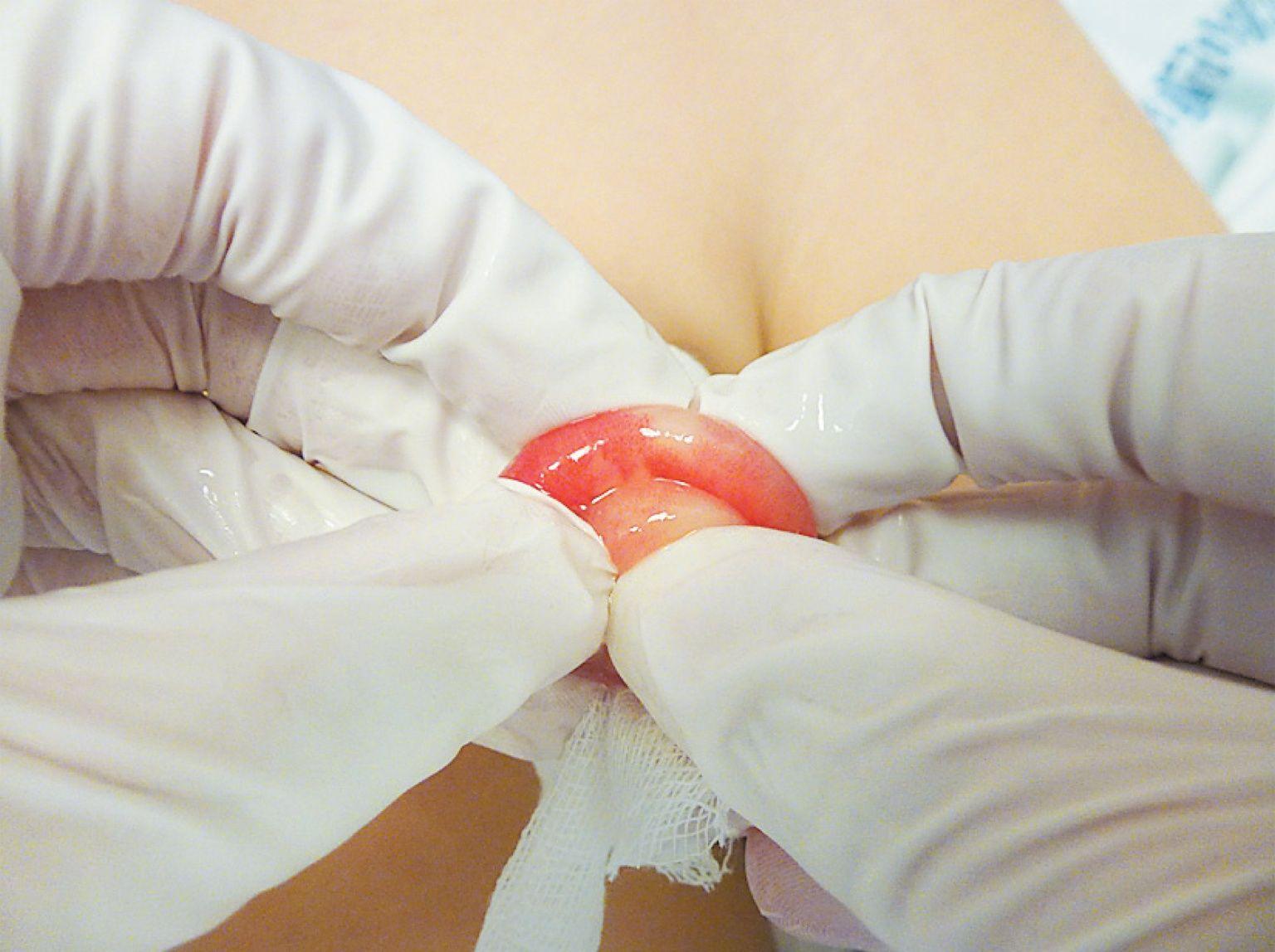
- Manual reduction: thumbs push glans proximally while fingers pull foreskin distally [6][28]
- If manual reduction fails: puncture technique (multiple small punctures in edematous prepuce to express fluid) [29]
- If all else fails: dorsal slit followed by elective circumcision [5-6]
The following figure from the NEJM demonstrates the manual reduction technique for paraphimosis:
17. Disposition
Discharge criteria (majority of cases)
- Uncomplicated physiological phimosis — discharge with reassurance and education [3]
- Phimosis started on topical steroids — outpatient follow-up in 4–8 weeks
- Successfully reduced paraphimosis — discharge with urology follow-up for elective circumcision
Admission/urgent referral criteria
- Irreducible paraphimosis requiring dorsal slit [5]
- Acute urinary retention
- Suspected penile malignancy → urgent urology/oncology referral [22]
- Severe BXO with meatal stenosis causing obstructive symptoms [11]
Urology consultation triggers
- Failed topical steroid therapy (after 1–2 courses)
- Pathological phimosis with scarring/BXO features
- Recurrent paraphimosis
- Recurrent UTIs in setting of phimosis
- Any suspicious penile lesion
18. Follow Up / Return Precautions
Follow-up timing
- Topical steroid therapy: reassess at 4–8 weeks for treatment response [1][4]
- Post-circumcision: 2–4 weeks for wound check
- BXO/LS: long-term surveillance recommended given premalignant potential [7-8]
Return precautions (counsel patients/parents)
- Foreskin stuck behind the glans and unable to be replaced → paraphimosis — seek immediate care [5]
- Increasing pain, swelling, or color change of the glans
- Inability to urinate or significant decrease in urinary stream
- Fever, purulent discharge, or spreading redness (infection)
- New penile lesion, ulcer, or mass
Patient counseling
- Physiological phimosis in young boys is normal and usually resolves without intervention [1][3]
- Avoid forceful retraction — this causes scarring and worsens phimosis [1]
- Gentle retraction during bathing for hygiene once foreskin becomes retractable [3][20]
- Expected recovery after circumcision: 2–4 weeks; avoid strenuous activity and sexual intercourse during healing
References
1. Topical Corticosteroids for Treating Phimosis in Boys. — Moreno G, Ramirez C, Corbalán J, et al. The Cochrane Database of Systematic Reviews. 2024.
2. Prevalence of Phimosis in Males of All Ages: Systematic Review. — Morris BJ, Matthews JG, Krieger JN. Urology. 2020.
3. Pathologic and Physiologic Phimosis: Approach to the Phimotic Foreskin. — McGregor TB, Pike JG, Leonard MP. Canadian Family Physician Medecin De Famille Canadien. 2007.
4. Efficacy of Topical Steroid Treatment in Children With Severe Phimosis in China: A Long-Term Single Centre Prospective Study. — Zhou G, Jiang M, Yang Z, Xu W, Li S. Journal of Paediatrics and Child Health. 2021.
5. Paraphimosis: Current Treatment Options. — Choe JM. American Family Physician. 2000.
6. Paraphimosis in Elderly Men. — Williams JC, Morrison PM, Richardson JR. The American Journal of Emergency Medicine. 1995.
7. Recent Advances in Understanding and Managing Lichen Sclerosus. — Kwok R, Shah TT, Minhas S. F1000Research. 2020.
8. Balanitis Xerotica Obliterans: A Review of Diagnosis and Management. — Charlton OA, Smith SD. International Journal of Dermatology. 2019.
9. Noninfectious Penile Lesions. — Teichman JMH, Mannas M, Elston DM. American Family Physician. 2018.
10. Malignant Neoplasms of the Penis With Radiologic and Pathologic Correlation. — Lubner MG, Marko J, Hu R, et al. Radiographics : A Review Publication of the Radiological Society of North America, Inc. 2023.
11. Balanitis Xerotica Obliterans: An Update for Clinicians. — Nguyen ATM, Holland AJA. European Journal of Pediatrics. 2020.
12. Topical Steroids Are Effective Even in Severe Phimosis: Evidence From a Multicenter Cohort. — Campos JM, Ceballos V, Torres AF, et al. Journal of Pediatric Surgery. 2026.
13. Predictive Power of Objectivation of Phimosis Grade on Outcomes of Topical 0.1% Betamethasone Treatment of Phimosis. — Kuehhas FE, Miernik A, Sevcenco S, et al. Urology. 2012.
14. Topical Triamcinolone for Persistent Phimosis. — Letendre J, Barrieras D, Franc-Guimond J, Abdo A, Houle AM. The Journal of Urology. 2009.
15. Topical Corticosteroids for Phimosis in Children: A Network Meta-Analysis of Randomized Clinical Trials. — Sridharan K, Sivaramakrishnan G. Pediatric Surgery International. 2021.
16. Sex-Related Variations in Comorbidities in Lichen Sclerosus: A Systematic Review and Meta-Analysis. — Šuler Baglama Š, Jemec GBE, Zmazek J, Trčko K. Acta Dermato-Venereologica. 2024.
17. Clinical Features, Complications and Autoimmunity in Male Lichen Sclerosus. — Kantere D, Alvergren G, Gillstedt M, Pujol-Calderon F, Tunbäck P. Acta Dermato-Venereologica. 2017.
18. Lichen Sclerosus and Phimosis - Discrepancies Between Clinical and Pathological Diagnosis and Its Consequences. — Czajkowski M, Żawrocki A, Czajkowska K, et al. Urology. 2021.
19. Incidence of Preputial Lichen Sclerosus in Adults: Histologic Study of Circumcision Specimens. — Aynaud O, Piron D, Casanova JM. Journal of the American Academy of Dermatology. 1999.
20. Foreskin Care: Hygiene, Importance of Counselling, and Management of Common Complications. — Leeson C, Vigil H, Witherspoon L. Canadian Family Physician Medecin De Famille Canadien. 2025.
21. European Association of Urology-American Society of Clinical Oncology Collaborative Guideline on Penile Cancer: 2023 Update. — Brouwer OR, Albersen M, Parnham A, et al. European Urology. 2023.
22. Penile Cancer. — Updated 2025-11-12. National Comprehensive Cancer Network.
23. Prepuce: Phimosis, Paraphimosis, and Circumcision. — Hayashi Y, Kojima Y, Mizuno K, Kohri K. TheScientificWorldJournal. 2011.
24. Foreskin Morbidity in Uncircumcised Males. — Sneppen I, Thorup J. Pediatrics. 2016.
25. Comparison of Outcomes for Pediatric Paraphimosis Reduction Using Topical Anesthetic Versus Intravenous Procedural Sedation. — Burstein B, Paquin R. The American Journal of Emergency Medicine. 2017.
26. Ultrasound-Guided Dorsal Penile Nerve Block for ED Paraphimosis Reduction. — Flores S, Herring AA. The American Journal of Emergency Medicine. 2015.
27. Treatment Options for Paraphimosis. — Little B, White M. International Journal of Clinical Practice. 2005.
28. Reduction of Paraphimosis in Boys. — Vunda A, Lacroix LE, Schneider F, Manzano S, Gervaix A. The New England Journal of Medicine. 2013.
29. Modified Puncture Technique for Reduction of Paraphymosis. — Kumar V, Javle P. Annals of the Royal College of Surgeons of England. 2001.