Straddle Fracture
A straddle fracture refers to bilateral fractures of the superior and inferior pubic rami (i.e., fractures of all four pubic rami), creating a free-floating anterior pelvic segment.[1-2] The term d…
A straddle fracture refers to bilateral fractures of the superior and inferior pubic rami (i.e., fractures of all four pubic rami), creating a free-floating anterior pelvic segment.[1-2] The term derives from the classic mechanism of falling astride a hard object (e.g., fence, beam, bicycle crossbar), though it also occurs in high-energy trauma such as motor vehicle collisions and falls from height.[3-4] Because the pelvis is a ring structure, an isolated anterior ring fracture almost always implies a concomitant posterior ring injury — CT studies show posterior pelvic ring lesions in up to 96.8% of patients with pubic rami fractures.[5-6]
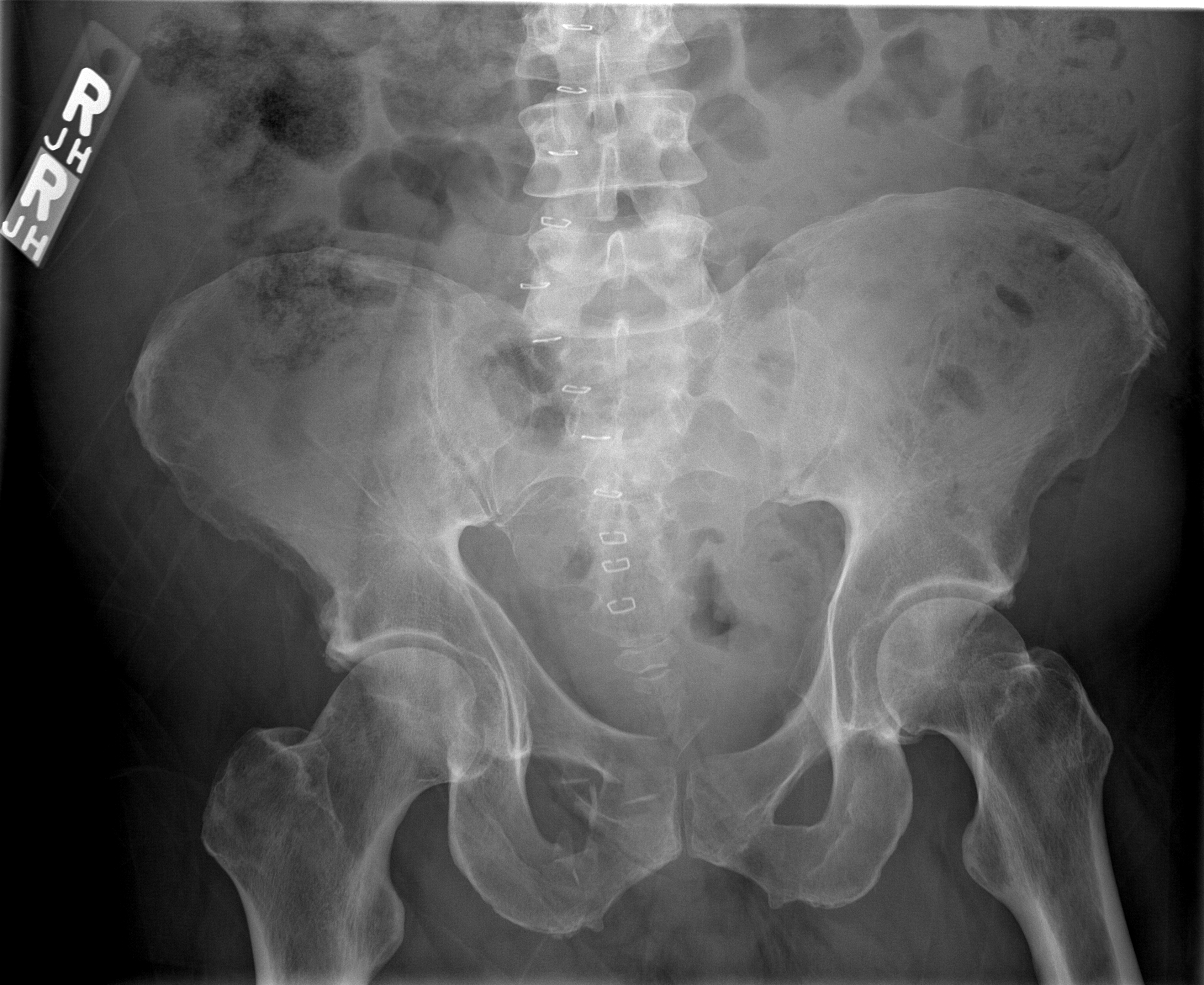
The following figure illustrates a comminuted pubic rami fracture with symphyseal offset, demonstrating the principle that displaced anterior ring fractures should prompt evaluation for a second disruption in the posterior ring:
1. History
- Mechanism: fall astride a hard object (classic "straddle" mechanism), MVC, motorcycle collision, pedestrian struck, fall from height, or crush injury[3]
- Pain location: groin, perineum, suprapubic region, low back, or hip
- Ability to ambulate or bear weight — inability to mobilize is a key concern for occult instability[8]
- Voiding symptoms: hematuria, inability to void, blood at urethral meatus (suggests urethral or bladder injury)[9]
- Timing: acute onset with trauma vs. insidious onset (consider insufficiency fracture in elderly/osteoporotic patients)[10]
- Anticoagulant use — increases hemorrhagic risk even in "stable" fractures[11]
2. Alarm Features
- Hemodynamic instability (SBP <90, HR >120): suggests significant pelvic hemorrhage — mortality up to 32% in unstable pelvic fractures[12]
- Blood at the urethral meatus, perineal butterfly hematoma, inability to void → suspect urethral injury[9]
- Gross hematuria → suspect bladder injury[9]
- Open fracture (perineal laceration, rectal/vaginal wound) — dramatically increases mortality and infection risk[13]
- Progressive abdominal distension or peritonitis → intraperitoneal bladder rupture or associated abdominal injury
- Neurologic deficits in lower extremities → lumbosacral plexus injury
- Delayed hemodynamic deterioration in elderly patients — corona mortis artery avulsion can present 48–72 hours after seemingly benign pubic rami fractures[11]
3. Medications
- Analgesics: Acetaminophen, NSAIDs (if no contraindication), opioids for acute pain management
- VTE prophylaxis: LMWH (enoxaparin 30 mg BID or 40 mg daily) is the most commonly used agent; aspirin (81 mg BID) shown noninferior to LMWH for prevention of death from any cause in the PREVENT CLOT trial. Initiate within 24 hours if hemodynamically stable[14-16]
- Anticoagulants to review: Patients on warfarin, DOACs, or antiplatelet agents are at increased hemorrhagic risk and may need reversal
- Avoid: Foley catheter insertion if urethral injury suspected — obtain retrograde urethrogram first[9]
4. Diet
- NPO if hemodynamically unstable or surgical intervention anticipated
- Adequate calcium and vitamin D supplementation in elderly/osteoporotic patients for long-term bone health[10]
- Ensure adequate hydration and nutrition to support fracture healing
- High-fiber diet or stool softeners to prevent straining during recovery
5. Review of Systems
- GU: Hematuria, dysuria, inability to void, blood at meatus, vaginal bleeding
- GI: Rectal bleeding (5% of urethral injuries have concomitant rectal injury), abdominal pain, distension[9]
- Neurologic: Lower extremity weakness, numbness, saddle anesthesia (lumbosacral plexus or cauda equina)
- Vascular: Signs of DVT (leg swelling, calf pain) — up to 34% develop proximal DVT[15]
- MSK: Low back pain (posterior ring injury), hip pain, inability to bear weight
6. Collateral History and Family History
- Witnesses to mechanism (speed, height of fall, position of impact)
- Pre-injury ambulatory status and functional baseline (especially in elderly)
- History of osteoporosis, prior fragility fractures, pelvic irradiation, rheumatoid arthritis, chronic corticosteroid use — all predispose to insufficiency fractures of the pubic rami[10][17]
- Anticoagulant/antiplatelet medication use
- Family history of osteoporosis or bleeding disorders
7. Risk Factors
- High-energy trauma: MVC, motorcycle collision, pedestrian struck, fall from height[3]
- Low-energy/insufficiency fractures: Osteoporosis, advanced age, female sex, rheumatoid arthritis, chronic corticosteroid use, pelvic irradiation, renal failure, post-hip surgery mechanical changes[10][17]
- Stress fractures: Long-distance runners, female athletes (pubic ramus stress fractures near symphysis)[18]
- Anticoagulation therapy increases hemorrhagic complications[11]
- Male sex increases risk of associated urethral injury (urethral injury in 6.1% of males vs. 0.5% of females with pelvic fractures)[19]
8. Differential Diagnosis
- Unstable pelvic ring disruption (APC-II/III, LC-II/III, vertical shear) — must rule out with CT[3][20]
- Acetabular fracture — found in 28.8% of patients with pubic rami fractures on CT[5]
- Hip fracture (femoral neck, intertrochanteric) — overlapping presentation with groin pain and inability to ambulate[21]
- Pubic symphysis diastasis ("open book" injury)
- Sacral fracture (often occult on plain films) — present in nearly all patients with pubic rami fractures[5-6]
- Insufficiency fracture of pubic rami in elderly — may mimic traumatic fracture[10]
- Pubic ramus stress fracture in athletes[18]
- Osteitis pubis or athletic pubalgia
9. Past Medical History
- Prior pelvic fractures or pelvic surgery
- Osteoporosis, osteopenia, metabolic bone disease
- Rheumatoid arthritis, chronic steroid use[17]
- Prior pelvic irradiation[10]
- History of DVT/PE
- Anticoagulant use
- Chronic kidney disease (affects bone quality)
10. Physical Exam
- Vitals: Tachycardia and hypotension suggest hemorrhage — pelvic fractures can cause massive retroperitoneal bleeding[12]
- Inspection: Perineal ecchymosis (butterfly hematoma), scrotal/labial swelling, blood at urethral meatus, open wounds[9]
- Palpation: Tenderness over pubic symphysis, pubic rami, sacroiliac joints; avoid repeated manual pelvic compression/distraction testing — it is neither sensitive nor specific and may worsen hemorrhage[22]
- Rectal/vaginal exam: Assess for open fracture, rectal tone, high-riding prostate (low sensitivity/specificity for urethral injury)[9]
- Neurovascular: Lower extremity motor/sensory exam, pedal pulses
- Functional: Ability to bear weight — inability to mobilize suggests occult instability in 42% of geriatric patients with seemingly isolated rami fractures[8]
11. Lab Studies
- CBC: Serial hemoglobin/hematocrit to monitor for hemorrhage
- Type and screen/crossmatch: Essential in unstable fractures; activate massive transfusion protocol if hemodynamically unstable[15][23]
- Coagulation studies: PT/INR, PTT (especially if on anticoagulants)
- BMP/CMP: Renal function, electrolytes
- Lactate: Marker of tissue hypoperfusion/occult hemorrhage
- Urinalysis: Hematuria screening — present in most bladder injuries[9]
- Anti-Factor Xa levels: If on LMWH, to ensure adequate prophylactic dosing (many pelvic fracture patients are underdosed)[16]
12. Imaging
- AP pelvis radiograph: First-line screening; identifies pubic rami fractures, symphyseal diastasis, and gross displacement. However, posterior ring injuries are frequently occult on plain films[5][20-21]
- CT pelvis with contrast: Gold standard for full characterization of pelvic ring injury, detection of posterior ring fractures (sacral fractures, SI joint disruption), acetabular fractures, and active hemorrhage (contrast extravasation)[15][24]
- Retrograde urethrogram (RUG): Indicated before Foley placement if blood at meatus or suspected urethral injury[9][25]
- CT cystogram: If bladder injury suspected (gross hematuria with pelvic fracture)[9]
- MRI: Useful for detecting occult posterior ring injuries (sacral insufficiency fractures) not visible on CT, particularly in geriatric patients who fail to mobilize[8]
- Bone scintigraphy: Historically used for occult fractures; largely replaced by MRI[10]
13. Special Tests
- Young-Burgess Classification: Categorizes pelvic ring injuries by mechanism — APC (I–III), LC (I–III), vertical shear, combined mechanism. Useful for predicting instability, transfusion needs, and guiding surgical decision-making[20][26]
- Tile/AO Classification: Type A (stable), Type B (rotationally unstable), Type C (rotationally and vertically unstable)[2]
- FAST/E-FAST: Rapid bedside assessment for intraabdominal free fluid and pubic symphyseal widening[15][27]
- Stress radiographs under fluoroscopy: Can identify dynamic instability in patients with seemingly isolated rami fractures who fail to mobilize[8]
- Diagnostic peritoneal aspiration (DPA): Alternative to FAST in hemodynamically unstable patients to rule out intraabdominal hemorrhage[15]
14. ECG
- Not specific to straddle fractures, but should be obtained in:
- Elderly patients (baseline cardiac assessment)
- Polytrauma patients
- Patients with hemodynamic instability to rule out cardiac causes
- Pre-operative assessment
- Monitor for signs of right heart strain (S1Q3T3, new RBBB) if PE suspected in the setting of VTE
15. Assessment
- A straddle fracture represents a bilateral anterior pelvic ring disruption that, by the ring principle, almost always involves a concomitant posterior injury.[5-6] Severity ranges widely:
- Low-energy (elderly/insufficiency): Often minimally displaced, may appear benign but carries significant morbidity — 16% 90-day mortality in geriatric patients with rami fractures and inability to mobilize. Occult posterior ring instability is present in ~42%[8]
- High-energy (trauma): Associated with life-threatening hemorrhage (mortality up to 32%), genitourinary injuries (4.5–7.7%), and polytrauma[12][19][28]
- Complications: hemorrhage, urethral/bladder injury, DVT/PE, infection (open fractures), chronic pain, sexual dysfunction, malunion/nonunion
16. Treatment Plan
Initial stabilization (ED)
- ATLS primary survey; apply pelvic binder at the level of the greater trochanters with legs internally rotated[15][22-23]
- If hemodynamically unstable: activate massive transfusion protocol (1:1:1 pRBC:FFP:platelets or whole blood), consider TXA[23]
- Establish urinary drainage — if no signs of urethral injury, place Foley; if urethral injury suspected, obtain RUG first → suprapubic catheter for complete PFUI[9][29]
Hemorrhage control (unstable patients)
- FAST/DPA to rule out intraabdominal hemorrhage[15]
- If intraabdominal hemorrhage → OR for laparotomy + consider external fixation
- If no intraabdominal source → pelvic external fixation + preperitoneal packing → angioembolization if still unstable[15]
- REBOA may be considered in extremis[12][23]
Definitive management
- Stable fractures (APC-I, LC-I): Conservative management with weight-bearing as tolerated, pain control, early mobilization, and physical therapy[2-3]
- Unstable fractures (APC-II/III, LC-II/III, VS): Surgical fixation — anterior plating of symphysis for diastasis >2.5 cm; retrograde transpubic screws or anterior plating for straddle fractures; posterior fixation as indicated[1][3]
- VTE prophylaxis: Initiate LMWH or aspirin as soon as hemodynamically stable, ideally within 24 hours[14-16]
17. Disposition
- Admit (ICU or monitored bed): Hemodynamic instability, need for transfusion, unstable fracture pattern, associated injuries (urethral, bladder, vascular), polytrauma, inability to mobilize, need for surgical fixation[30-31]
- Admit (floor): Stable straddle fracture with inability to bear weight, elderly patients requiring pain management and PT evaluation, patients on anticoagulation requiring monitoring
- Observation: Hemodynamically stable patients with minimally displaced fractures who need serial hemoglobin checks and functional assessment
- Discharge (rare for true straddle fractures): Only if minimally displaced, hemodynamically stable, able to ambulate with assistive device, adequate pain control, reliable follow-up, and no GU injury
- Consult triggers: Orthopedic surgery (all pelvic ring injuries), urology (blood at meatus, hematuria, voiding difficulty), interventional radiology (active hemorrhage on CT), trauma surgery (polytrauma, hemodynamic instability)[30]
18. Follow Up / Return Precautions
- Orthopedic follow-up: Within 1–2 weeks with repeat imaging to assess alignment and healing
- Urology follow-up: Essential for all patients with urethral or bladder injury — timing of definitive urethroplasty typically 3–6 months post-injury[32]
- VTE prophylaxis duration: Continue for up to 4 weeks post-injury for high-risk patients; consider extended prophylaxis with aspirin or LMWH post-discharge[33]
- Return precautions: Instruct patients to return immediately for increasing pain, inability to urinate, blood in urine, fever, leg swelling, shortness of breath, lightheadedness/syncope, or worsening inability to bear weight
- Expected recovery: Stable fractures typically heal in 6–12 weeks with conservative management; elderly patients may have prolonged recovery and increased 90-day mortality (16%). Stress fractures in athletes require cessation of running until symptom-free[8][18]
- Long-term considerations: Monitor for chronic pelvic pain, sexual dysfunction, urethral stricture (in PFUI patients), and post-traumatic arthritis
References
1. Is Anterior Plating Superior to the Bilateral Use of Retrograde Transpubic Screws for Treatment of Straddle Pelvic Ring Fractures? A Biomechanical Investigation. — Lodde MF, Katthagen JC, Schopper CO, et al. Journal of Clinical Medicine. 2021.
2. Acute Pelvic Fractures: I. Causation and Classification. — Tile M. The Journal of the American Academy of Orthopaedic Surgeons. 1996.
3. Pelvic Trauma: WSES Classification and Guidelines. — Coccolini F, Stahel PF, Montori G, et al. World Journal of Emergency Surgery : WJES. 2017.
4. Fractures of the Pelvis. — Eid AM. Postgraduate Medical Journal. 1983.
5. Detection of Posterior Pelvic Injuries in Fractures of the Pubic Rami. — Scheyerer MJ, Osterhoff G, Wehrle S, et al. Injury. 2012.
6. A Clinical and Experimental Investigation of Occult Injuries of the Pelvic Ring. — Chenoweth DR, Cruickshank B, Gertzbein SD, Goldfarb P, Janosick J. Injury. 1980.
7. Geriatric Patients Presenting With Isolated Pubic Rami Fractures and Inability to Mobilize May Have Occult Lateral Compression Pelvic Ring Injuries With Dynamic Instability. — Tucker NJ, Scott B, Mauffrey C, Parry JA. Journal of Orthopaedic Trauma. 2023.
8. Best Practices Guidelines Management of Gentiunrinary Injuries. — Niels Johnsen, Hunter Wessells, Krystal Archer-Arroyo, et al American College of Surgeons (2025). 2025.
9. Insufficiency Fractures of the Pubic Ramus. — Schapira D, Militeanu D, Israel O, Scharf Y. Seminars in Arthritis and Rheumatism. 1996.
10. Corona Mortis Artery Avulsion Due to a Stable Pubic Ramus Fracture. — Garrido-Gómez J, Pena-Rodríguez C, Martín-Noguerol T, Hernández-Cortes P. Orthopedics. 2012.
11. Association Between Hemorrhage Control Interventions and Mortality in US Trauma Patients With Hemodynamically Unstable Pelvic Fractures. — Anand T, El-Qawaqzeh K, Nelson A, et al. JAMA Surgery. 2023.
12. Pelvic Fractures: Part 1. Evaluation, Classification, and Resuscitation. — Langford JR, Burgess AR, Liporace FA, Haidukewych GJ. The Journal of the American Academy of Orthopaedic Surgeons. 2013.
13. Aspirin or Low-Molecular-Weight Heparin for Thromboprophylaxis after a Fracture. — Major Extremity Trauma Research Consortium (METRC), O'Toole RV, Stein DM, et al. The New England Journal of Medicine. 2023.
14. Best Practices In The Management Of Orthopaedic Trauma. — Matthew L. Davis MD FACS, Gregory J. Della Rocca MD PhD FACS, Megan Brenner MD MS RPVI FACS, et al American College of Surgeons (2015). 2015.
15. Chemoprophylaxis for Venous Thromboembolism in Pelvic and/or Acetabular Fractures: A Systematic Review. — Shu HT, Yu AT, Lim PK, Scolaro JA, Shafiq B. Injury. 2022.
16. Stress Fractures of the Pubic Rami in Rheumatoid Arthritis. — Isdale AH. Annals of the Rheumatic Diseases. 1993.
17. Stress Fractures of the Pubic Ramus. A Report of Twelve Cases. — Pavlov H, Nelson TL, Warren RF, Torg JS, Burstein AH. The Journal of Bone and Joint Surgery. American Volume. 1982.
18. Fracture Types Influence the Likelihood of Lower Urinary Tract Injuries in Patients With Pelvic Fractures. — Zhao X, Lu S, Wang B, et al. Journal of Clinical Medicine. 2023.
19. Pelvic Ring Fractures: What the Orthopedic Surgeon Wants to Know. — Khurana B, Sheehan SE, Sodickson AD, Weaver MJ. Radiographics : A Review Publication of the Radiological Society of North America, Inc. 2014.
20. ACR Appropriateness Criteria Acute Hip Pain-Suspected Fracture. — Expert Panel on Musculoskeletal Imaging:, Ross AB, Lee KS, et al.' Journal of the American College of Radiology : JACR. 2019.
21. Prehospital Trauma Compendium: Evaluation and Management of Suspected Pelvis Fractures - An NAEMSP Position Statement and Resource Document. — Lyng JW, Corsa JG, Raetzke BD, et al. Prehospital Emergency Care. 2025.
22. Novel Resuscitation Strategies in Patients With a Pelvic Fracture. — Copp J, Eastman JG. Injury. 2021.
23. Pelvic Fractures and Associated Genitourinary and Vascular Injuries: A Multisystem Review of Pelvic Trauma. — Lee MJ, Wright A, Cline M, et al. AJR. American Journal of Roentgenology. 2019.
24. Urethral Injuries: Diagnostic and Management Strategies for Critical Care and Trauma Clinicians. — Patel AB, Osterberg EC, Satarasinghe PN, et al. Journal of Clinical Medicine. 2023.
25. Young-Burgess Classification of Pelvic Ring Fractures: Does It Predict Mortality, Transfusion Requirements, and Non-Orthopaedic Injuries?. — Manson T, O'Toole RV, Whitney A, et al. Journal of Orthopaedic Trauma. 2010.
26. Appropriateness of Initial Course of Action in the Management of Blunt Trauma Based on a Diagnostic Workup Including an Extended Ultrasonography Scan. — Planquart F, Marcaggi E, Blondonnet R, et al. JAMA Network Open. 2022.
27. Incidence of genitourinary injuries in pelvic fractures: A 12‐year single‐center retrospective study. — Kaneko T, Yanagida K, Matsui K, et al. Neurourology and Urodynamics. 2022.
28. Urotrauma Guideline 2020: AUA Guideline. — Morey AF, Broghammer JA, Hollowell CMP, McKibben MJ, Souter L. The Journal of Urology. 2021.
29. Team Approach: Evaluation and Management of Pelvic Ring Injuries. — Kazley JM, Potenza MA, Marthy AG, et al. JBJS Reviews. 2020.
30. Transferrals and Clinical Pathways of Unstable Pelvic Fractures Over the Last 10 Years - A Retrospective Analysis of the Trauma Register DGU®. — Weuster M, Pfeifer R, Seekamp A, et al. European Journal of Trauma and Emergency Surgery : Official Publication of the European Trauma Society. 2026.
31. Urethral Stricture Disease Guideline Amendment (2023). — Wessells H, Morey A, Souter L, Rahimi L, Vanni A. The Journal of Urology. 2023.
32. Updated Guidelines to Reduce Venous Thromboembolism in Trauma Patients: A Western Trauma Association Critical Decisions Algorithm. — Ley EJ, Brown CVR, Moore EE, et al. The Journal of Trauma and Acute Care Surgery. 2020.