Acalculous Cholecystitis
Acute acalculous cholecystitis (AAC) is acute inflammation of the gallbladder without gallstones, accounting for 5–10% of all acute cholecystitis cases. [1] It predominantly affects critically ill patients and carries a historically high mortality (~30%), though early diagnosis and intervention significantly improve outcomes. [2-3] The pathogenesis centers on gallbladder ischemia and bile stasis rather than stone obstruction, with nonobstructive mechanisms (ischemia-reperfusion injury, eicosanoid-mediated inflammation) being the central drivers. [3-4]
History
- RUQ pain: Onset, radiation (to right shoulder/scapula), relationship to meals, severity, and progression
- Fever, nausea, vomiting: Classic triad alongside RUQ pain [1]
- ICU context: Duration of critical illness, recent surgery/trauma, prolonged fasting, TPN use, mechanical ventilation, vasopressor requirement [2][5]
- Outpatient context: Recent viral illness (especially in children — EBV, hepatitis A), autoimmune/rheumatic disease [6-7]
- Important negatives: Absence of prior gallstone history, no biliary colic episodes, no prior cholecystectomy
- Pearl: In sedated/intubated ICU patients, AAC often presents as unexplained sepsis, rising WBC, or new organ dysfunction rather than classic biliary symptoms [4][8]
Alarm Features
- Unexplained sepsis or septic shock in a critically ill patient — must consider AAC [4]
- Peritoneal signs (guarding, rigidity) — suggests perforation or gangrenous cholecystitis
- Rapid clinical deterioration despite broad-spectrum antibiotics
- Gangrenous cholecystitis occurs at a significantly higher rate in AAC vs. calculous cholecystitis (31.2% vs. 5.6%) [9]
- Gallbladder perforation risk is high if diagnosis is delayed [8]
- Myocardial infarction/CHF is the only independent risk factor for in-hospital death in AAC [10]
Medications
- Contributors: Opioids (promote bile stasis), vasopressors (splanchnic ischemia), TPN [3]
- Empiric antibiotics:
- Contraindicated: Avoid hepatotoxic agents; aminoglycoside courses should be brief due to increased nephrotoxicity risk during cholestasis [12]
Diet
- NPO during acute illness and pending surgical decision
- Avoid TPN if possible — TPN is a recognized risk factor for AAC; transition to enteral feeding as soon as clinically feasible [3][5]
- Long-term: Low-fat diet if gallbladder preserved; standard diet post-cholecystectomy
- Adequate hydration to support hemodynamics and prevent further splanchnic ischemia
Review of Systems
- GI: Nausea, vomiting, anorexia, abdominal distension, jaundice, change in stool color
- Constitutional: Fever, chills, rigors, malaise
- Cardiovascular: Hypotension, tachycardia (signs of sepsis/shock)
- Respiratory: Dyspnea, pleuritic right-sided chest pain (referred pain or reactive effusion)
- Neurologic: Altered mental status (may be only sign in ICU patients)
- Infectious: Recent viral illness, HIV status, immunosuppression
Collateral History and Family History
- Collateral: ICU nursing observations (abdominal distension, feeding intolerance, rising vasopressor requirements), medication administration record (opioids, TPN duration)
- Family history: Generally not contributory; no strong hereditary component
- Social context: Immunosuppression (HIV/AIDS — AAC associated with CMV, Cryptosporidium cholangiopathy), alcohol use, IV drug use
Risk Factors
- Critical illness: Trauma, major surgery, burns, sepsis, shock, cardiac arrest [2-3]
- Prolonged ICU stay: Mechanical ventilation, vasopressor use, TPN, prolonged fasting [5]
- Comorbidities: Diabetes mellitus, CHF, atherosclerotic disease, cerebrovascular accident, malignancy, abdominal vasculitis, cholesterol embolization [3][9]
- Age: Risk increases significantly in patients >50 years (92.8% of AAC patients in one series) [9]
- Immunosuppression: HIV/AIDS, bone marrow transplant, chemotherapy [7]
- Pediatric: Viral infections (EBV, hepatitis A, enteroviruses) are the most common trigger [7]
Differential Diagnosis
- Acute calculous cholecystitis — most common mimic; distinguished by presence of gallstones on imaging
- Ascending cholangitis — Charcot's triad (fever, jaundice, RUQ pain); CBD dilation on imaging
- Hepatic abscess — fever, RUQ pain; CT with rim-enhancing collection
- Acute pancreatitis — epigastric pain radiating to back, elevated lipase
- Peptic ulcer disease/perforation — epigastric pain, free air on imaging
- Right lower lobe pneumonia — referred RUQ pain, cough, CXR findings
- Fitz-Hugh-Curtis syndrome — RUQ pain in young women, perihepatitis
- Mesenteric ischemia — pain out of proportion to exam in vascular patients; lactate elevation
- Appendicitis (high-riding cecum) — can mimic RUQ pathology
- Pearl: In ICU patients, the differential for unexplained sepsis is broad — AAC should be considered when other sources are excluded [4]
Past Medical History
- Prior episodes of cholecystitis or biliary disease
- Recent surgery, trauma, or ICU admission
- Chronic illnesses: DM, CHF, CKD, CVA, malignancy, HIV/AIDS [3][9]
- Vascular disease (atherosclerosis, vasculitis)
- Prior cholecystectomy (rules out diagnosis)
- Immunosuppressive medications or transplant history
Physical Exam
- Vital signs: Fever, tachycardia, hypotension (sepsis)
- RUQ tenderness with or without guarding
- Murphy's sign: Positive (inspiratory arrest with RUQ palpation) — but often difficult to elicit in sedated/intubated patients [8][13]
- Abdominal distension, decreased bowel sounds
- Jaundice: Present in minority; suggests concomitant biliary obstruction or severe inflammation
- Peritoneal signs: Rebound, rigidity — suggest perforation or gangrenous cholecystitis
- Pearl: In ICU patients, physical exam is often unreliable; maintain a high index of suspicion based on clinical trajectory [2]
Lab Studies
- CBC: Leukocytosis with left shift (nonspecific but expected)
- CMP/LFTs: Mild transaminase elevation (cytolysis), possible mild bilirubin elevation; cholestasis pattern less common [5]
- Lipase: To rule out pancreatitis
- Lactate: Elevated in sepsis/ischemia
- Blood cultures: Before initiating antibiotics
- CRP/Procalcitonin: Inflammatory markers to trend
- Coagulation studies: Pre-procedural assessment
- NLR (neutrophil-to-lymphocyte ratio): Elevated NLR and lower lymphocyte counts correlate with higher severity (Grade III) [14]
- Pearl: Lab findings are nonspecific — the diagnosis rests on the combination of clinical suspicion + imaging [2]
Imaging
- First-line: Right upper quadrant ultrasound [1][13]
- Gallbladder wall thickening >3 mm (some reports up to 3.5–9 mm)
- Gallbladder distension (short-axis diameter >40 mm)
- Pericholecystic fluid
- Sludge without stones
- Sonographic Murphy's sign (limited utility in sedated patients)
- Sensitivity ~81%, specificity ~83% for acute cholecystitis [1]
- CT abdomen: Comparable accuracy; useful when ultrasound is equivocal or to evaluate complications (perforation, abscess) [4]
- Gold standard: Hepatobiliary iminodiacetic acid (HIDA) scan — non-filling of gallbladder at 4 hours; accuracy up to 95%. However, may not be practical in critically ill patients and can yield false positives in ICU patients on TPN or fasting [1-2][13]
- When imaging is unnecessary: If clinical picture is clear and patient is going to OR
- Pearl: Many sonographic features of AAC (wall thickening, distension, sludge) may be routinely present in ICU patients without cholecystitis — serial imaging and clinical correlation are essential [13]
Special Tests
- Tokyo Guidelines (TG18) Severity Grading: [10][15]
- Grade I (mild): No organ dysfunction, mild inflammatory changes
- Grade II (moderate): Elevated WBC >18,000, palpable RUQ mass, symptom duration >72 hours, marked local inflammation
- Grade III (severe): Organ dysfunction (cardiovascular, neurologic, respiratory, renal, hepatic, hematologic)
- Charlson Comorbidity Index (CCI): CCI ≥4 identifies poor surgical candidates [15]
- ASA Physical Status: ASA ≥3 suggests high perioperative risk [15]
- Point-of-care ultrasound (POCUS): Can provide additional bedside information, though the Society of Critical Care Medicine suggests intensivists should not perform the definitive exam for cholecystitis diagnosis [13]
- Bile culture: Obtained at time of cholecystostomy or cholecystectomy to guide antibiotic therapy
ECG
- Indications: All patients with suspected AAC, particularly given the association with MI/CHF as a risk factor and cause of death [10]
- Evaluate for acute coronary syndrome as a mimic or precipitant of AAC
- ST changes, arrhythmias, or signs of right heart strain may be present in critically ill patients
- Pearl: MI/CHF was the only independent risk factor for in-hospital death in a large AAC series [10]
Assessment
AAC is a life-threatening condition with a high risk of gangrenous cholecystitis (31% vs. 6% in calculous disease) and perforation if untreated. [8-9] It represents a systemic disease manifestation rather than a local gallbladder process. [16] Two distinct populations are affected:
- ICU patients: Critically ill with ischemia/stasis-driven pathology; subtle presentation; high mortality
- Outpatient/community patients: Often viral or autoimmune etiology; may have a more benign course amenable to conservative management [6]
Severity stratification per Tokyo Guidelines helps guide management decisions. [10][15] Overall in-hospital mortality is approximately 2.5% in contemporary series, though historically reported as high as 30% in critically ill cohorts. [3][10]
Treatment Plan
Initial stabilization
- IV fluid resuscitation, electrolyte correction
- NPO status
- Broad-spectrum IV antibiotics (see Medications section above) [11]
- Analgesics (avoid opioids if possible given bile stasis risk)
Definitive treatment
- Laparoscopic cholecystectomy is the gold standard, ideally within 1–3 days of diagnosis [1][10]
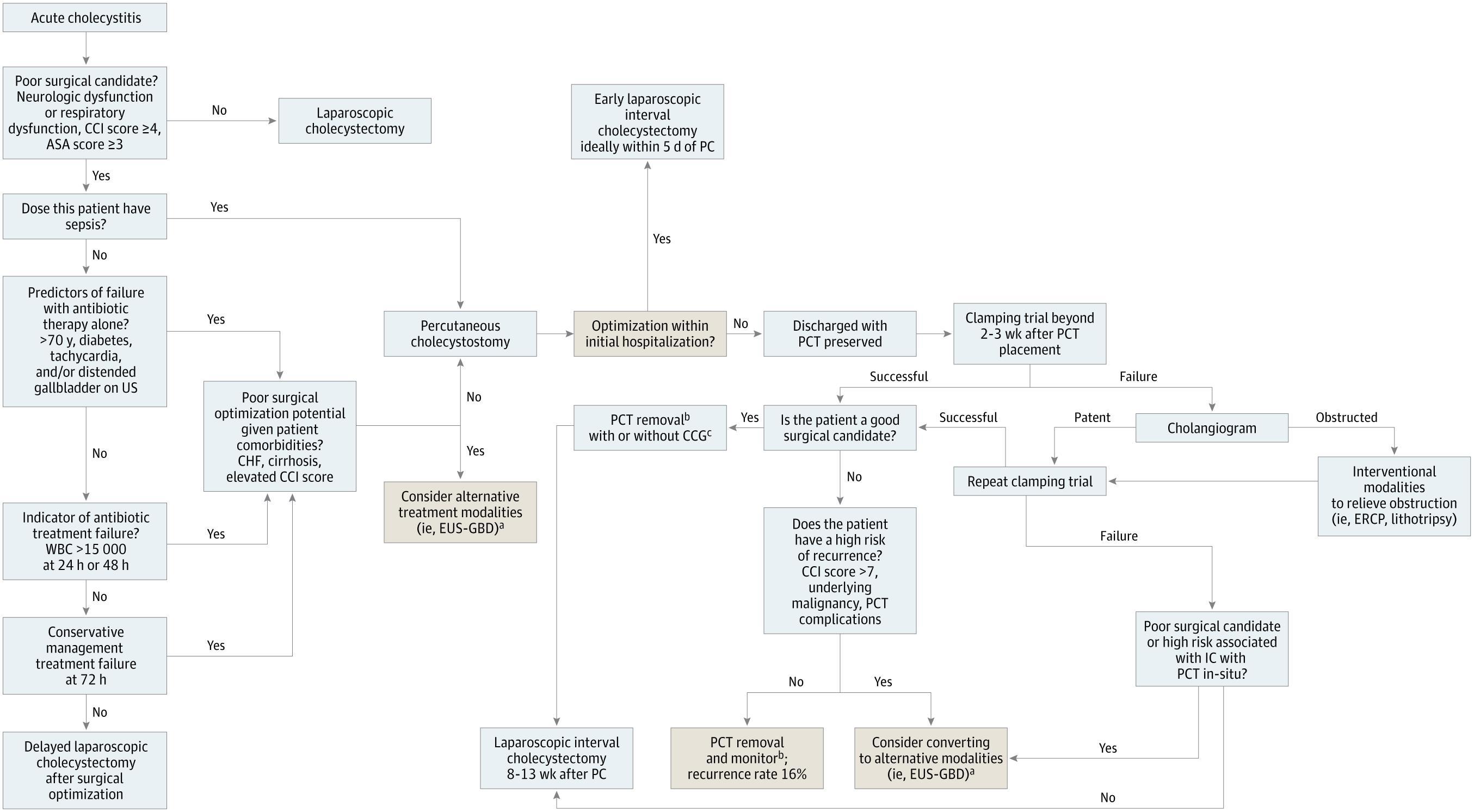
- Percutaneous cholecystostomy (PC): Reserved for patients who are severely ill and poor surgical candidates (ASA ≥3, CCI ≥4, septic shock) [1][15]
- Controls disease in ~85% of patients [3]
- Higher complication rate than cholecystectomy (65% vs. 12% in one RCT) [1]
- Can serve as bridge to interval cholecystectomy (8–13 weeks) or as definitive therapy if patient remains a non-surgical candidate [15]
- Interval cholecystectomy may not be necessary in AAC survivors if stones are absent and precipitating condition resolves [3][17]
- EUS-guided gallbladder drainage: Emerging option for non-surgical candidates; avoids external drain complications [8]
- Conservative management (antibiotics alone): May be appropriate for viral or rheumatic disease-associated AAC; recurrence rate ~9.8% over long-term follow-up [6][18]
The following figure from a JAMA Surgery systematic review illustrates a proposed algorithm for managing acute cholecystitis, including percutaneous cholecystostomy indications and management:
Disposition
- Admit all patients with confirmed or suspected AAC — this is not an outpatient diagnosis
- ICU admission: Sepsis, hemodynamic instability, organ dysfunction (TG18 Grade III), need for vasopressors [5][10]
- Surgical floor: Hemodynamically stable patients awaiting cholecystectomy
- Surgical consultation: Mandatory in all cases [1]
- Interventional radiology consultation: If percutaneous cholecystostomy is indicated
- GI consultation: Consider for EUS-guided drainage in non-surgical candidates [8]
- Discharge criteria: Post-cholecystectomy — tolerating diet, pain controlled, afebrile, no signs of complications
Follow Up / Return Precautions
- Post-cholecystectomy: Surgical follow-up in 2–4 weeks; wound check, pathology review
- Post-cholecystostomy tube: Close follow-up for tube management; interval cholecystectomy typically at 8–13 weeks if patient becomes a surgical candidate; clamping trial preferred over cholangiogram for tube removal decisions [15]
- Conservatively managed patients: Follow-up imaging in 4–6 weeks; recurrence rate ~9.8% over ~5.7 years; low threshold for re-evaluation [18]
- Return precautions: Fever, worsening abdominal pain, jaundice, nausea/vomiting, inability to tolerate PO, signs of wound infection
- Expected course: If treated early, rapid clinical improvement expected; delayed diagnosis carries risk of gangrenous cholecystitis, perforation, and multiorgan failure [5][8]
- Pearl: Unlike calculous cholecystitis, interval cholecystectomy may not be necessary in AAC patients treated with cholecystostomy if the precipitating condition resolves and stones remain absent [3][17][19]
References
1. Acute Cholecystitis: A Review. — Gallaher JR, Charles A. The Journal of the American Medical Association. 2022.
2. Acute Acalculous Cholecystitis: A Review. — Huffman JL, Schenker S. Clinical Gastroenterology and Hepatology : The Official Clinical Practice Journal of the American Gastroenterological Association. 2010.
3. Acute Acalculous Cholecystitis. — Barie PS, Eachempati SR. Current Gastroenterology Reports. 2003.
4. Acalculous Cholecystitis in the Critically Ill: Evolving Insights Into Diagnosis and Management. — Munir MM, Khan S, Huerta S. Current Opinion in Critical Care. 2026.
5. Acute Acalculous Cholecystitis in Hospitalized Patients in Intensive Care Unit: Study of 5 Cases. — Mossaab G, Ben Khlifa M, Karim N, et al. Heliyon. 2022.
6. Is Conservative Management a Safe Approach for Patients With Acute Acalculous Cholecystitis Presenting With an Acute Abdomen?. — Chang C, Wang Y, Shi W, et al. Medicine. 2023.
7. Acute Acalculous Cholecystitis in Children. — Poddighe D, Sazonov V. World Journal of Gastroenterology. 2018.
8. Recent Advances in Management of Acalculous Cholecystitis. — Balmadrid B. F1000Research. 2018.
9. Risk Factors and Therapeutic Outcomes of Acute Acalculous Cholecystitis. — Gu MG, Kim TN, Song J, et al. Digestion. 2014.
10. Current Status and Therapeutic Strategy of Acute Acalculous Cholecystitis: Japanese Nationwide Survey in the Era of the Tokyo Guidelines. — Morikawa T, Akada M, Shimizu K, et al. Journal of Hepato-Biliary-Pancreatic Sciences. 2024.
11. Clinical Update on Acute Cholecystitis and Biliary Pancreatitis: Between Certainties and Grey Areas. — Fugazzola P, Podda M, Tian BW, et al. EClinicalMedicine. 2024.
12. Biliary Tract Infections: A Guide to Drug Treatment. — Westphal JF, Brogard JM. Drugs. 1999.
13. Guidelines for the Appropriate Use of Bedside General and Cardiac Ultrasonography in the Evaluation of Critically Ill Patients-Part I: General Ultrasonography. — Frankel HL, Kirkpatrick AW, Elbarbary M, et al. Critical Care Medicine. 2015.
14. Clinical Outcomes of Percutaneous Cholecystostomy in Patients With Acute Cholecystitis Classified According to the Tokyo Guidelines, 2018: A Single-Center, Retrospective, Observational Study. — Kahraman YS, Yildiz YA, Taşkent İ, Gün F, Şentürk A. C Medicine. 2026.
15. Indications for and Optimal Management of Percutaneous Cholecystectomy Drainage. — Sadaka AH, Tseng JF, Itani KMF. JAMA Surgery. 2025.
16. Advances in the Study of Acute Acalculous Cholecystitis: A Comprehensive Review. — Fu Y, Pang L, Dai W, Wu S, Kong J. Digestive Diseases. 2021.
17. Acalculous Cholecystitis: Is an Elective Interval Cholecystectomy Necessary. — Abbas SH, Ghazanfar MA, Gordon-Weeks AN, et al. Digestive Surgery. 2017.
18. Long-Term Outcomes of Acute Acalculous Cholecystitis Treated by Non-Surgical Management. — Kim SB, Gu MG, Kim KH, Kim TN. Medicine. 2020.
19. Outcomes Following Percutaneous Cholecystostomy Tube Placement for Acalculous Versus Calculous Cholecystitis. — Chen SY, Huang R, Kallini J, et al. World Journal of Surgery. 2022.
Figure
More from Deep Dive
Reviewed by Dr. Lucas Mastropaolo. SymptomDx is an educational tool for medical professionals — it does not replace clinical judgment. Verify all clinical data and drug dosages with authoritative sources.