Acute Kidney Injury (ATN)
Acute tubular necrosis is the most common cause of intrinsic AKI, resulting from ischemic or nephrotoxic injury to renal tubular epithelial cells. [1-2] It is a clinical-pathologic diagnosis often made presumptively based on history, urine studies, and exclusion of prerenal/postrenal causes. [2] The following is a comprehensive clinical summary organized for emergency medicine and primary care workflows.
History
- Key HPI questions: Onset and duration of decreased urine output; recent hypotensive episodes, surgeries, or ICU stays; exposure to nephrotoxic drugs or contrast agents; symptoms of volume depletion (vomiting, diarrhea, hemorrhage); dark or cola-colored urine (rhabdomyolysis/hemolysis) [1][3]
- Symptom characterization: Oliguria or anuria is the hallmark; may be nonoliguric in ~30–40% of cases (especially nephrotoxic ATN). Patients may report fatigue, nausea, edema, or dyspnea from volume overload [4-5]
- Timing: Ischemic ATN typically develops 24–72 hours after the insult; aminoglycoside-related ATN may appear 5–10 days into therapy; contrast-induced ATN peaks at 48–72 hours [5-6]
- Important negatives: Absence of obstructive symptoms (hesitancy, retention), absence of rash/fever/eosinophilia (argues against AIN), absence of hematuria/proteinuria (argues against glomerulonephritis) [3][5]
Alarm Features
- Anuria or oliguria (<0.3 mL/kg/h for ≥24 hours) [7-8]
- Refractory hyperkalemia with ECG changes (peaked T waves, widened QRS, sine wave) [9-10]
- Severe metabolic acidosis (pH <7.20, bicarb <12) unresponsive to medical therapy [9]
- Pulmonary edema/volume overload refractory to diuretics [7][9]
- Uremic symptoms: encephalopathy, pericarditis, bleeding diathesis [9]
- Unstable vital signs or signs of ongoing shock/sepsis [7]
Medications
- Common nephrotoxic causes of ATN:
- Medications to discontinue immediately: NSAIDs, ACE inhibitors/ARBs, diuretics, aminoglycosides, and any other nephrotoxins [6][13][16]
- Dose-adjust renally cleared drugs: antibiotics, anticoagulants, opioids, digoxin [6]
- Treatments for complications:
Diet
- Acute phase: Restrict potassium intake (<2 g/day), restrict sodium (<2 g/day), limit phosphorus [10]
- Fluid management: In oliguric patients, restrict daily intake to ~400 mL + previous day's urine output unless volume-depleted [10]
- Protein: Adequate but not excessive protein intake (0.8–1.0 g/kg/day in non-dialysis patients; higher if on CRRT) to avoid worsening azotemia [17]
- Long-term: Nutritional consultation recommended for critically ill patients; enteral nutrition preferred over parenteral [18]
Review of Systems
- Cardiovascular: Chest pain (uremic pericarditis), dyspnea, orthopnea, edema (volume overload)
- Neurologic: Confusion, asterixis, seizures (uremic encephalopathy)
- GI: Nausea, vomiting, anorexia (uremia), diarrhea or GI bleeding (precipitant or complication)
- Musculoskeletal: Muscle pain, weakness, dark urine (rhabdomyolysis as cause)
- Hematologic: Easy bruising, bleeding (uremic platelet dysfunction)
- Infectious: Fever, chills (sepsis as precipitant — sepsis causes 30–70% of ATN-related deaths) [18]
Collateral History and Family History
- Collateral: Medication reconciliation (OTC NSAIDs, herbal supplements), recent procedures/surgeries, contrast exposure, baseline creatinine from prior records, recent hospitalizations
- Family history: Generally not a major contributor to ATN; however, family history of CKD or polycystic kidney disease may affect baseline renal reserve
- Social context: Occupational heat exposure, substance use (cocaine → rhabdomyolysis), alcohol use (hepatorenal considerations), access to follow-up care
Risk Factors
- Pre-existing CKD (strongest risk factor for non-recovery) [19-20]
- Advanced age [21]
- Sepsis/septic shock (most common cause of ATN in ICU — 47% of severe AKI cases) [2][22]
- Major surgery (especially cardiac, vascular) [22]
- Hypovolemia/hemorrhage [2]
- Heart failure/cardiogenic shock [16]
- Cirrhosis [13]
- Diabetes mellitus [3]
- Multiple nephrotoxin exposure (polypharmacy) — 22% of inpatient AKI events meet nephrotoxic AKI criteria [15]
- Rhabdomyolysis (crush injury, statins, seizures, immobilization) [23-24]
Differential Diagnosis
- Prerenal azotemia (most common AKI cause overall) — responds to volume resuscitation within 24–48 hours; FENa <1%, urine osmolality >500 mOsm/kg [2][5]
- Acute interstitial nephritis (AIN) — fever, rash, eosinophilia; WBC casts on urine microscopy; drug-related (beta-lactams, PPIs, checkpoint inhibitors) [5]
- Postrenal obstruction — bilateral hydronephrosis on ultrasound; BPH, malignancy, nephrolithiasis [2-3]
- Glomerulonephritis — RBC casts, dysmorphic RBCs, heavy proteinuria; FENa <1% [5][25]
- Hepatorenal syndrome (HRS-AKI) — in cirrhosis; FENa <0.1%, bland sediment; responds to vasoconstrictors + albumin [13][26]
- Thrombotic microangiopathy (TMA/HUS/TTP) — schistocytes, thrombocytopenia, elevated LDH [5]
- Atheroembolic disease — post-procedure, livedo reticularis, eosinophilia, blue toes [5]
Past Medical History
- Prior AKI episodes (recurrent AKI within 12 months occurs in ~25% of patients) [20]
- Baseline CKD stage (affects recovery likelihood and management thresholds)
- Diabetes, hypertension, heart failure, liver disease
- Prior kidney transplant
- History of nephrolithiasis or urologic surgery (postrenal risk)
- Chronic NSAID or nephrotoxin use
Physical Exam
- Vital signs: Hypotension (ischemic cause), tachycardia (hypovolemia), hypertension with pulmonary edema (volume overload), fever (sepsis/infection) [7]
- Volume status assessment (critical first step):
- Skin: Livedo reticularis (atheroemboli), maculopapular rash (AIN), track marks (rhabdomyolysis from drug use)
- Abdomen: Palpable bladder (obstruction), ascites (cirrhosis), flank tenderness
- Cardiac: Pericardial friction rub (uremic pericarditis), murmurs
- Neurologic: Asterixis, altered mental status (uremic encephalopathy)
Lab Studies
The FENa calculator can help differentiate prerenal from intrinsic AKI:
Imaging
- First-line: Renal ultrasound — assess kidney size (normal or enlarged in ATN), echogenicity, and rule out hydronephrosis (postrenal obstruction) [3][16]
- Normal-sized kidneys with increased cortical echogenicity is typical of ATN
- Small kidneys suggest underlying CKD rather than acute process
- CT abdomen/pelvis without contrast if obstruction suspected but ultrasound equivocal
- Imaging is unnecessary when the clinical picture clearly points to ATN (e.g., post-septic shock with muddy brown casts and FENa >2%) and there are no risk factors for obstruction
Special Tests
- Urine microscopy scoring: Muddy brown granular casts and renal tubular epithelial cells — a urine sediment score ≥3 has high specificity for ATN [5][18]
- KDIGO staging for AKI severity (Stages 1–3 based on creatinine rise and urine output) [8][16]
- Novel biomarkers (limited clinical availability): NGAL (predicts severe AKI; may distinguish ATN from HRS in cirrhosis), TIMP-2 × IGFBP-7 (FDA-approved for AKI prediction), CCL14 (persistence of severe AKI) [20]
- Renal biopsy: Reserved for unclear etiology, suspected glomerulonephritis, AIN not responding to drug withdrawal, or prolonged AKI without recovery [2][5]
ECG
- Indications: Obtain in all patients with AKI and hyperkalemia or suspected uremic pericarditis
- Hyperkalemia progression:
- Uremic pericarditis: Diffuse ST elevation, PR depression (though often absent in uremic pericarditis)
- Any ECG changes from hyperkalemia = emergent treatment required (IV calcium gluconate first) [10]
Assessment
- ATN is a clinical diagnosis made by identifying a precipitating ischemic or nephrotoxic insult, excluding prerenal and postrenal causes, and finding characteristic urine findings (muddy brown casts, FENa >1%, urine osmolality <350) [5][18]
- Severity stratification per KDIGO: Stage 1 (Cr ↑ ≥0.3 mg/dL or 1.5×), Stage 2 (Cr ↑ 2–2.9×), Stage 3 (Cr ↑ ≥3× or ≥4 mg/dL or need for RRT) [8][16]
- Oliguric ATN carries worse prognosis than nonoliguric ATN and is more likely to require dialysis [16]
- Complications: Hyperkalemia, metabolic acidosis, volume overload, uremia, infection (sepsis causes 30–70% of ATN deaths), CKD progression [4][10][18]
- Prognosis: >85% of patients with ATN on previously normal kidneys recover sufficient renal function within 6–12 months; in-hospital mortality remains ~50% in severe cases requiring RRT, largely driven by the underlying illness [19][21][30]
Treatment Plan
Initial stabilization
- Assess and optimize volume status — isotonic crystalloid for hypovolemia; diuretics for overload [3][16]
- Discontinue all nephrotoxins (NSAIDs, aminoglycosides, contrast, ACEi/ARBs) [6][13][16]
- Treat the underlying cause (antibiotics for sepsis, fluids for rhabdomyolysis, relieve obstruction) [3][23]
Supportive management
- Maintain euvolemia; avoid both hypovolemia and fluid overload [16-17]
- Correct electrolyte abnormalities (hyperkalemia, hyperphosphatemia, acidosis) [10]
- Adjust all medication doses for renal function [6]
- Avoid unnecessary IV lines, catheters, and ventilators to reduce infection risk [18]
- Nutritional support: enteral preferred; adequate calories with appropriate protein [17-18]
Renal replacement therapy (RRT)
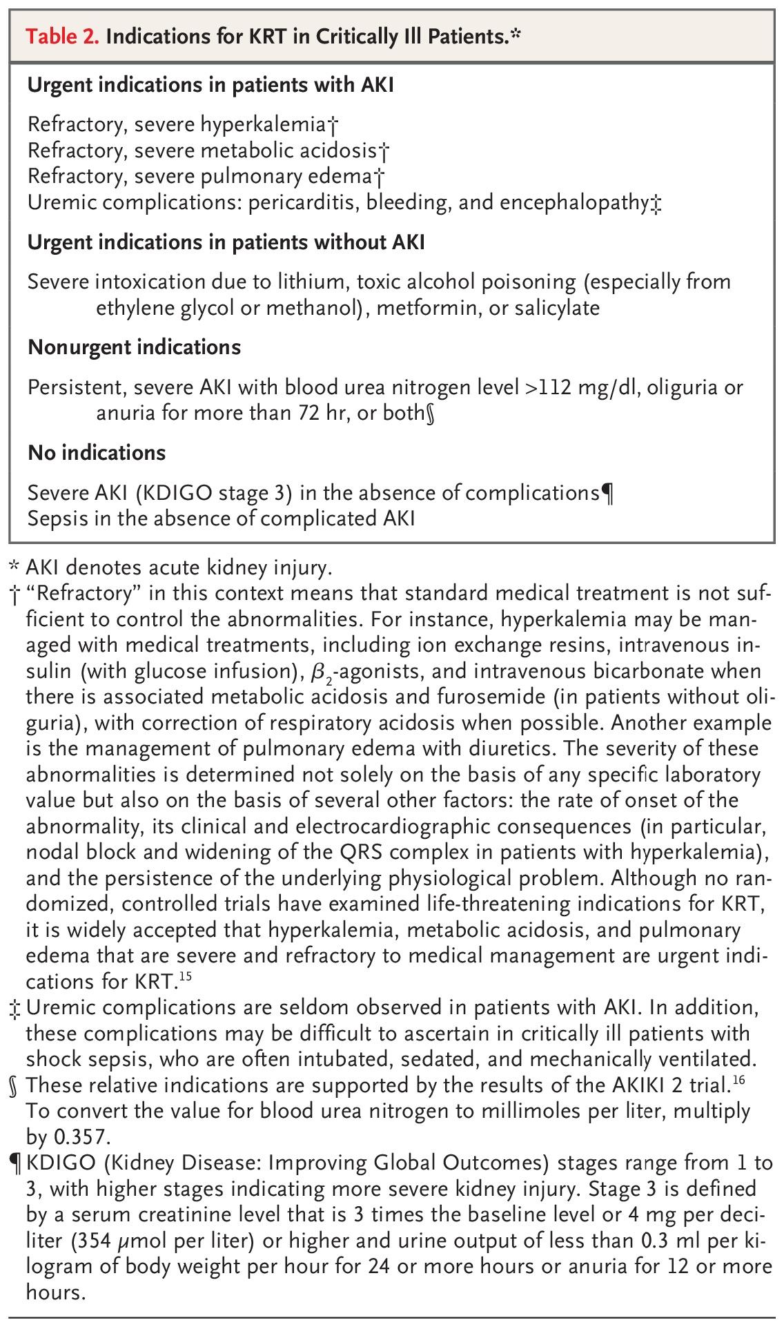
- Urgent indications: Refractory hyperkalemia, refractory metabolic acidosis, refractory pulmonary edema, uremic complications (pericarditis, encephalopathy, bleeding) [9]
- Nonurgent indications: BUN >112 mg/dL, oliguria/anuria >72 hours [9][20]
- Timing: In the absence of urgent indications, a watchful waiting approach is supported by the STARRT-AKI, AKIKI, and IDEAL-ICU trials — early initiation does not improve mortality and ~40–50% of deferred patients never require RRT [16][20][31]
- CRRT preferred in hemodynamically unstable patients; intermittent HD for stable patients [16][23]
The following table from the NEJM summarizes indications for kidney-replacement therapy:
Disposition
- Admission criteria:
- Nephrology consultation triggers: Stage 3 AKI, unclear etiology, need for RRT, preexisting CKD stage 4+, inadequate response to supportive care, suspected glomerulonephritis or vasculitis [3]
- Observation: Mild AKI (Stage 1) with clear reversible cause (e.g., NSAID use in otherwise healthy patient) may be managed with close outpatient follow-up if creatinine is trending down and electrolytes are stable
Follow Up / Return Precautions
- Post-discharge follow-up: Recheck creatinine and electrolytes within 1–2 weeks; nephrology follow-up within 1–3 months for Stage 2–3 AKI [20]
- Long-term monitoring: AKI survivors have increased risk of CKD, cardiovascular events, and recurrent AKI (25% recurrence within 12 months). Monitor creatinine, proteinuria, and blood pressure at 3 months and annually thereafter [20]
- Recovery timeline: If prerenal component is corrected early, creatinine may improve within 24–48 hours; established ATN typically requires several days to weeks for recovery; most recover within 6–12 months [6][19][30]
- Return precautions for patients:
- Decreased urine output or no urine output
- Swelling in legs/face, sudden weight gain, shortness of breath
- Confusion, excessive drowsiness
- Nausea/vomiting preventing oral intake
- Chest pain or palpitations
- Counseling: Avoid NSAIDs and nephrotoxins long-term; stay hydrated; inform all providers of AKI history; future contrast studies require pre-hydration protocols [6]
References
1. Acute Kidney Injury: Medical Causes and Pathogenesis. — Turgut F, Awad AS, Abdel-Rahman EM. Journal of Clinical Medicine. 2023.
2. Acute Kidney Injury: An Increasing Global Concern. — Lameire NH, Bagga A, Cruz D, et al. Lancet. 2013.
3. Acute Kidney Injury: Diagnosis and Management. — Mercado MG, Smith DK, Guard EL. American Family Physician. 2019.
4. Diuretics for Preventing and Treating Acute Kidney Injury. — Hashimoto H, Yamada H, Murata M, Watanabe N. The Cochrane Database of Systematic Reviews. 2025.
5. Acute Renal Failure. — Thadhani R, Pascual M, Bonventre JV. The New England Journal of Medicine. 1996.
6. Normotensive Ischemic Acute Renal Failure. — Abuelo JG. The New England Journal of Medicine. 2007.
7. The Primary Care Management of Chronic Kidney Disease (CKD) (2025). — Jonathan Casey Brown DO MPH, Wendy Caesar-Gibbs RD, Cynthia Delgado MD FASN FNKF, et al Department of Veterans Affairs. 2025.
8. ACR Manual on Contrast Media 2025. — ACR Committee on Drugs and Contrast Media American College of Radiology. 2025.
9. Extracorporeal Kidney-Replacement Therapy for Acute Kidney Injury. — Gaudry S, Palevsky PM, Dreyfuss D. The New England Journal of Medicine. 2022.
10. Acute Renal Failure. — Lameire N, Van Biesen W, Vanholder R. Lancet. 2005.
11. Drug-Induced Acute Kidney Injury: Diverse Mechanisms of Tubular Injury. — Perazella MA. Current Opinion in Critical Care. 2019.
12. Mechanisms of Antimicrobial-Induced Nephrotoxicity in Children. — Downes KJ, Hayes M, Fitzgerald JC, et al. The Journal of Antimicrobial Chemotherapy. 2020.
13. Acute Kidney Injury in Patients with Cirrhosis. — Nadim MK, Garcia-Tsao G. The New England Journal of Medicine. 2023.
14. Acute Kidney Injury in Patients with Cancer. — Rosner MH, Perazella MA. The New England Journal of Medicine. 2017.
15. Nephrotoxin Exposure and Acute Kidney Injury in Adults. — Griffin BR, Wendt L, Vaughan-Sarrazin M, et al. Clinical Journal of the American Society of Nephrology : CJASN. 2023.
16. Contemporary Management of Severe Acute Kidney Injury and Refractory Cardiorenal Syndrome: JACC Council Perspectives. — Jentzer JC, Bihorac A, Brusca SB, et al. Journal of the American College of Cardiology. 2020.
17. Updates in Management and Timing of Dialysis in Acute Kidney Injury. — Yu MK, Kamal F, Chertow GM. Journal of Hospital Medicine. 2019.
18. Diagnosis and Treatment of Acute Tubular Necrosis. — Esson ML, Schrier RW. Annals of Internal Medicine. 2002.
19. Renal Recovery After Severe Acute Renal Injury. — Schiffl H. European Journal of Medical Research. 2008.
20. Acute Kidney Injury. — Ostermann M, Lumlertgul N, Jeong R, et al. Lancet. 2025.
21. Long-Term Outcome of Acute Tubular Necrosis: A Contribution to Its Natural History. — Liaño F, Felipe C, Tenorio MT, et al. Kidney International. 2007.
22. Fenoldopam for Preventing and Treating Acute Kidney Injury. — Esezobor CI, Bhatt GC, Effa EE, Hodson EM. The Cochrane Database of Systematic Reviews. 2024.
23. Management of Acute Kidney Injury/Renal Replacement Therapy in the Intensive Care Unit. — Shaikhouni S, Yessayan L. The Surgical Clinics of North America. 2022.
24. Rhabdomyolysis and Acute Kidney Injury. — Bosch X, Poch E, Grau JM. The New England Journal of Medicine. 2009.
25. Differential Diagnosis of Acute Renal Failure. — Espinel CH, Gregory AW. Clinical Nephrology. 1980.
26. Diagnosis, Evaluation, and Management of Ascites, Spontaneous Bacterial Peritonitis and Hepatorenal Syndrome: 2021 Practice Guidance by the American Association for the Study of Liver Diseases. — Biggins SW, Angeli P, Garcia-Tsao G, et al. Hepatology. 2021.
27. Diagnostic Performance of Fractional Excretion of Sodium for the Differential Diagnosis of Acute Kidney Injury: A Systematic Review and Meta-Analysis. — Abdelhafez M, Nayfeh T, Atieh A, et al. Clinical Journal of the American Society of Nephrology : CJASN. 2022.
28. Evaluation of Simple Diagnostic Parameters in Acute Kidney Injury in Hospitalized Patients-Diagnostic Recommendations for Non-Nephrologists. — Buckenmayer A, Siebler N, Haas CS. Internal and Emergency Medicine. 2023.
29. Urinary Diagnostic Indices in Acute Renal Failure: A Prospective Study. — Miller TR, Anderson RJ, Linas SL, et al. Annals of Internal Medicine. 1978.
30. Renal Recovery From Acute Tubular Necrosis Requiring Renal Replacement Therapy: A Prospective Study in Critically Ill Patients. — Schiffl H. Nephrology, Dialysis, Transplantation : Official Publication of the European Dialysis and Transplant Association - European Renal Association. 2006.
31. Timing of Initiation of Renal-Replacement Therapy in Acute Kidney Injury. — STARRT-AKI Investigators, Canadian Critical Care Trials Group, Australian and New Zealand Intensive Care Society Clinical Trials Group, et al. The New England Journal of Medicine. 2020.
Figure
More from Deep Dive
Reviewed by Dr. Lucas Mastropaolo. SymptomDx is an educational tool for medical professionals — it does not replace clinical judgment. Verify all clinical data and drug dosages with authoritative sources.