Acute Pancreatitis
Acute pancreatitis is the most common gastrointestinal-related reason for hospitalization in the United States, with approximately 300,000 ED visits annually. [1-2] Diagnosis requires 2 of 3 revised Atlanta criteria: (1) abdominal pain consistent with the disease, (2) serum amylase and/or lipase >3× the upper limit of normal, and (3) characteristic findings on cross-sectional imaging. [1][3] Approximately 80% of cases can be diagnosed on clinical and laboratory findings alone without imaging. [1]
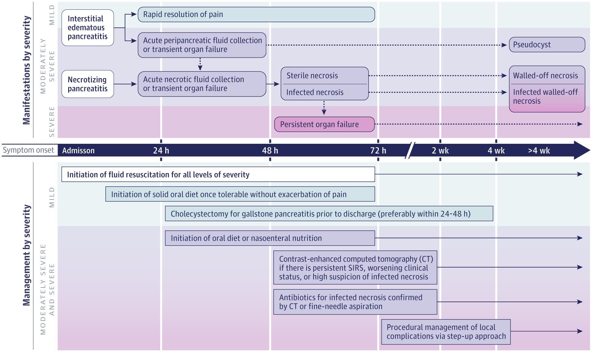
The following figure from JAMA illustrates the timeline, manifestations, and management of acute pancreatitis stratified by severity:
History
- Pain characterization: Constant epigastric or LUQ pain, often radiating to the back, chest, or flanks; exacerbated by eating, drinking, or lying supine [1][3]
- Dull, colicky, or lower abdominal pain is not consistent with acute pancreatitis and suggests an alternative diagnosis [3]
- Pain intensity does not correlate with severity [3]
- Accompanying symptoms: nausea, vomiting, low- to moderate-grade fever [1]
- Timing: Onset typically acute; ask about prior episodes, recent heavy meals, alcohol binge
- Key HPI questions:
- History of gallstones, biliary colic, or prior cholecystectomy
- Alcohol use: quantity, duration (>5 years of heavy use for alcohol-related AP) [3-4]
- Recent ERCP or EUS (5–10% and <1% risk, respectively) [1]
- New medications within the past weeks to months [1][5]
- Recent infections, trauma, insect bites [1]
- Hypertriglyceridemia history, hypercalcemia, pregnancy
Alarm Features
- Persistent organ failure >48 hours (defines severe AP): SBP <90 mmHg, PaO₂ <60 mmHg, creatinine >2 mg/dL, GI bleeding >500 mL/24h [3]
- SIRS at admission — highly predictive of organ failure and severe disease [3]
- Altered mental status [3]
- Hemoconcentration (HCT ≥44%) or rising BUN (≥20 mg/dL) within 24 hours [3]
- Pleural effusions or infiltrates on CXR [3]
- Signs of abdominal compartment syndrome (tense distension, oliguria, respiratory failure)
- Grey Turner sign (flank ecchymosis) or Cullen sign (periumbilical ecchymosis) — rare but suggest hemorrhagic pancreatitis
- Fever >101°F with clinical deterioration after 7–10 days — suspect infected necrosis [3]
Medications
Causative medications (>30 drugs with definite association): [1][4]
- Highest evidence: azathioprine, 6-mercaptopurine, didanosine, valproic acid, ACE inhibitors (enalapril, perindopril), mesalamine/sulfasalazine
- Other notable agents: furosemide, tetracycline, TMP-SMX, statins (simvastatin, rosuvastatin, pravastatin), carbamazepine, estrogens, metronidazole, isoniazid
- Drug-induced pancreatitis is usually mild and resolves with drug discontinuation [4]
- GLP-1 receptor agonists: recent data do not support a causal role [4]
Treatment medications
- Analgesia: Multimodal approach per WHO pain ladder; opioids are appropriate and may decrease supplementary analgesic need. No particular analgesic is superior [6]
- IV fluids: Lactated Ringer solution preferred [1][3]
- Antiemetics: Ondansetron, prochlorperazine as needed
- Prophylactic antibiotics are NOT recommended routinely in predicted severe AP or necrotizing pancreatitis [7]
Contraindicated/caution
- Avoid NSAIDs if renal impairment or hemodynamic instability
- Discontinue any suspected causative medication immediately
Diet
- Early oral feeding (within 24 hours) as tolerated is recommended — no need to wait for pain resolution or enzyme normalization [2][7]
- Low-fat solid diet is preferred over clear liquids (no benefit to gradual advancement) [2]
- If oral feeding not tolerated: nasogastric or nasojejunal enteral feeding preferred over parenteral nutrition [2][7]
- Parenteral nutrition reserved for patients who cannot tolerate enteral feeding
- Long-term: Alcohol cessation is critical; low-fat diet for hypertriglyceridemia-related AP; fibrate therapy for TG >1000 mg/dL
Review of Systems
- GI: Nausea, vomiting, anorexia, abdominal distension, diarrhea/steatorrhea (suggests chronic component)
- Pulmonary: Dyspnea, pleuritic chest pain (pleural effusion, ARDS)
- Cardiovascular: Chest pain, palpitations (ECG changes can mimic ACS) [8-9]
- Renal: Decreased urine output (hypovolemia, AKI)
- Neurologic: Altered mental status (component of BISAP; suggests severe disease) [1]
- Constitutional: Fevers, chills, diaphoresis, weight loss
Collateral History and Family History
- Alcohol use: Confirm quantity and duration with family/friends; >50 g/day for >5 years required for alcohol-related AP [3-4]
- Family history: Hereditary pancreatitis (PRSS1, SPINK1, CFTR mutations) — consider if recurrent AP without identifiable cause [1]
- Social context: Smoking (independent risk factor and cofactor with alcohol); recreational drug use (cannabis has been implicated) [1][3]
- Prior episodes of pancreatitis or known chronic pancreatitis
Risk Factors
- Gallstones (40–70% of cases) — most common cause [3]
- Alcohol (25–35%) — requires prolonged heavy use; lifetime risk in heavy drinkers only 2–5% [3-4]
- Hypertriglyceridemia (2–5%) — typically TG >1000 mg/dL; up to 56% of AP in pregnancy [3]
- Obesity — independent risk factor for severe disease [1]
- Older age [1]
- Smoking [3]
- Post-ERCP (5–10%) [1]
- Medications (<5% of cases) [4]
- Hypercalcemia, autoimmune disease, infections, trauma, genetic predisposition [1][6]
- Pancreatic cancer should be suspected in any patient >40 years with idiopathic AP [3]
Differential Diagnosis
- Perforated peptic ulcer — sudden onset, peritoneal signs, free air on imaging [1]
- Acute cholecystitis/cholangitis — RUQ pain, Murphy sign, Charcot triad
- Mesenteric ischemia — pain out of proportion to exam, lactic acidosis, atrial fibrillation
- Small bowel obstruction — colicky pain, distension, vomiting, prior surgery
- Acute myocardial infarction — AP can mimic inferior STEMI on ECG; obtain troponin if chest pain present [8]
- Ruptured/dissecting aortic aneurysm — tearing pain, hemodynamic instability
- Diabetic ketoacidosis — can cause elevated lipase and abdominal pain without true pancreatitis
- Ectopic pregnancy (reproductive-age women)
Past Medical History
- Prior episodes of pancreatitis (recurrence risk: biliary > alcohol > HTG) [10]
- Known gallstone disease or prior cholecystectomy
- Chronic pancreatitis (alcohol-related AP often represents flare on chronic disease) [4]
- Hypertriglyceridemia, hypercalcemia, hyperparathyroidism
- Autoimmune conditions (IgG4-related disease)
- HIV status (increased risk; also drug-related causes) [1]
- Pancreatic cysts, tumors, or structural anomalies (pancreas divisum)
- Prior abdominal surgery
Physical Exam
- Vitals: Tachycardia, hypotension (hypovolemia), fever, tachypnea
- Abdomen: Epigastric tenderness, guarding, distension, decreased bowel sounds; rebound tenderness is uncommon [1]
- Skin: Jaundice (biliary obstruction); Grey Turner sign (flank ecchymosis) and Cullen sign (periumbilical ecchymosis) — rare, late findings of hemorrhagic pancreatitis
- Lungs: Decreased breath sounds at bases (pleural effusion, especially left-sided)
- Cardiovascular: Assess volume status — capillary refill, JVP, peripheral perfusion
- Mental status: Altered mentation is a BISAP component and red flag [1]
Lab Studies
Diagnostic
- Serum lipase (preferred over amylase) — >3× ULN is diagnostic. Amylase may be normal in alcohol-induced or hypertriglyceridemia-related AP [3]
- Comprehensive metabolic panel: BUN, creatinine (volume status, renal function), glucose, calcium
- Hepatic panel: ALT, AST, alkaline phosphatase, total/direct bilirubin — elevated ALP/direct bilirubin suggests choledocholithiasis [1]
- Serum triglycerides — obtain on all patients; >1000 mg/dL suggests HTG as etiology [3]
- CBC: Leukocytosis; HCT ≥44% suggests hemoconcentration [3]
Severity markers
- BUN ≥20 mg/dL at admission or rising BUN at 24h — associated with increased mortality (OR 4.6 and 4.3, respectively) [1]
- CRP >150 mg/L at 48–72h predicts necrosis [3][11]
- Lactate if concern for shock or mesenteric ischemia
Other
- IgG4 if autoimmune pancreatitis suspected [1]
- Fasting TG level 1 month post-discharge if HTG suspected [3]
- Genetic testing for recurrent idiopathic AP [1]
Imaging
- Transabdominal ultrasound — obtain on all patients to evaluate for gallstones; first-line imaging [3][6]
- CT abdomen/pelvis with IV contrast — not required for diagnosis if clinical/lab criteria met; indicated for:
- MRI/MRCP — useful for evaluating bile duct stones, pancreatic duct anatomy, and in patients with contrast allergy or renal insufficiency
- Chest X-ray — assess for pleural effusion (BISAP component), ARDS [3]
- Imaging is unnecessary in straightforward mild AP with clear etiology and clinical improvement
Special Tests
Scoring systems (all have similar predictive accuracy; none is clearly superior): [1][12]
- BISAP (Bedside Index for Severity in Acute Pancreatitis) — most practical for ED use; calculated within 24 hours: [1]
- BUN >25 mg/dL
- Impaired mental status
- SIRS
- Age >60
- Pleural effusion
- Score ≥3: OR 7.4 for organ failure, OR 12.7 for persistent organ failure, >20% mortality [1]
- Ranson criteria — requires 48 hours to complete; 11 variables [1]
- APACHE II — comprehensive but cumbersome; 12 variables [1]
- Modified Marshall Score — defines organ failure (respiratory, renal, cardiovascular) [1][3]
- CT Severity Index — useful after 72 hours for assessing necrosis extent [1]
Point-of-care
- Bedside ultrasound for gallstones, free fluid, IVC assessment for volume status
- SIRS assessment at admission — highly sensitive for predicting severe disease [2-3]
ECG
- ~50% of patients with AP have ECG abnormalities [9][13]
- Most common: sinus tachycardia, T-wave flattening, ST-segment depression, nonspecific repolarization changes [9][13]
- Can mimic acute MI: ST-elevation (most commonly inferior leads), peaked T waves — reported in 34 cases without true coronary thrombosis [8][14]
- QTc prolongation seen in ~58% of patients; associated with higher mortality [15]
- Mechanisms: hypovolemia, electrolyte derangements (hypocalcemia, hypokalemia, hypomagnesemia), coronary vasospasm, direct myocyte injury from pancreatic enzymes [8-9]
- Obtain ECG in all patients with chest pain, hemodynamic instability, or significant comorbidities to rule out concurrent ACS
- Obtain troponin if ECG changes are present — true concurrent MI can occur [16]
Assessment
Severity classification (Revised Atlanta): [1][3][6]
- Mild (~80%): No organ failure, no local/systemic complications. Self-limiting; discharge typically within 1 week
- Moderately severe: Transient organ failure (<48h) and/or local complications (fluid collections, necrosis)
- Severe: Persistent organ failure >48 hours ± local complications. Mortality significant
Morphologic classification
- Interstitial edematous pancreatitis — usually mild
- Necrotizing pancreatitis — sterile vs. infected; infected necrosis carries highest morbidity/mortality [3]
Etiology-specific severity: Hypertriglyceridemia-induced AP has the highest complication rate, followed by alcoholic AP, then biliary AP. [10]
Treatment Plan
Initial stabilization (ED)
- IV fluid resuscitation: Begin immediately with lactated Ringer solution; goal-directed at 5–10 mL/kg/h until targets met (HR <120, MAP 65–85 mmHg, UOP >0.5–1 mL/kg/h) [1][3][6]
- Most effective in the first 6–12 hours; after 24–48 hours or in established severe disease, aggressive hydration may be harmful [3]
- Monitor BUN and HCT — do not allow these to rise in the first 24–48 hours [3]
- Caution in patients with heart failure or CKD (risk of pulmonary edema, abdominal compartment syndrome) [1]
- Analgesia: Multimodal — acetaminophen, opioids as needed; no evidence one analgesic is superior [6]
- NPO is NOT required: Early oral feeding (low-fat diet) within 24 hours as tolerated [2][7]
- Antiemetics as needed
Etiology-directed interventions
- Gallstone pancreatitis with cholangitis: Urgent ERCP within 24 hours [7]
- Gallstone pancreatitis without cholangitis: ERCP is NOT routinely indicated [7]
- Same-admission cholecystectomy for mild biliary pancreatitis; within 2–4 weeks if unsafe during admission [1][17]
- Hypertriglyceridemia: Insulin infusion ± heparin to lower TG; consider plasmapheresis if TG >1000 with severe metabolic acidosis [18]
- Alcohol-related: Brief alcohol intervention during hospitalization [7]
Infected necrosis (typically >7–10 days)
- Antibiotics (carbapenems have best pancreatic penetration)
- Step-up approach: Percutaneous/endoscopic drainage first → minimally invasive necrosectomy if needed → open surgery as last resort [2][18]
Disposition
Admission criteria — virtually all patients with confirmed AP require admission:
- All patients meeting diagnostic criteria
- Inability to tolerate oral intake
- Need for IV fluid resuscitation and pain control
- Persistent organ failure (SBP <90, PaO₂ <60, Cr >2 after rehydration)
- Persistent SIRS, hemodynamic instability requiring vasopressors
- BISAP ≥3 or APACHE II ≥8
- Respiratory failure, altered mental status, anuria
Intermediate care/step-down: [19]
Discharge criteria: [1][6][20]
- Pain controlled on oral analgesics
- Tolerating oral diet
- Stable or improving labs (BUN trending down, no rising WBC)
- No signs of organ failure or complications
- Mild AP patients typically discharged within 3–7 days [6]
Specialist consultation triggers
- GI: Biliary pancreatitis (ERCP consideration), recurrent/idiopathic AP, suspected autoimmune pancreatitis
- Surgery: Cholecystectomy planning, necrosectomy, abdominal compartment syndrome
- Interventional radiology: Percutaneous drainage of collections
- ICU/critical care: Organ failure, hemodynamic instability
Follow Up / Return Precautions
Follow-up timing
- GI or surgery follow-up within 2–4 weeks post-discharge [1]
- If cholecystectomy deferred: schedule within 2–4 weeks to prevent recurrence [1]
- Repeat fasting TG level at 1 month if hypertriglyceridemia suspected [3]
- Monitor for pancreatic exocrine insufficiency (develops in up to 35%) — check fecal elastase if diarrhea/steatorrhea [1]
- Screen for type 3c (pancreatogenic) diabetes after severe/necrotizing AP [1]
Return precautions — instruct patients to return immediately for:
- Recurrent severe abdominal pain
- Fever, chills, rigors (infected necrosis)
- Persistent vomiting, inability to eat or drink
- Jaundice or dark urine
- Lightheadedness, dizziness, decreased urine output
- Shortness of breath or chest pain
Patient counseling
- Absolute alcohol cessation — critical to prevent recurrence and progression to chronic pancreatitis [1][7]
- Smoking cessation [3]
- Low-fat diet; medication compliance for lipid-lowering therapy if applicable
- Expected recovery: Mild AP typically resolves within 1–2 weeks; moderately severe/severe disease may take weeks to months [1]
- Readmission rate for mild AP is approximately 13%, mainly due to recurrent or ongoing AP [21]
References
1. Acute Pancreatitis: A Review. — Mederos MA, Reber HA, Girgis MD. The Journal of the American Medical Association. 2021.
2. Acute Pancreatitis: Rapid Evidence Review. — Oppenlander KE, Chadwick C, Carman K. American Family Physician. 2022.
3. American College of Gastroenterology Guidelines: Management of Acute Pancreatitis. — Tenner S, Vege SS, Sheth SG, et al. The American Journal of Gastroenterology. 2024.
4. Acute Pancreatitis. — Forsmark CE, Vege SS, Wilcox CM. The New England Journal of Medicine. 2016.
5. Drug-Induced Pancreatitis: A Real-World Analysis of the FDA Adverse Event Reporting System and Network Pharmacology. — Xie H, Jiang L, Peng J, et al. Frontiers in Pharmacology. 2025.
6. Acute Pancreatitis. — Boxhoorn L, Voermans RP, Bouwense SA, et al. Lancet. 2020.
7. American Gastroenterological Association Institute Guideline On Initial Management of Acute Pancreatitis. — Crockett SD, Wani S, Gardner TB, Falck-Ytter Y, Barkun AN. Gastroenterology. 2018.
8. Acute Pancreatitis and Myocardial Infarction: A Narrative Review. — Khan U, Petrechko O, Sagheer S, et al. Cardiology. 2023.
9. Cardiovascular Manifestations of Acute Pancreatitis. — Yegneswaran B, Kostis JB, Pitchumoni CS. Journal of Critical Care. 2011.
10. Assessment of the Course of Acute Pancreatitis in the Light of Aetiology: A Systematic Review and Meta-Analysis. — Bálint ER, Fűr G, Kiss L, et al. Scientific Reports. 2020.
11. Acute Pancreatitis: Diagnosis and Treatment. — Szatmary P, Grammatikopoulos T, Cai W, et al. Drugs. 2022.
12. Management of Gallstone Pancreatitis: A Review. — McDermott J, Kao LS, Keeley JA, Nahmias J, de Virgilio C. JAMA Surgery. 2024.
13. Electrocardiographic Abnormalities in Patients With Acute Pancreatitis. — Rubio-Tapia A, García-Leiva J, Asensio-Lafuente E, Robles-Díaz G, Vargas-Vorácková F. Journal of Clinical Gastroenterology. 2005.
14. Acute Pancreatitis Presented With Diffuse ST-segment Elevation: A Case Report and Literature Review. — Hsieh YL, Wu SH, Liu CY, et al. Medicine. 2024.
15. Diastolic Dysfunction, Prolonged QTc Interval and Pericardial Effusion as Predictors of Mortality in Acute Pancreatitis. — Nadkarni N, Bhasin DK, Rana SS, et al. Journal of Gastroenterology and Hepatology. 2012.
16. A Case of Acute Necrotizing Pancreatitis Complicated With Non ST Elevation Myocardial Infarction. — Ralapanawa U, Jayalath T, Senadhira D. BMC Research Notes. 2018.
17. Contemporary Management of Acute Pancreatitis: What You Need to Know. — Palumbo R, Schuster KM. The Journal of Trauma and Acute Care Surgery. 2024.
18. Interventional Management of Acute Pancreatitis and Its Complications. — Masood M, Vedamurthy A, Krishnamoorthi R, et al. Journal of Clinical Medicine. 2025.
19. IAP/APA Evidence-Based Guidelines for the Management of Acute Pancreatitis. — Pancreatology : Official Journal of the International Association of Pancreatology .... 2013.
20. Discharge Protocol in Acute Pancreatitis: An International Survey and Cohort Analysis. — Nagy R, Ocskay K, Sipos Z, et al. Scientific Reports. 2023.
21. Evaluation of a Predictive Model and Clinical Outcomes in Mild Acute Pancreatitis: Towards Evidence-Based Early Discharge. — Kant N, Spanier BWM, Witjes B, Doggen CJM. Digestive Diseases and Sciences. 2025.
Figure
More from Deep Dive
Reviewed by Dr. Lucas Mastropaolo. SymptomDx is an educational tool for medical professionals — it does not replace clinical judgment. Verify all clinical data and drug dosages with authoritative sources.