Acute Pericarditis
Acute pericarditis is inflammation of the pericardium, accounting for approximately 4.4% of ED visits for nonischemic chest pain, with higher prevalence in men. Most cases in North America and Western Europe are idiopathic/viral (80–90%). With appropriate treatment, 70–85% of patients have a benign course. [1-2]
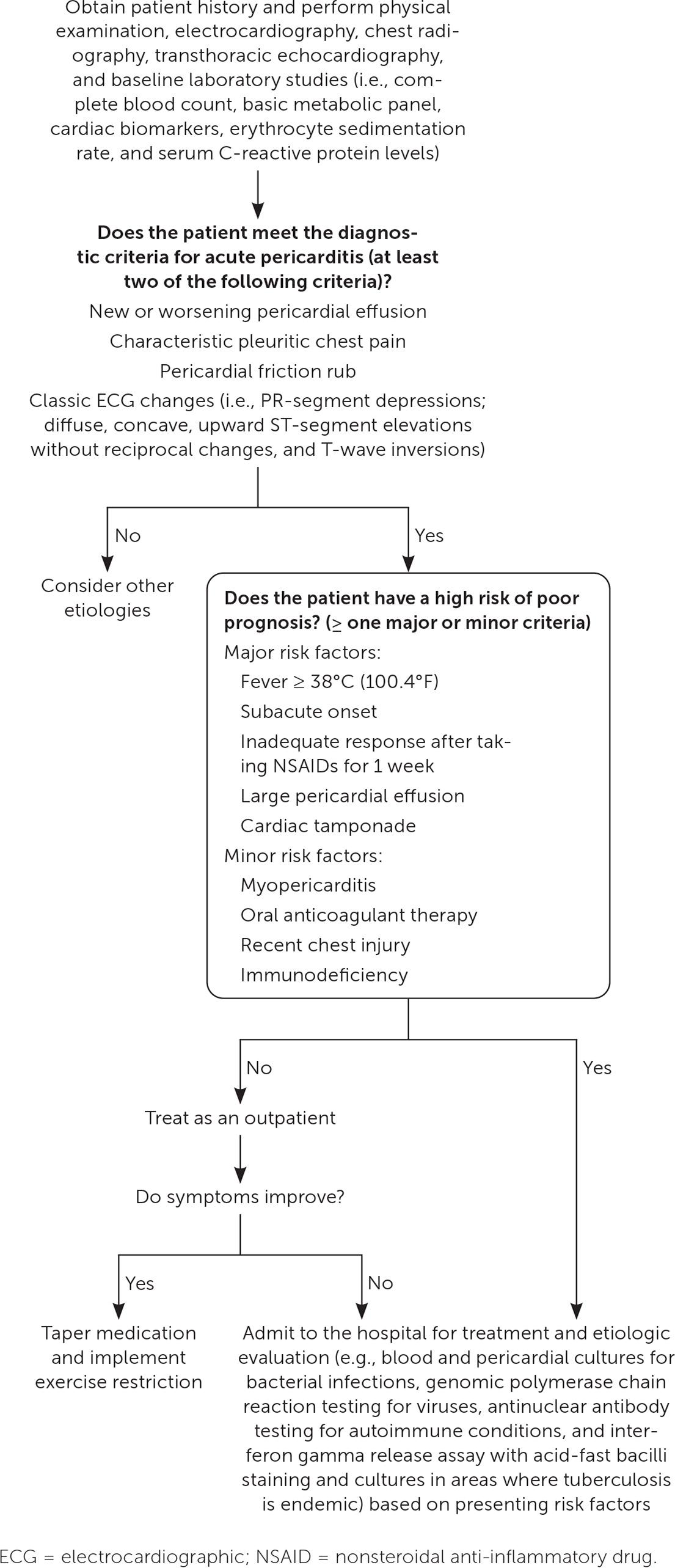
The following diagnostic and management algorithm from the AAFP provides a practical overview:
History
- Pain quality: Sharp, stabbing, pleuritic chest pain — worse when supine, improved by sitting up and leaning forward [1][3]
- Radiation to the trapezius ridge is virtually pathognomonic [3]
- Onset: Often abrupt; ask about preceding viral URI or gastroenteritis (1–3 weeks prior)
- Timing: Constant or intermittent; typically acute onset over hours
- Associated symptoms: Dyspnea, cough, fatigue, low-grade fever, malaise
- Important negatives: Exertional component (ACS), tearing quality (dissection), unilateral pleuritic pain with leg swelling (PE), dysphagia (esophageal), trauma history
- Ask about recent cardiac surgery or procedures (post-cardiac injury syndrome), prior pericarditis episodes, and autoimmune disease symptoms [1]
Alarm Features
- Fever >38.5°C — suggests specific etiology (bacterial, TB, autoimmune) [3-4]
- Subacute onset (symptoms developing over days without clear onset) [1]
- Large pericardial effusion (>2.0 cm) or signs of cardiac tamponade (Beck's triad: hypotension, JVD, muffled heart sounds; pulsus paradoxus) [1][3]
- Failure to respond to NSAIDs within 1 week [1]
- Concomitant myocarditis (elevated troponin with LV dysfunction) [5]
- Suspected malignancy or immunocompromised state [1]
- Hemodynamic instability — tachycardia, hypotension
Medications
First-line (dual therapy)
- Ibuprofen 600–800 mg PO q8h (1600–2400 mg/day), taper by 200–400 mg every 2–4 weeks after symptom resolution and CRP normalization [1][6]
- OR Aspirin 650–1000 mg PO TID (preferred if concomitant CAD) [5][7]
- PLUS Colchicine 0.5 mg BID (>70 kg) or 0.5 mg daily (≤70 kg) for 3 months (first episode) or ≥6 months (recurrence) [1]
- Proton pump inhibitor for gastroprotection with NSAIDs [1][5]
Second-line
Third-line (recurrent/refractory)
- IL-1 blockers[8]
Contraindicated/Cautions
- Avoid anticoagulants if possible (risk of hemorrhagic pericardial effusion)
- NSAIDs: caution in CKD, HF, peptic ulcer disease, bleeding diathesis [5]
- Colchicine: dose-reduce with P-gp/CYP3A4 inhibitors; GI intolerance is common [5]
Diet
- No specific dietary triggers
- Adequate hydration, especially with NSAID use (renal protection)
- Avoid alcohol during acute illness (may exacerbate inflammation and interact with medications)
Review of Systems
- Cardiovascular: Chest pain character, dyspnea, orthopnea, syncope/presyncope, palpitations
- Respiratory: Cough, pleuritic pain, dyspnea (pleural effusion common)
- Constitutional: Fever, night sweats, weight loss (TB, malignancy)
- MSK/Rheumatologic: Joint pain, rashes, oral ulcers (autoimmune)
- GI: Dysphagia, epigastric pain (alternative diagnoses)
- Infectious: Recent URI, GI illness, travel history, TB exposure
Collateral History and Family History
- Family history of pericarditis — may prompt genetic evaluation (familial Mediterranean fever), though yield is low [1]
- Family history of autoimmune disease (SLE, RA)
- Social context: Immigration from TB-endemic areas, incarceration, homelessness, HIV status [1]
- Recent cardiac surgery/catheterization (post-cardiac injury syndrome) [5]
- Medication history: Hydralazine, procainamide, isoniazid (drug-induced lupus/pericarditis)
Risk Factors
- Male sex, young to middle age (most common demographic) [2]
- Recent viral illness (most common identifiable trigger)
- Post-cardiac surgery or intervention (Dressler syndrome / post-cardiac injury)
- Autoimmune disease (SLE, RA, systemic sclerosis)
- Malignancy (lung, breast, lymphoma, leukemia)
- Tuberculosis exposure (leading cause in endemic areas) [1]
- Uremia / end-stage renal disease
- Immunosuppression / HIV
- Chest radiation therapy
- Risk factors for recurrence: Lack of NSAID response, early corticosteroid use, persistently elevated CRP, severe pericardial LGE on CMR [5]
Differential Diagnosis
- Acute coronary syndrome / STEMI — regional ST elevation with reciprocal changes, convex ST morphology, troponin rise with wall motion abnormalities [2-3]
- Myocarditis — chest pain with troponin elevation and LV dysfunction; nonspecific ST-T changes; ~15% have concomitant myopericarditis [2][5]
- Pulmonary embolism — pleuritic pain, dyspnea, tachycardia, right heart strain on ECG [2]
- Aortic dissection — tearing pain radiating to back, pulse/BP differential
- Pneumonia / pleuritis — fever, productive cough, focal findings
- Pneumothorax — sudden onset, unilateral decreased breath sounds
- Costochondritis — reproducible chest wall tenderness
- GERD / esophageal spasm — burning quality, meal-related
- Cardiac tamponade (complication of pericarditis itself)
Key distinguishing features of pericarditis vs. STEMI: diffuse concave ST elevation, PR depression, no reciprocal ST depression (except aVR/V1), ST/T ratio >0.25 in V6 [2-3][9]
Past Medical History
- Prior episodes of pericarditis (recurrence rate 15–30% after first episode, up to 50% after first recurrence) [5]
- Autoimmune conditions (SLE, RA, IBD, vasculitis)
- Recent cardiac surgery, MI, or catheterization
- Malignancy (especially thoracic)
- CKD/ESRD (uremic pericarditis)
- Prior chest radiation
- Hypothyroidism (myxedema pericarditis — rare)
Physical Exam
- Pericardial friction rub: High-pitched, scratchy, triphasic (atrial systole, ventricular systole, early diastole); best heard with diaphragm at left lower sternal border with patient leaning forward; present in <30% of cases and may be transient [1-2]
- Vital signs: Sinus tachycardia, low-grade fever common; high fever (>38.5°C) is a red flag [3]
- Pulsus paradoxus (>10 mmHg drop in SBP with inspiration) — suggests tamponade [3]
- JVD, muffled heart sounds, hypotension (Beck's triad — tamponade)
- Kussmaul sign (JVP rise with inspiration — constrictive physiology)
- Assess for signs of systemic disease: joint swelling, rashes, lymphadenopathy
Lab Studies
Initial workup (all patients)
- CRP (elevated in ~78%; key for monitoring treatment response and guiding taper) [1]
- ESR
- CBC with differential (neutrophilic leukocytosis supports inflammatory phenotype) [1]
- Troponin (elevated in ~30%; if elevated without LV dysfunction = myopericarditis, same management; not a negative prognostic marker) [1]
- BMP (baseline renal function before NSAIDs; rule out uremia)
- BNP/NT-proBNP (generally normal in isolated pericarditis; elevated suggests myocardial involvement or HF)
Selective/high-risk workup
- ANA — only if clinical suspicion for autoimmune disease (low-level titers are nonspecific) [1]
- TB testing (PPD/IGRA) — if risk factors present [1]
- Blood cultures — if bacterial pericarditis suspected
- HIV testing — if risk factors present
- TSH — if hypothyroidism suspected
Monitoring: Serial CRP to guide NSAID taper and detect recurrence [5]
Imaging
First-line
- Transthoracic echocardiography (TTE) — recommended in all patients; evaluates effusion size, tamponade physiology, LV function, constrictive features [1][5]
- Chest X-ray — evaluate for cardiomegaly (effusion >300 mL), pulmonary pathology, malignancy, TB [1]
- Point-of-care ultrasound (POCUS) — can rapidly identify hemodynamically significant effusion with RV diastolic collapse in the ED [1]
Second-line
- Cardiac MRI (CMR) — recommended for complicated, recurrent, or indeterminate cases; identifies pericardial LGE (inflammation), edema, thickening, and myocardial involvement [5][10]
- Cardiac CT — preferred for pericardial calcification (constrictive pericarditis); not recommended for routine assessment [5]
When imaging is unnecessary: Routine cardiac CT is not recommended for uncomplicated acute pericarditis [5]
Special Tests
- Pericardiocentesis — indicated for tamponade, suspected bacterial/TB/malignant effusion; fluid sent for cell count, Gram stain, culture, cytology, and AFB. Light criteria should NOT be applied to pericardial fluid [1]
- ST/T ratio in lead V6: >0.25 favors pericarditis over early repolarization [2]
- Spodick sign: Downsloping TP segment — suggestive but not pathognomonic
ECG
ECG changes occur in 25–60% of cases and evolve through 4 classic stages: [1-2][4]
- Stage I (acute): Diffuse concave ST-segment elevation + PR-segment depression (most prominent in leads II, aVF, V3–V6); reciprocal ST depression and PR elevation in aVR
- Stage II: ST segments normalize, T waves flatten
- Stage III: Diffuse T-wave inversions
- Stage IV: ECG normalizes
Key distinguishing features from STEMI
- Diffuse (not territorial) ST elevation
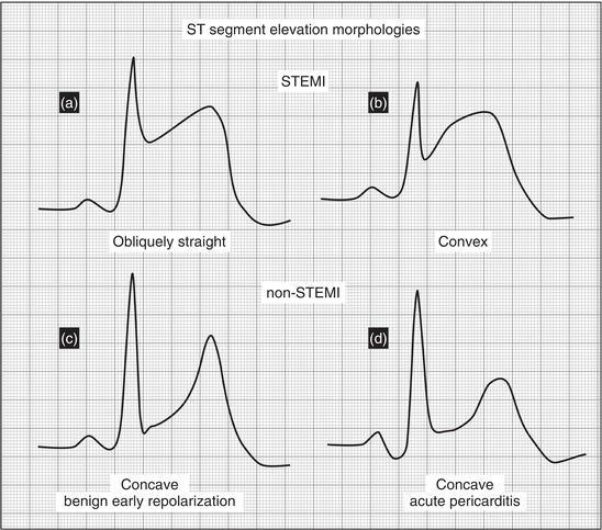
- Concave ("saddleback") ST morphology vs. convex in STEMI
- No reciprocal ST depression (except aVR/V1)
- PR-segment depression (uncommon in STEMI)
- No pathologic Q waves
- ST/T ratio >0.25 in V6 [2][9]
Other ECG findings: Sinus tachycardia; ~4% present with atrial fibrillation/flutter; low voltage or electrical alternans if large effusion [1]
The following figure illustrates the morphological differences between ST elevation in STEMI vs. pericarditis:
Assessment
- Epidemiology: ~4.4% of nonischemic chest pain in the ED; most common in men aged 16–65 [1-2]
- Etiology: Idiopathic/viral (80–90%); post-cardiac injury; autoimmune; TB (endemic areas); malignancy; uremia; drug-induced [1]
- Phenotypes: Inflammatory (80–90%, elevated CRP) vs. noninflammatory (10–20%, often autoimmune-associated) [5]
- Complications: Recurrence (15–30%), cardiac tamponade (<3%), constrictive pericarditis (<0.5%) [1]
- Concomitant myocarditis (~15%) — elevated troponin ± LV dysfunction [5]
- Prognosis: Benign in 70–85% with appropriate treatment; recurrence risk increases to 50% after first recurrence [1][5]
Treatment Plan
Initial stabilization
- ABCs, IV access, continuous monitoring if hemodynamically unstable
- Emergent pericardiocentesis for tamponade
Pharmacotherapy (first episode)
- Ibuprofen 600–800 mg PO q8h + Colchicine 0.5 mg BID (or 0.5 mg daily if ≤70 kg) [1]
- PPI for gastroprotection
- Taper NSAIDs by 200–400 mg every 2–4 weeks once pain resolves and CRP normalizes [6]
- Continue colchicine for 3 months (first episode) [1]
Exercise restriction: ≥1 month, keeping maximal heart rate <100 bpm until clinical remission [5]
Refractory/recurrent
- Corticosteroids (prednisone 0.2–0.5 mg/kg/day) if NSAID/colchicine failure [5]
- IL-1 blockers (anakinra, rilonacept) for multiple recurrences with inflammatory phenotype refractory to colchicine/steroids [8]
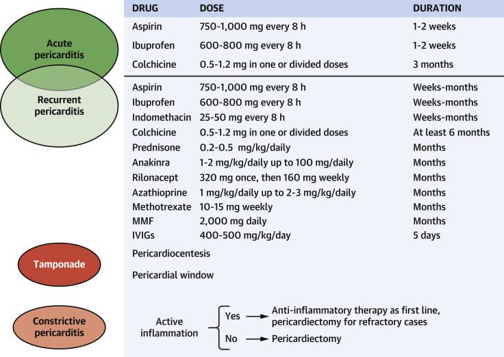
The following treatment algorithm summarizes the stepwise approach:
Disposition
Discharge criteria (low-risk, outpatient management)
- No major or minor risk factors
- Responsive to initial NSAID therapy
- Small or no pericardial effusion
- Hemodynamically stable
- Reliable follow-up available
- Fever >38°C
- Subacute course (insidious onset)
- Large pericardial effusion (>2.0 cm) or tamponade
- Failure to respond to NSAIDs after 1 week
- Suspected malignancy or bacterial etiology
- Concomitant myocarditis with LV dysfunction
- Oral anticoagulant use (risk of hemorrhagic effusion)
- Immunodeficiency
- Recent chest trauma
Specialist consultation triggers
- Cardiology: tamponade, large effusion, myopericarditis, recurrent disease, constrictive physiology
- Rheumatology: suspected autoimmune etiology
- Infectious disease: TB or bacterial pericarditis
- Cardiothoracic surgery: refractory constrictive pericarditis requiring pericardiectomy
Follow Up / Return Precautions
Follow-up timing
- 1–2 weeks after ED discharge for clinical reassessment and repeat CRP [5]
- Serial CRP to guide NSAID taper — do not taper until CRP normalizes
- Repeat echocardiography if effusion was present at diagnosis
- Follow-up at completion of colchicine course (3 months for first episode)
Return precautions — instruct patients to return immediately for:
- Worsening chest pain unresponsive to medications
- New or worsening dyspnea, orthopnea
- Lightheadedness, syncope, or near-syncope
- Fever >38°C
- Leg swelling or new edema
Patient counseling
- Exercise restriction for ≥1 month (no vigorous activity; keep HR <100 bpm) [5]
- Take medications as prescribed; do not stop colchicine early (reduces recurrence by ~50%) [1]
- Avoid NSAIDs on empty stomach; take with food
- Expected recovery: most cases resolve within 1–2 weeks of treatment, but full course of colchicine is essential to prevent recurrence
References
1. Diagnosis, Risk Stratification, and Treatment of Pericarditis: A Review. — Cremer PC, Klein AL, Imazio M. The Journal of the American Medical Association. 2024.
2. Acute Pericarditis: Rapid Evidence Review. — Peterson TA, Turner SP, Dolezal KA. American Family Physician. 2024.
3. Acute Pericarditis. — LeWinter MM. The New England Journal of Medicine. 2014.
4. Evaluation and Treatment of Pericarditis: A Systematic Review. — Imazio M, Gaita F, LeWinter M. The Journal of the American Medical Association. 2015.
5. 2025 Concise Clinical Guidance: An ACC Expert Consensus Statement on the Diagnosis and Management of Pericarditis: A Report of the American College of Cardiology Solution Set Oversight Committee. — Wang TKM, Klein AL, Cremer PC, et al. Journal of the American College of Cardiology. 2025.
6. Recurrent Pericarditis and Paradigm Shift in Cardiovascular Imaging and Targeted Therapeutics. — Karmali R, Kafil TS, Bayat A, et al. JACC. Advances. 2024.
7. Advances in Imaging and Targeted Therapies for Recurrent Pericarditis: A Review. — Kumar S, Khubber S, Reyaldeen R, et al. JAMA Cardiology. 2022.
8. Inflammation and Cardiovascular Disease: 2025 ACC Scientific Statement: A Report of the American College of Cardiology. — Mensah GA, Arnold N, Prabhu SD, Ridker PM, Welty FK. Journal of the American College of Cardiology. 2025.
9. Fourth Universal Definition of Myocardial Infarction (2018). — Thygesen K, Alpert JS, Jaffe AS, et al. Journal of the American College of Cardiology. 2018.
10. 2021 AHA/ACC/ASE/CHEST/SAEM/SCCT/SCMR Guideline for the Evaluation and Diagnosis of Chest Pain: A Report of the American College of Cardiology/American Heart Association Joint Committee on Clinical Practice Guidelines. — Gulati M, Levy PD, Mukherjee D, et al. Journal of the American College of Cardiology. 2021.
11. Electrocardiographic Differential Diagnosis of ST Segment Elevation. — Megan Starling, William J. Brady The Electrocardiagram in Emergency and Acute Care. 2023.
12. Management of Acute and Recurrent Pericarditis: JACC State-of-the-Art Review. — Chiabrando JG, Bonaventura A, Vecchié A, et al. Journal of the American College of Cardiology. 2020.
Figures
More from Deep Dive
Reviewed by Dr. Lucas Mastropaolo. SymptomDx is an educational tool for medical professionals — it does not replace clinical judgment. Verify all clinical data and drug dosages with authoritative sources.