Adrenal Insufficiency (Addisonian Crisis)
Adrenal crisis is a life-threatening endocrine emergency defined by hemodynamic instability (hypotension/shock) due to cortisol deficiency, requiring immediate parenteral hydrocortisone and aggressive fluid resuscitation — treatment must never be delayed for diagnostic workup. [1-3] Up to 50% of patients with primary adrenal insufficiency present with adrenal crisis as their first manifestation. [3]
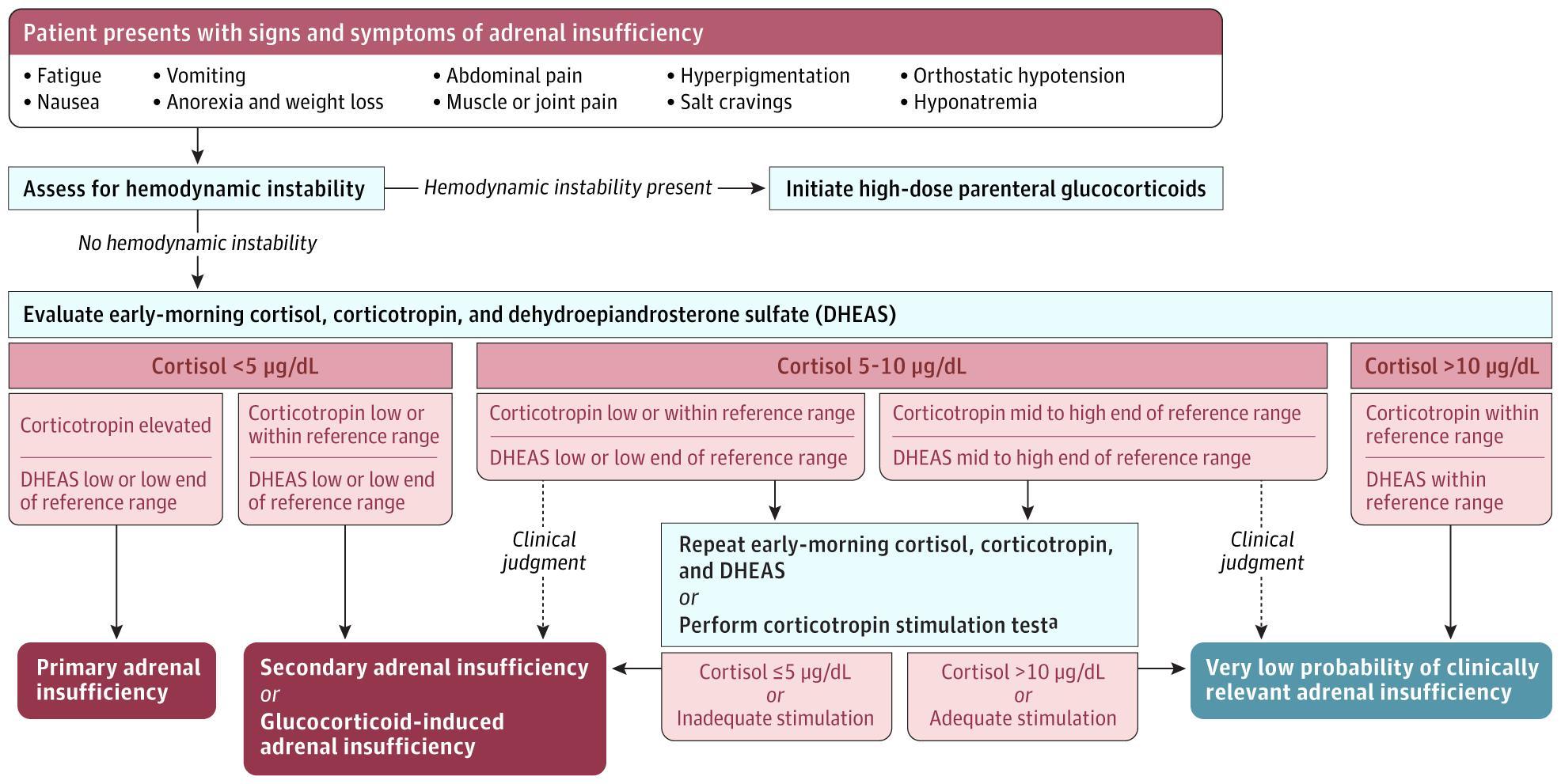
The following diagnostic algorithm from a 2025 JAMA review outlines the approach to suspected adrenal insufficiency based on hemodynamic stability and biochemical testing:
History
- Key HPI questions: Ask about chronic fatigue, anorexia, weight loss, nausea/vomiting, abdominal pain, salt cravings, dizziness/syncope, and skin darkening [1-2]
- Timing/triggers: Identify precipitating events — recent infection (especially GI illness), surgery, trauma, emotional stress, dental procedures, strenuous exercise, or recent steroid taper/discontinuation [2][4]
- Medication history is critical: Any current or recent glucocorticoid use (oral, inhaled, topical, intra-articular), opioids, immune checkpoint inhibitors, ketoconazole, etomidate, mitotane, or CYP3A4 inducers (rifampin, phenytoin, carbamazepine) [1][4]
- Symptom characterization: Fatigue (50%–95%), nausea/vomiting (20%–62%), anorexia/weight loss (43%–73%), orthostatic hypotension (68% in primary AI), hyperpigmentation (74% in primary AI), muscle/joint pain (40%) [1]
- Important negatives: Absence of hyperpigmentation suggests secondary or glucocorticoid-induced AI rather than primary; absence of hyperkalemia also favors secondary AI [1-2]
Alarm Features
- Hypotension/shock refractory to fluids and vasopressors — hallmark of adrenal crisis [4-5]
- Altered mental status: confusion, delirium, obtundation, seizures, coma [2]
- Severe hyponatremia, hyperkalemia, or hypoglycemia
- Abdominal pain mimicking acute abdomen (peritoneal irritation can occur) [6]
- Fever without clear infectious source
- Acute reversible heart failure (decreased myocardial contractility from cortisol deficiency) [5]
- Circulatory collapse or syncope
Medications
- Causative/contributing medications:
- Exogenous glucocorticoids (most common cause of AI overall) — abrupt withdrawal or taper below physiological doses [1][7]
- Opioids (suppress ACTH) [1]
- Immune checkpoint inhibitors (hypophysitis/adrenalitis, risk <1%) [2]
- Adrenal enzyme inhibitors: ketoconazole, metyrapone, etomidate, mitotane [8]
- CYP3A4 inducers accelerating glucocorticoid metabolism: rifampin, phenytoin, carbamazepine, phenobarbital, apalutamide, enzalutamide [4]
- Treatment medications: Hydrocortisone (preferred), prednisolone, dexamethasone (least preferred); fludrocortisone for primary AI maintenance [2][8]
- Caution: Initiating levothyroxine in undiagnosed AI can precipitate crisis by increasing cortisol clearance [2-3]
Diet
- Patients with primary AI may have salt cravings due to aldosterone deficiency — liberal salt intake is recommended during maintenance [1]
- During acute illness: electrolyte-containing fluids as tolerated when managing at home [8]
- Hydration is critical — dehydration from vomiting/diarrhea is a common crisis precipitant and impairs oral medication absorption [2]
Review of Systems
- Constitutional: Fatigue, malaise, weight loss, anorexia, fever
- GI: Nausea, vomiting, abdominal pain, diarrhea
- Cardiovascular: Orthostatic dizziness, syncope, palpitations
- Neuropsychiatric: Confusion, mood changes, depression, anxiety, cognitive deficits [3][6]
- Musculoskeletal: Myalgias, arthralgias, muscle cramps
- Dermatologic: Hyperpigmentation (buccal mucosa, palmar creases, scars — primary AI only)
- Endocrine: Amenorrhea, decreased libido (DHEAS deficiency)
Collateral History and Family History
- Confirm medication compliance — nonadherence to glucocorticoid replacement is a common crisis precipitant [2][6]
- Check for medical alert identification (bracelet, necklace, steroid emergency card) [8]
- Ask about prior adrenal crises (strongest risk factor for future crises) [4]
- Family history: Autoimmune conditions (type 1 diabetes, autoimmune thyroid disease, vitiligo, pernicious anemia) — autoimmune polyendocrine syndromes [8]
- Social context: Ability to self-inject hydrocortisone, access to emergency care, caregiver availability
Risk Factors
- History of prior adrenal crisis (single strongest predictor) [4]
- Current or recent glucocorticoid use (any route — oral, inhaled, topical, intra-articular) [1][4]
- Age >65 years or adolescence/transition of care [4]
- Higher comorbidity burden [4]
- Autoimmune adrenalitis (Addison disease) — most common cause of primary AI in developed countries [1]
- Coexisting autoimmune conditions: type 1 diabetes, autoimmune thyroid disease, vitiligo, celiac disease [8]
- Infections: tuberculosis (leading cause in developing countries), HIV, fungal infections [1]
- Pituitary pathology: tumors, surgery, radiation, hemorrhage (Sheehan syndrome), hypophysitis [1]
- Bilateral adrenalectomy, adrenal metastases, adrenal hemorrhage (Waterhouse-Friderichsen syndrome)
Differential Diagnosis
- Septic shock — most important mimic; adrenal crisis may coexist with sepsis
- Hypovolemic shock from GI losses, hemorrhage
- Cardiogenic shock / acute heart failure — adrenal crisis itself can cause reversible cardiomyopathy [5]
- Anaphylaxis — hypotension, GI symptoms overlap
- Diabetic ketoacidosis — nausea, vomiting, abdominal pain, dehydration
- Acute abdomen (appendicitis, perforation) — abdominal pain with guarding can mimic surgical pathology [6]
- Myxedema coma — hypothyroidism with altered mental status and hypotension
- Hypoglycemia from other causes (insulinoma, liver failure)
- Distinguishing feature: Shock disproportionate to the severity of the trigger and refractory to fluids/vasopressors should raise suspicion for adrenal crisis [4-5]
Past Medical History
- Known adrenal insufficiency (primary, secondary, or glucocorticoid-induced)
- Previous adrenal crises and precipitants
- Autoimmune conditions (polyendocrine syndromes)
- Pituitary surgery, radiation, or known pituitary pathology
- History of chronic glucocorticoid use for any indication (asthma, COPD, rheumatologic disease, transplant)
- Bilateral adrenalectomy
- Congenital adrenal hyperplasia
- Cancer with adrenal metastases or checkpoint inhibitor therapy [2]
Physical Exam
- Vital signs: Hypotension (SBP <100 mmHg or ≥20 mmHg below baseline), tachycardia, orthostatic changes, fever [2]
- Skin: Hyperpigmentation of palmar creases, buccal mucosa, scars, areolae, axillae (primary AI only — due to elevated ACTH/MSH) [2-3]
- Abdomen: Tenderness, guarding (can mimic acute abdomen) [6]
- Neurologic: Altered mental status ranging from lethargy to coma [2]
- General: Cachexia, dehydration, poor skin turgor
- Absence of hyperpigmentation does not rule out AI — secondary and glucocorticoid-induced AI will not have this finding [1]
Lab Studies
- Draw before treatment if possible, but never delay treatment: [6][8]
- BMP: Hyponatremia (84% of undiagnosed AI), hyperkalemia (34%, primary AI only), elevated creatinine/BUN (prerenal), hypoglycemia [4][6]
- CBC: Eosinophilia, lymphocytosis, mild normocytic anemia [2]
- Additional: Aldosterone/renin (low aldosterone, high renin in primary AI), calcium (mild hypercalcemia possible), TSH (may be elevated) [6][8]
- Etiologic workup (once stable): 21-hydroxylase antibodies (autoimmune), very long-chain fatty acids (adrenoleukodystrophy in males), 17-hydroxyprogesterone (CAH) [1]
Imaging
- Not required for acute diagnosis — adrenal crisis is a clinical diagnosis [4]
- CT abdomen: Consider if etiology unclear after labs — may show adrenal enlargement (infection, hemorrhage, metastases) or calcification (TB, prior hemorrhage) [1]
- Pituitary MRI: Indicated for suspected secondary AI (pituitary tumor, hemorrhage, hypophysitis, infiltrative disease) [1]
- Chest X-ray: May show small heart size (chronic volume depletion); rule out infectious precipitant
Special Tests
- Cosyntropin (ACTH) stimulation test — gold standard for confirming AI in stable patients: [8]
- Administer 250 µg cosyntropin IV/IM → measure cortisol at 0 and 60 min
- Peak cortisol <18 µg/dL confirms AI (assay-dependent; newer assays may use lower thresholds ~15–17 µg/dL) [1][3]
- Sensitivity 64%, specificity 93% for secondary AI — a normal result does not fully exclude recent-onset secondary AI [1]
- Not needed if baseline cortisol is unequivocally low with elevated ACTH (primary AI) [8]
- Point-of-care glucose: Check immediately — hypoglycemia common, especially in children [8]
- VBG/ABG: Assess acid-base status in shock
ECG
- Hyperkalemia findings: Peaked T waves, widened QRS, prolonged PR, sine wave pattern — particularly in primary AI with mineralocorticoid deficiency [2]
- Sinus tachycardia from hypovolemia/shock
- Low voltage QRS (chronic volume depletion)
- Cardiac monitoring recommended during acute crisis management [8]
- Rule out acute MI as a precipitant of crisis [6]
Assessment
Adrenal crisis is defined as hypotension or hypovolemic shock plus at least one of: nausea/vomiting, severe fatigue, fever, or impaired consciousness. [4] The shock is characteristically out of proportion to the severity of the trigger and resistant to conventional vasopressors and fluids until glucocorticoids are administered. [4-5]
Key distinctions:
- Primary AI (Addison disease): Deficiency of cortisol + aldosterone + androgens → hyperpigmentation, hyperkalemia, salt wasting [1]
- Secondary AI: Cortisol deficiency only (aldosterone preserved) → no hyperpigmentation, no hyperkalemia [1]
- Glucocorticoid-induced AI: Most common form overall; crisis may be the first manifestation [4]
Treatment Plan
Immediate stabilization (do not delay for labs): [2-3][8]
- Hydrocortisone 100 mg IV bolus immediately (IM if no IV access)
- IV fluids: 1 L of 0.9% normal saline in the first hour
- D50 or D10 for hypoglycemia (dextrose 0.5–1 g/kg)
Ongoing management
- Hydrocortisone 200 mg/24 hours — preferably as continuous IV infusion, or 50 mg IV q6h [2-3][8]
- Continue IV crystalloid resuscitation guided by hemodynamics and electrolytes [6]
- After 24 hours: reduce to hydrocortisone 100 mg/day [3]
- Once clinically stable and tolerating PO: transition to oral hydrocortisone at 2–3× usual dose, taper to maintenance over 2–3 days [2]
If hydrocortisone unavailable: Prednisolone 25 mg bolus then 50 mg/day, or dexamethasone 4 mg/day (least preferred) [2][8]
Additional mineralocorticoid (fludrocortisone) is not needed during crisis — high-dose hydrocortisone provides sufficient mineralocorticoid activity. Resume fludrocortisone (0.05–0.3 mg daily) once on maintenance hydrocortisone for primary AI [2-3]
Treat the precipitant: Antibiotics for infection, surgical management if indicated [2]
Sick-day rules for known AI patients: [1][8]
- Fever >38°C: double oral hydrocortisone dose
- Fever >39°C: triple oral hydrocortisone dose
- Vomiting/unable to take PO: IM hydrocortisone 100 mg and present to ED
Disposition
- Admit all patients with adrenal crisis — ICU or high-dependency unit for hemodynamic monitoring [6]
- Consider ICU for: persistent hypotension despite initial treatment, altered mental status, severe electrolyte derangements, or significant comorbidities
- Observation may be appropriate for incipient crisis (symptomatic AI without frank hypotension) that responds rapidly to stress-dose steroids [2]
- Endocrinology consultation for all new diagnoses, recurrent crises, or patients with HPA axis recovery not achieved within 1 year [4]
- Persistent shock despite adequate hydrocortisone and fluids should prompt investigation for alternative causes of hypotension [2]
Follow Up / Return Precautions
- Before discharge: Ensure patient has emergency hydrocortisone injection kit (100 mg vial + syringes), steroid emergency card, and medical alert identification [6][8]
- Education: Teach sick-day rules, self-injection technique, and ensure a caregiver can also administer IM hydrocortisone [6][8]
- Follow-up: Endocrinology within 1–2 weeks of discharge; reinforce education at every visit [8]
- Vaccinations: Annual influenza; pneumococcal vaccine if >60 years [6]
- Return immediately for: Persistent vomiting/diarrhea preventing oral medication, fever not responding to stress dosing, dizziness/near-syncope, confusion, or any symptoms resembling prior crisis
- Expected recovery: With prompt treatment, hemodynamic improvement typically occurs within hours; oral transition usually feasible within 24–48 hours; full taper to maintenance over 2–3 days [2]
- Monitor for glucocorticoid-induced AI recovery: Periodic morning cortisol measurement (after holding glucocorticoids for 24 hours) to assess HPA axis recovery [1]
References
1. Adrenal Insufficiency in Adults. — Vaidya A, Findling J, Bancos I. The Journal of the American Medical Association. 2025.
2. Adrenal Crisis. — Rushworth RL, Torpy DJ, Falhammar H. The New England Journal of Medicine. 2019.
3. Clinical Features, Investigation, and Management of Addison's Disease. — Dong J, Hahner S, Bancos I, Tomlinson JW. The Lancet. Diabetes & Endocrinology. 2026.
4. European Society of Endocrinology and Endocrine Society Joint Clinical Guideline: Diagnosis and Therapy of Glucocorticoid-Induced Adrenal Insufficiency. — Beuschlein F, Else T, Bancos I, et al. The Journal of Clinical Endocrinology and Metabolism. 2024.
5. An Unexpected Cause of Hypotension. — Heath JR, Chang A, Finkbeiner M. JAMA Internal Medicine. 2025.
6. Adrenal Insufficiency. — Husebye ES, Pearce SH, Krone NP, Kämpe O. Lancet. 2021.
7. What Is Adrenal Insufficiency?. — Voelker R. The Journal of the American Medical Association. 2026.
8. Diagnosis and Treatment of Primary Adrenal Insufficiency: An Endocrine Society Clinical Practice Guideline. — Bornstein SR, Allolio B, Arlt W, et al. The Journal of Clinical Endocrinology and Metabolism. 2016.
Figure
More from Deep Dive
Reviewed by Dr. Lucas Mastropaolo. SymptomDx is an educational tool for medical professionals — it does not replace clinical judgment. Verify all clinical data and drug dosages with authoritative sources.