Bacterial Endocarditis
History
- Onset and tempo: Acute vs. subacute presentation — acute IE (typically S. aureus) presents with sudden high fever, rigors, and rapid deterioration; subacute IE (viridans streptococci, enterococci) evolves over weeks to months with nonspecific symptoms [1-2]
- Key HPI questions:
- Fever pattern, duration, and response to antipyretics
- Rigors, chills, night sweats, diaphoresis
- Dyspnea, orthopnea, lower extremity edema (heart failure symptoms)
- Neurologic symptoms: focal weakness, vision changes, headache, confusion (embolic stroke, mycotic aneurysm)
- Back pain, arthralgias, myalgias
- Weight loss, fatigue, malaise, anorexia
- Hematuria (renal emboli or glomerulonephritis)
- Painful lesions on fingers/toes (Osler nodes) or painless lesions on palms/soles (Janeway lesions)
- Timing: Ask about recent dental procedures, invasive procedures, hospitalizations, or IV line placement within prior weeks [3-4]
- Important negatives: No recent antibiotic use (affects blood culture yield), no prior cardiac surgery, no injection drug use
Alarm Features
- Acute heart failure: New dyspnea, pulmonary edema, cardiogenic shock from acute valvular regurgitation — requires emergency surgery within 24 hours [5-6]
- Neurologic events: Sudden focal deficits (embolic stroke in ~20–40% of left-sided IE), intracranial hemorrhage, meningitis, brain abscess [3]
- Septic shock / hemodynamic instability
- Persistent bacteremia >5–7 days despite appropriate antibiotics — indicates uncontrolled infection [5]
- New conduction abnormalities (heart block) — suggests perivalvular abscess extending into the conduction system [5]
- Splenic or renal infarction, mycotic aneurysm, septic pulmonary emboli (right-sided IE)
- Large mobile vegetations >10 mm, especially on the anterior mitral leaflet — high embolic risk [5][7]
Medications
Empiric therapy
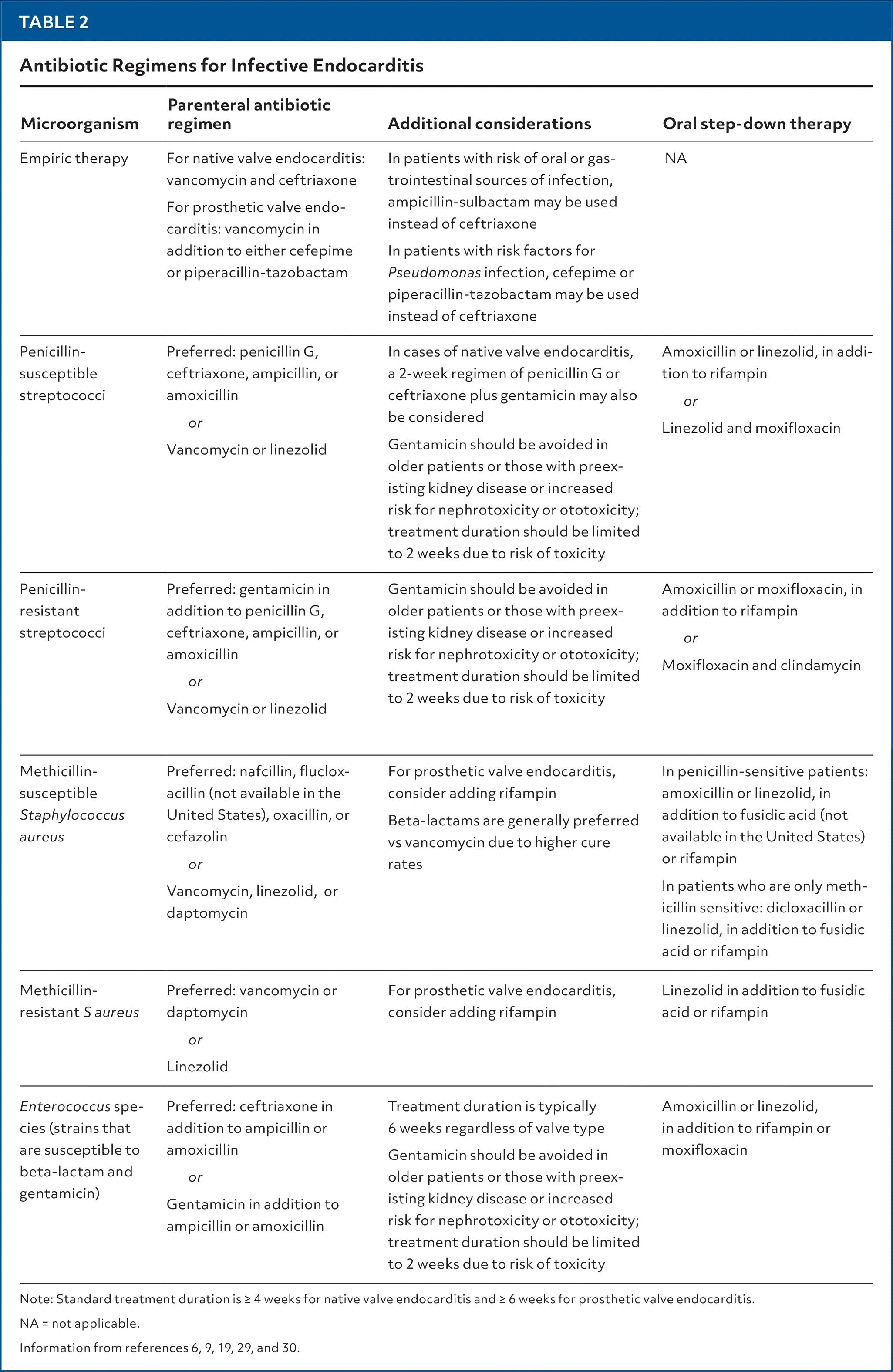
- Native valve: Vancomycin + ceftriaxone is a reasonable empiric regimen [1][4]
- Prosthetic valve: Vancomycin + cefepime or piperacillin-tazobactam (antipseudomonal coverage) [4]
- If GI/odontogenic source suspected: ampicillin-sulbactam may replace ceftriaxone [4]
Definitive therapy (organism-directed)
- MSSA: Nafcillin/oxacillin 12 g/day IV ÷ 4–6 doses × 4–6 weeks; cefazolin 6 g/day ÷ 3 doses is an alternative. Beta-lactams preferred over vancomycin for higher cure rates [1][4][7]
- MRSA: Vancomycin (target AUC 400–600) or daptomycin 10 mg/kg/day × 6 weeks [1]
- Viridans streptococci (penicillin MIC ≤0.12): Penicillin G or ceftriaxone × 4 weeks; 2-week short course with gentamicin is an option in select patients [1]
- Enterococci: Ampicillin + ceftriaxone × 6 weeks (preferred for high-level aminoglycoside resistance or renal risk); ampicillin + gentamicin is an alternative [1]
Duration: ≥4 weeks for native valve IE; ≥6 weeks for prosthetic valve IE and enterococcal IE [4]
Medication cautions
- Aminoglycosides: Avoid in elderly, renal disease, or concurrent nephrotoxic agents; limit to ≤2 weeks [1]
- Rifampin: Reserve for definitive therapy in prosthetic valve IE; do not use empirically [8]
- Daptomycin: Inactivated by pulmonary surfactant — do not use for pneumonia/pulmonary infections
- Combination nafcillin + gentamicin or rifampin for MSSA NVE does not improve outcomes and increases toxicity [1]
The following table summarizes antibiotic regimens by pathogen:
Diet
- No specific dietary triggers or restrictions unique to endocarditis
- Adequate nutrition is critical during prolonged hospitalization and IV antibiotic therapy — consider nutritional support for malnourished or cachectic patients
- Hydration: Maintain adequate hydration, particularly with nephrotoxic antibiotics (vancomycin, gentamicin)
- Patients with heart failure from valvular dysfunction should follow sodium and fluid restriction
Review of Systems
- Cardiovascular: Dyspnea, orthopnea, PND, palpitations, chest pain, syncope
- Neurologic: Headache, focal weakness, vision changes, confusion, seizures
- Musculoskeletal: Back pain (spondylodiscitis), arthralgias, myalgias
- Dermatologic: Petechiae, painful finger/toe nodules, painless palmar/plantar lesions, splinter hemorrhages
- Renal/GU: Hematuria, flank pain, decreased urine output
- Pulmonary: Cough, hemoptysis, pleuritic chest pain (right-sided IE with septic pulmonary emboli)
- Ophthalmologic: Visual changes (Roth spots)
- Constitutional: Fever, weight loss, night sweats, fatigue
Collateral History and Family History
- Collateral: Injection drug use practices (frequency, injection sites, needle sharing, water source), recent dental work, recent hospitalization or procedures, indwelling catheters, prior episodes of IE
- Social context: Housing status (homelessness associated with Bartonella quintana), farm animal exposure (Coxiella burnetii) [1]
- Family history: Congenital heart disease, rheumatic heart disease
- Medication adherence history: Critical for planning outpatient parenteral antibiotic therapy (OPAT) or oral step-down
Risk Factors
- Cardiac: Prior IE (strongest risk factor), prosthetic heart valve, valvular heart disease (degenerative, rheumatic, bicuspid aortic valve), congenital heart disease, hypertrophic cardiomyopathy, CIEDs, ventricular assist devices [1-3]
- Non-cardiac: IV drug use, chronic hemodialysis, indwelling intravascular catheters, poor dentition/dental pathology, immunosuppression, diabetes, chronic liver disease, malignancy, recent hospitalization >48 hours [1-2]
- Demographic: Age >60, male sex [2]
- Microbiologic: S. aureus bacteremia carries ~25–30% risk of concurrent IE
Differential Diagnosis
- Sepsis from other sources (pneumonia, UTI, intra-abdominal abscess) — most common mimic; distinguished by echocardiographic findings and persistent bacteremia
- Non-bacterial thrombotic (marantic) endocarditis — associated with malignancy, SLE (Libman-Sacks); culture-negative, no fever
- Rheumatic fever — migratory polyarthritis, erythema marginatum, Sydenham chorea; Jones criteria; ASO titers
- Systemic vasculitis (polyarteritis nodosa, granulomatosis with polyangiitis) — can mimic embolic phenomena
- Atrial myxoma — intracardiac mass on echo, embolic events, constitutional symptoms; distinguished by tissue pathology
- Antiphospholipid syndrome — thrombotic events, valve thickening
- Fever of unknown origin with culture-negative bacteremia — consider Coxiella, Bartonella, Tropheryma whipplei, fungi [1]
Past Medical History
- Prior endocarditis — recurrence risk is significant
- Valvular disease: Rheumatic heart disease, mitral valve prolapse, degenerative aortic stenosis, bicuspid aortic valve
- Prosthetic valves or cardiac devices — early PVE (<12 months) vs. late PVE have different microbiology
- Congenital heart disease (VSD, unrepaired cyanotic lesions)
- Chronic kidney disease / hemodialysis
- Diabetes, liver cirrhosis, HIV/immunosuppression
- History of IV drug use — duration, current status, prior treatment
- Recent surgical or dental procedures
Physical Exam
- Vitals: Fever (≥38°C in ~78% of cases), tachycardia, hypotension (septic/cardiogenic shock), tachypnea [3]
- Cardiac: New or changing murmur (64.5% of cases) — particularly new aortic regurgitation murmur; signs of heart failure (JVD, S3, pulmonary crackles, peripheral edema) [3-4]
- Skin/extremities:
- Petechiae (conjunctival, oral mucosa, extremities)
- Janeway lesions — painless erythematous macules on palms/soles (septic emboli)
- Osler nodes — painful nodules on finger/toe pads (immune complex)
- Splinter hemorrhages (linear subungual)
- Eyes: Roth spots on fundoscopy (white-centered retinal hemorrhages)
- Abdomen: Splenomegaly (present in ~15–30% of subacute IE), LUQ tenderness (splenic infarct/abscess)
- Neurologic: Focal deficits (embolic stroke), meningismus, altered mental status
- Injection sites: Track marks, skin abscesses, cellulitis in PWID
Lab Studies
- Blood cultures: The cornerstone of diagnosis — obtain ≥3 sets from separate venipuncture sites, ideally 30 minutes apart, before starting antibiotics. ~90–95% of native valve IE is culture-positive [1]
- CBC: Leukocytosis (acute), anemia of chronic disease (subacute), thrombocytopenia (DIC)
- Inflammatory markers: Elevated ESR, CRP, procalcitonin
- BMP/CMP: Creatinine (renal emboli, drug nephrotoxicity, glomerulonephritis), electrolytes
- Urinalysis: Hematuria, proteinuria, RBC casts (immune-complex glomerulonephritis)
- Rheumatoid factor: Positive in ~50% of subacute IE (minor Duke criterion)
- Complement levels: May be low (immune complex consumption)
- Coagulation studies: PT/INR, aPTT (DIC screening)
- Lactate: If septic shock suspected
- Culture-negative workup: PCR for Coxiella burnetii, Bartonella spp., Tropheryma whipplei; Coxiella phase I IgG titer; Bartonella IFA IgG/IgM [1][4][9]
- Monitoring: Repeat blood cultures every 24–48 hours until clearance; vancomycin AUC/trough levels; weekly CBC, BMP for drug toxicity
Imaging
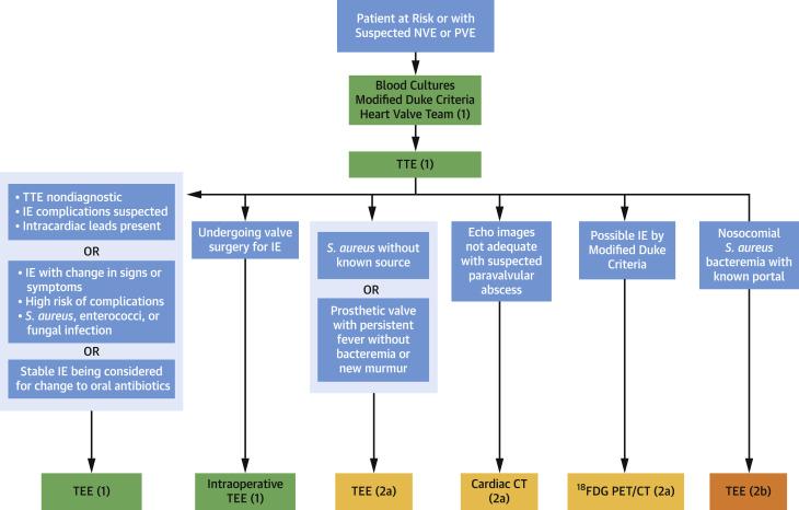
- First-line: Transthoracic echocardiography (TTE) — sensitivity ~70% for native valves, ~50% for prosthetic valves; specificity ~94% [4][7]
- Transesophageal echocardiography (TEE): Sensitivity ~90–96% for both native and prosthetic valves. Indicated when: [4-5][7]
- TTE is negative or equivocal but clinical suspicion remains high
- Prosthetic valve or intracardiac device
- Suspected complications (abscess, fistula, perforation)
- S. aureus bacteremia
- Cardiac CT: Useful for detecting abscesses, pseudoaneurysms, and surgical planning; less sensitive for small (<4 mm) vegetations [4]
- ¹⁸F-FDG PET/CT: Incorporated into 2023 Duke-ISCVID criteria; particularly valuable for prosthetic valve IE, CIED infections, and culture-negative cases [3][9]
- Brain MRI: For neurologic symptoms — detects silent cerebral emboli in up to 60–80% of left-sided IE even without symptoms
- CT abdomen/pelvis: Splenic abscess/infarct, renal infarct, mycotic aneurysm
- CT chest: Septic pulmonary emboli in right-sided IE
- When imaging is unnecessary: If TTE is clearly positive with uncomplicated native valve IE and low clinical suspicion for complications, TEE may not add value
The following figure illustrates the diagnostic algorithm for IE:
Special Tests
- Modified Duke Criteria / 2023 Duke-ISCVID Criteria: The standard diagnostic framework — classifies IE as definite, possible, or rejected based on major criteria (positive blood cultures, imaging evidence of endocardial involvement) and minor criteria (predisposition, fever, vascular phenomena, immunologic phenomena, supportive microbiology) [3][7][9]
- Definite IE: 2 major criteria, or 1 major + 3 minor, or 5 minor criteria
- Possible IE: 1 major + 1 minor, or 3 minor criteria
- Intraoperative inspection: Added as a new major clinical criterion in the 2023 Duke-ISCVID update [9]
- Point-of-care ultrasound (POCUS): Can identify large vegetations, pericardial effusion, and gross valvular dysfunction at bedside in the ED
- Dental evaluation: Assess for dental abscess or periodontal disease as source
ECG
- Indications: Obtain on all patients with suspected IE
- New PR prolongation or heart block: Highly concerning for perivalvular/aortic root abscess extending into the conduction system — this is a surgical emergency [3][5]
- ST changes: May indicate coronary embolization causing myocardial infarction (rare)
- Atrial fibrillation: May develop secondary to atrial dilation from valvular regurgitation
- Serial ECGs recommended to monitor for evolving conduction abnormalities
Assessment
Severity stratification
- In-hospital mortality: ~15–20% overall; higher with S. aureus (~25–40%), prosthetic valve IE, and left-sided IE [3]
- 1-year mortality: ~30% [3]
- Typical presentation: Fever + new murmur + positive blood cultures with a typical organism
- Atypical presentations: Subacute with only fatigue and weight loss (elderly, immunocompromised); initial presentation as stroke, renal infarct, or spondylodiscitis without obvious cardiac symptoms; culture-negative IE [4]
- Complications to anticipate: Heart failure (most common indication for surgery), embolic events (stroke, splenic/renal infarcts), perivalvular abscess, mycotic aneurysm, renal failure, septic shock [3]
Treatment Plan
Initial stabilization
- ABCs, IV access, hemodynamic monitoring
- Obtain blood cultures before antibiotics, then start empiric IV antibiotics promptly in acute presentations; in subacute presentations, may wait for culture results if hemodynamically stable [3]
- Fluid resuscitation and vasopressors for septic shock
Empiric antibiotics
- Native valve: Vancomycin + ceftriaxone [1][4]
- Prosthetic valve: Vancomycin + cefepime or piperacillin-tazobactam [4]
- Narrow to definitive therapy once cultures and sensitivities return [4]
Source control
- Remove infected intravascular catheters, lines, and devices
- Evaluate for abscess drainage [4]
Surgical indications (Class I): [5-6]
- Heart failure from valve dysfunction
- Perivalvular abscess, heart block, fistula, destructive penetrating lesions
- Persistent bacteremia/fever >5–7 days on appropriate antibiotics
- Fungal or highly resistant organisms
- Recurrent emboli with persistent vegetations despite antibiotics
- Emergency (within 24 hours): Refractory pulmonary edema or cardiogenic shock
- Urgent (within 3–5 days): Uncontrolled infection, large vegetations with embolic events
- Elective: After 1–2 weeks of antibiotics in stable patients
Oral step-down (POET trial): In stable patients who are afebrile with negative blood cultures and no surgical indications, transition to oral antibiotics during the continuation phase (after ≥7–10 days IV) may be considered [3-4]
Multidisciplinary team: Infectious disease, cardiology, and cardiothoracic surgery should be involved early — associated with improved survival [4][11]
Disposition
- All patients with confirmed or suspected IE require hospital admission — typically to a telemetry or ICU bed depending on hemodynamic status
- ICU admission criteria: Septic shock, cardiogenic shock, acute heart failure, new neurologic deficits, need for vasopressors or mechanical circulatory support
- Transfer to tertiary center: If surgical capability is not available and surgical indications are present or anticipated [12]
- Discharge criteria: Afebrile, blood cultures negative, hemodynamically stable, no surgical indications, reliable outpatient IV access (PICC line) and OPAT infrastructure in place, or meeting criteria for oral step-down
- Observation: Not appropriate — IE requires inpatient management
Specialist consultation triggers
- Infectious disease: All cases [4]
- Cardiothoracic surgery: All complicated IE, large vegetations, prosthetic valve IE
- Cardiology: Echocardiographic assessment, hemodynamic management
- Neurology/neurosurgery: Stroke, intracranial hemorrhage, mycotic aneurysm
- Addiction medicine: PWID
Follow Up / Return Precautions
- Follow-up timing: Clinical and laboratory reassessment within 1–2 weeks of discharge; repeat echocardiography at completion of antibiotic therapy to establish new baseline; follow-up at 1, 3, 6, and 12 months [11]
- Symptoms requiring immediate reassessment:
- Recurrent fever or chills
- New neurologic symptoms (weakness, speech difficulty, severe headache)
- Worsening dyspnea, chest pain, or edema
- Signs of line infection (erythema, drainage at PICC site)
- Patient counseling:
- Complete the full antibiotic course — do not stop early
- Antibiotic prophylaxis before dental procedures is recommended for patients with a history of IE [4]
- Maintain meticulous oral and skin hygiene to reduce reinfection risk [4]
- Avoid IV drug use; connect with addiction medicine and harm reduction services if applicable
- Expected recovery: Clinical improvement typically within 5–7 days of effective antibiotics; full antibiotic course is 4–6 weeks; recurrence risk is ~2–6% per year
- Long-term monitoring: Surveillance for late valve dysfunction, heart failure, and recurrent IE
References
1. Native-Valve Infective Endocarditis. — Chambers HF, Bayer AS. The New England Journal of Medicine. 2020.
2. Management Considerations in Infective Endocarditis: A Review. — Wang A, Gaca JG, Chu VH. The Journal of the American Medical Association. 2018.
3. Infective Endocarditis. — Li M, Kim JB, Sastry BKS, Chen M. Lancet. 2024.
4. Infective Endocarditis: Diagnosis and Treatment. — Nohria R, Romaine A, Garcia-Sampson G. American Family Physician. 2026.
5. Infective Endocarditis in Adults: Diagnosis, Antimicrobial Therapy, and Management of Complications: A Scientific Statement for Healthcare Professionals From the American Heart Association. — Baddour LM, Wilson WR, Bayer AS, et al. Circulation. 2015.
6. Challenges in Infective Endocarditis. — Cahill TJ, Baddour LM, Habib G, et al. Journal of the American College of Cardiology. 2017.
7. Diagnosis and Management of Infective Endocarditis in People Who Inject Drugs: JACC State-of-the-Art Review. — Yucel E, Bearnot B, Paras ML, et al. Journal of the American College of Cardiology. 2022.
8. Guidelines for Diagnosis and Management of Infective Endocarditis in Adults: A WikiGuidelines Group Consensus Statement. — McDonald EG, Aggrey G, Aslan AT, et al. JAMA Network Open. 2023.
9. The 2023 Duke-International Society for Cardiovascular Infectious Diseases Criteria for Infective Endocarditis: Updating the Modified Duke Criteria. — Fowler VG, Durack DT, Selton-Suty C, et al. Clinical Infectious Diseases : An Official Publication of the Infectious Diseases Society of America. 2023.
10. Best Practices for Imaging Cardiac Device-Related Infections and Endocarditis: A JACC: Cardiovascular Imaging Expert Panel Statement. — Dilsizian V, Budde RPJ, Chen W, et al. JACC. Cardiovascular Imaging. 2022.
11. Surgical Implications of the 2023 ESC Endocarditis Guidelines Endorsed by EACTS: Bridging Guidelines and Practice. — de la Cuesta M, Marin-Cuartas M, de Waha S, et al. European Journal of Cardio-Thoracic Surgery : Official Journal of the European Association for Cardio-Thoracic Surgery. 2025.
12. Staphylococcus Aureus Infective Endocarditis: JACC Patient Pathways. — Grapsa J, Blauth C, Chandrashekhar YS, et al. Journal of the American College of Cardiology. 2022.
Figures
More from Deep Dive
Reviewed by Dr. Lucas Mastropaolo. SymptomDx is an educational tool for medical professionals — it does not replace clinical judgment. Verify all clinical data and drug dosages with authoritative sources.