Bladder Rupture
History
- Mechanism of injury: Blunt abdominal/pelvic trauma (most common — MVC, falls, crush injuries), penetrating trauma (GSW, stab wounds), or spontaneous rupture [1-2]
- Key HPI questions:
- Timing and mechanism of trauma; was the bladder distended at time of injury?
- Presence and character of hematuria (gross vs. microscopic)
- Ability to void since injury; urine output
- Abdominal pain — location, onset, progression
- Alcohol intoxication at time of injury (risk factor for both trauma and spontaneous rupture) [3-4]
- Prior pelvic radiation, bladder surgery, or neurogenic bladder
- Last void before injury (distended bladder increases risk of intraperitoneal rupture at the dome) [1]
- Symptom characterization: Abdominal pain (76% of spontaneous cases), progressive abdominal distension, oliguria/anuria, inability to void, suprapubic tenderness [1][3]
- Associated symptoms: Dyspnea (from abdominal distension), nausea, shoulder pain (referred from diaphragmatic irritation by urinary ascites)
Alarm Features
- Gross hematuria with pelvic fracture — bladder injury present in ~30% of these cases [2]
- Inability to void or markedly decreased urine output
- Progressive abdominal distension with peritoneal signs → suspect intraperitoneal rupture with urinary peritonitis [1][4]
- Rising creatinine/BUN disproportionate to clinical picture → "pseudo-AKI" from peritoneal reabsorption of urine ("reverse peritoneal dialysis") [4]
- Hemodynamic instability with pelvic fracture
- Signs of sepsis or frank peritonitis
- Pneumoperitoneum on imaging without obvious hollow viscus injury [5]
Medications
- Anticoagulants/antiplatelets: May worsen associated hemorrhage; assess need for reversal
- Antibiotics: Broad-spectrum coverage indicated for intraperitoneal rupture (risk of peritonitis/urosepsis); typically a fluoroquinolone or cephalosporin perioperatively
- Avoid nephrotoxic agents: Creatinine may be falsely elevated from urinary reabsorption — avoid unnecessary dose adjustments or dialysis before confirming true renal function [4]
- Anticholinergics/bladder relaxants: May mask urinary retention symptoms
Diet
- NPO if surgical repair anticipated
- No specific dietary triggers; however, alcohol binge drinking is a major associated factor for spontaneous rupture (11% of cases) due to bladder overdistension with impaired sensation [3][6]
- Post-repair: Adequate hydration to maintain catheter patency
Review of Systems
- GU: Hematuria (gross or microscopic), dysuria, urinary retention, decreased output
- GI: Abdominal pain, distension, nausea/vomiting, ileus symptoms
- MSK: Pelvic/hip pain (associated pelvic fracture)
- Neuro: Perineal/saddle numbness (concomitant urethral or nerve injury)
- Constitutional: Fever, tachycardia (infection/sepsis)
Collateral History and Family History
- Witnesses to mechanism (especially in intoxicated patients)
- Pre-hospital fluid resuscitation and catheterization attempts
- No hereditary component, but relevant comorbidities include:
Risk Factors
- Pelvic fracture — 60–90% of extraperitoneal ruptures have associated pelvic fracture; 6–8% of pelvic fractures have bladder injury [2]
- Distended bladder at time of blunt force (seatbelt injury, assault) → intraperitoneal dome rupture [1]
- Alcohol intoxication — impaired voiding sensation + overdistension [3]
- Pelvic radiation — weakened bladder wall [3]
- Neurogenic bladder / chronic retention
- Bladder pathology: Diverticula, tumors, prior surgery, augmentation cystoplasty [1][7]
- Pediatric patients: More susceptible due to anatomy (bladder more abdominal than pelvic) [2]
Differential Diagnosis
- Renal injury (also presents with hematuria post-trauma)
- Ureteral injury (rare in blunt trauma; consider in deceleration injuries)
- Urethral injury (blood at meatus, high-riding prostate — must exclude before catheterization)
- Hollow viscus injury (bowel perforation — peritonitis, pneumoperitoneum)
- Intra-abdominal hemorrhage (solid organ injury — liver, spleen)
- Spontaneous bacterial peritonitis (in patients with ascites)
- Acute kidney injury (true vs. pseudo-AKI from urinary reabsorption — ascites/serum creatinine ratio >2 indicates urinary ascites) [4]
- Ruptured bladder diverticulum [7]
Past Medical History
- Prior pelvic/bladder surgery or radiation
- Neurogenic bladder, BPH, urethral stricture
- Chronic urinary retention or catheterization history
- Bladder augmentation, neobladder, or continent reservoir (complex anatomy increases complication risk) [1]
- Coagulopathy or anticoagulant use
- Prior episodes of bladder rupture
Physical Exam
- Vitals: Tachycardia, hypotension (hemorrhage or sepsis); fever (peritonitis)
- Abdomen: Suprapubic tenderness, distension, guarding, rebound (intraperitoneal rupture → peritonitis); diminished bowel sounds (ileus)
- Pelvis: Instability on compression (pelvic fracture); perineal ecchymosis
- GU: Blood at urethral meatus (urethral injury — do not catheterize until urethrogram performed); scrotal/perineal swelling (complex extraperitoneal extravasation)
- Rectal: High-riding prostate (urethral disruption); rectal injury
- Vaginal exam (if indicated): Assess for concomitant vaginal laceration [1]
Lab Studies
- Urinalysis: Gross hematuria is the cardinal finding; microhematuria alone with low-risk pelvic fracture generally does not warrant cystography [1-2]
- BMP/CMP: Elevated creatinine and BUN (may be pseudo-AKI from peritoneal reabsorption); hyperkalemia, hyponatremia [4]
- CBC: Anemia from hemorrhage; leukocytosis (infection)
- Coagulation studies: PT/INR, PTT
- Type and screen/crossmatch: If significant hemorrhage or surgical repair anticipated
- Lactate: Assess for shock/sepsis
- Ascitic fluid analysis (if paracentesis performed): Ascites/serum creatinine ratio >2 is highly suggestive of urinary ascites [4]
Imaging
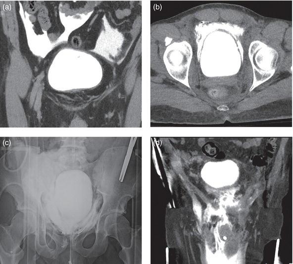
The following figure illustrates the key imaging findings distinguishing intraperitoneal from extraperitoneal bladder rupture on CT and fluoroscopic cystography:
- Gold standard: Retrograde CT cystography — sensitivity 95%, specificity ~100% [1][9-10]
- Requires active retrograde filling via Foley catheter with ≥300 mL diluted contrast; passive filling/delayed CT is inadequate (high false-negative rate) [2]
- Intraperitoneal rupture: Contrast outlines bowel loops, fills paracolic gutters
- Extraperitoneal rupture: Flame-shaped or "molar tooth" contrast extravasation confined to perivesical/pelvic space; may track to scrotum/perineum in complex injuries
- Fluoroscopic cystography: Similar sensitivity/specificity; used when CT unavailable [9]
- CT abdomen/pelvis with IV contrast (standard trauma CT): May show free fluid but cannot reliably diagnose bladder rupture without retrograde filling [2][11]
- Retrograde urethrogram (RUG): Must be performed before catheterization if urethral injury suspected [9]
- When imaging is unnecessary: Microhematuria alone without pelvic fracture or high-risk features [1][9]
Special Tests
- AAST Bladder Injury Grading Scale: [12]
- Grade 1: Contusion or partial-thickness laceration
- Grade 2: Extraperitoneal laceration <2 cm
- Grade 3: Extraperitoneal >2 cm or intraperitoneal <2 cm
- Grade 4: Intraperitoneal >2 cm
- Grade 5: Laceration extending into bladder neck or ureteric orifice
- Ascites/serum creatinine ratio: >2 confirms urinary ascites (spontaneous rupture) [4]
- Intraoperative methylene blue or indigo carmine instillation: Useful to identify injury site during laparotomy [2]
- E-FAST: May detect free fluid but cannot differentiate urine from blood; low specificity for bladder injury
ECG
- ECG indicated if hyperkalemia is present (pseudo-AKI from urinary reabsorption can cause K⁺ >6.0 mmol/L) [4]
- Look for peaked T waves, widened QRS, sine wave pattern
- Also indicated in polytrauma patients for cardiac contusion screening
Assessment
Bladder rupture is classified as extraperitoneal (most common, ~60–90% of traumatic cases, strongly associated with pelvic fractures) or intraperitoneal (~15–25% of traumatic cases, typically at the dome from sudden intravesical pressure rise). [1-2] Combined injuries occur in <5–12%. [2] Spontaneous rupture is rare but carries 15% mortality and is misdiagnosed in 64% of cases. [3] Key complications include urinary peritonitis, sepsis, pseudo-AKI, electrolyte derangements (hyperkalemia, hyponatremia), fistula formation, and abscess. [4][10]
Treatment Plan
Intraperitoneal rupture
- Operative repair is strongly recommended (strong recommendation per EAST, AUA, ACS, WSES-AAST) [1][10][13]
- Midline laparotomy; two-layer closure with absorbable 2-0 or 3-0 suture [1]
- Inspect for concomitant extraperitoneal injury via transvesical approach; inspect ureteral orifices [1]
- Place closed-suction pelvic drain; Foley catheter drainage postoperatively [1]
- 100% successful closure rate with operative repair [10]
Uncomplicated extraperitoneal rupture
- Nonoperative management with large-bore urethral catheter (≥18 Fr) drainage for minimum 7 days (typically 2–3 weeks) [1][10][13]
- Only 2.4% ultimately require operative intervention [10]
- Follow-up cystography to confirm healing before catheter removal [13]
Complicated extraperitoneal rupture (bladder neck injury, bone fragments in lumen, concomitant rectal/vaginal injury, pelvic hardware planned):
Penetrating injuries: Generally require operative repair [1]
Spontaneous rupture: Surgical repair with evacuation of urinary ascites; electrolytes and creatinine normalize rapidly (within 48 hours) post-repair [4]
Disposition
- Admit all confirmed bladder ruptures
- ICU if hemodynamically unstable, septic, or significant polytrauma
- OR urgently for intraperitoneal rupture, complicated extraperitoneal rupture, or penetrating injury [1][10]
- Floor/observation for uncomplicated extraperitoneal rupture managed with catheter drainage
- Urology consultation for all bladder injuries; trauma surgery if polytrauma
- Consider tertiary referral for patients with complex bladder anatomy (neobladder, augmentation cystoplasty) [1]
Follow Up / Return Precautions
- Follow-up cystography at minimum 7 days post-repair (may extend to 3–4 weeks for complex injuries) to confirm healing before catheter removal [1]
- For nonoperative management: cystography at 7–14 days; if persistent leak, continue drainage and repeat; consider operative repair if not healed by 4 weeks [13]
- Return precautions: Fever, worsening abdominal pain/distension, decreased urine output, hematuria recurrence, signs of wound infection
- Expected recovery: Most uncomplicated injuries heal within 2–3 weeks; catheter removed after confirmed cystographic healing [13]
- Long-term: Monitor for urinary incontinence, recurrent UTI, or fistula formation in complex injuries
References
1. Best Practices Guidelines Management of Gentiunrinary Injuries. — Niels Johnsen, Hunter Wessells, Krystal Archer-Arroyo, et al American College of Surgeons (2025). 2025.
2. Kidney and Uro-Trauma: WSES-AAST Guidelines. — Coccolini F, Moore EE, Kluger Y, et al. World Journal of Emergency Surgery : WJES. 2019.
3. Spontaneous (Idiopathic) Rupture of the Urinary Bladder: A Systematic Review of Case Series and Reports. — Reddy D, Laher AE, Lawrentschuk N, Adam A. BJU International. 2023.
4. Spontaneous Bladder Rupture Induced Pseudo-Acute Kidney Injury: A Case Report and Literature Review. — Yu T, Cai C, Shi J, Jiang YJ. Medicine. 2025.
5. Intraperitoneal Urinary Bladder Rupture as a Cause of Pneumoperitoneum. — Parvez M D, Supreet K, Ajay S, Subodh K. The American Surgeon. 2023.
6. An Arterial Perihepatic Enhancement Caused by Spontaneous Bladder Rupture. — Harada T, Suyama Y, Hiratsuka Y, Shimizu T. Urology. 2017.
7. Spontaneous Rupture of Bladder Diverticulum With Pseudo Renal Failure:A Case Report and Literature Review. — Zhang Z, Shen J, He Q, Nie H. The American Journal of Emergency Medicine. 2024.
8. Bladder Injuries. — Yosuke Nakajima A Clinical Guide to Urologic Emergencies. 2021.
9. ACR Appropriateness Criteria® Minor Blunt Trauma. — Expert Panel on Polytrauma Imaging, Hoff CN, Hajibonabi F, et al. Journal of the American College of Radiology : JACR. 2026.
10. Management of Blunt Force Bladder Injuries: A Practice Management Guideline From the Eastern Association for the Surgery of Trauma. — Yeung LL, McDonald AA, Como JJ, et al. The Journal of Trauma and Acute Care Surgery. 2019.
11. ACR Appropriateness Criteria® Major Blunt Trauma. — Shyu JY, Khurana B, Soto JA, et al. Journal of the American College of Radiology : JACR. 2020.
12. ACR Appropriateness Criteria® Penetrating Trauma-Lower Abdomen and Pelvis. — Expert Panel on Urological Imaging, Heller MT, Oto A, et al. Journal of the American College of Radiology : JACR. 2019.
13. Urotrauma Guideline 2020: AUA Guideline. — Morey AF, Broghammer JA, Hollowell CMP, McKibben MJ, Souter L. The Journal of Urology. 2021.
Figure
More from Deep Dive
Reviewed by Dr. Lucas Mastropaolo. SymptomDx is an educational tool for medical professionals — it does not replace clinical judgment. Verify all clinical data and drug dosages with authoritative sources.