Decompression Sickness
Decompression sickness results from bubble formation in blood and tissues when dissolved inert gas (usually nitrogen) comes out of solution during a reduction in ambient pressure, most commonly after scuba diving, but also in aviation and spaceflight. [1-2] The incidence in recreational scuba diving is 0.4–1 per 10,000 dives, but risk increases substantially with deeper, longer, or repetitive dives and decompression violations. [1]
History
- Dive profile details: depth, bottom time, ascent rate, number of dives that day/week, gas mixture used, compliance with dive tables or computer
- Decompression violations: missed safety stops, rapid ascent, out-of-air emergency
- Symptom onset timing: 73% of mild and 98% of severe cases present within 1 hour of surfacing; 99% within 6 hours. CNS cases present fastest — 56% within 10 minutes [1-2]
- Symptom characterization: deep boring joint pain ("the bends"), tingling/numbness (nondermatomal), weakness, vertigo, cognitive changes, rash, dyspnea, fatigue/malaise [1]
- Post-dive altitude exposure: commercial flight or mountain driving after diving can precipitate or worsen DCS — a common scenario in vacation divers [1][3]
- Important negatives: symptoms during descent or at depth are never DCS unless preceded by recent diving [2]
Alarm Features
- Spinal cord involvement: paraparesis/paraplegia, urinary retention, girdle pain (premonitory for cord DCS) [1][4]
- Cerebral symptoms: altered consciousness, confusion, seizure, focal deficits — consider concurrent arterial gas embolism (AGE) [2]
- "The chokes": dyspnea, cough, retrosternal pain — indicates massive pulmonary vascular bubble load [1][4]
- Cardiovascular collapse: hemoconcentration, shock, coagulopathy — rare but life-threatening [1]
- Inner ear DCS: acute vertigo, nystagmus, hearing loss — more common after deep dives [1]
- Rapid symptom progression: mild symptoms can evolve into severe neurological DCS within hours [2]
- Cutis marmorata: reticular, cyanotic truncal rash — almost pathognomonic for DCS and signals systemic involvement [2]
Medications
- First-line treatment: 100% oxygen via nonrebreather or demand valve — 14% complete symptom relief, 51% symptom reduction in one large series [1]
- Adjunctive NSAIDs: tenoxicam reduced median recompression sessions from 3 to 2 in an RCT [1][5]
- IV lidocaine: considered in severe neurological AGE cases; neuroprotective in animal models but not standard of care [1-2]
- DVT prophylaxis: fractionated heparin for immobile patients with spinal cord DCS [1]
- Contraindicated: Nitrous oxide — causes bubble expansion by inward diffusion and is absolutely contraindicated [2]
- Avoid: glucose-containing IV fluids (risk of hyperglycemia worsening neurological injury), hypotonic fluids (promote intracellular edema) [2]
- Aspirin: previously advocated for antiplatelet effects but not formally studied and not recommended [2]
- Steroids: historically used but lack evidence; not standard of care [5]
Diet
- Hydration is critical: divers are often dehydrated from immersion diuresis; predive hydration reduces postdive venous gas emboli [1]
- Acute: oral noncarbonated, noncaffeinated, nonalcoholic fluids with sodium and glucose if conscious; water acceptable if nothing else available [1]
- Avoid: alcohol and carbonated beverages acutely
- Long-term: adequate hydration before and after all dives; dehydration increases DCS severity in animal models [2]
Review of Systems
- Neurological: weakness, numbness, tingling (nondermatomal), gait instability, cognitive changes, visual disturbances, urinary retention, bowel/bladder dysfunction [1]
- Musculoskeletal: deep joint pain (shoulder, elbow, hip, knee) — present in 50–65% of cases [1]
- Vestibular/cochlear: vertigo, nausea, nystagmus, hearing loss, tinnitus [1]
- Cutaneous: rash, pruritus, mottled skin [1]
- Cardiopulmonary: dyspnea, cough, chest pain [1]
- Constitutional: fatigue, malaise, headache — present in 20–40%, often described as "viral illness" feeling [1]
- Lymphatic: truncal swelling, particularly upper chest and shoulders [1]
Collateral History and Family History
- Dive buddy information: corroborate dive profile, ascent rate, any in-water problems, symptom onset timing
- Dive computer data: download if available — provides objective depth-time profile
- Prior DCS episodes: history of DCS is a risk factor for recurrence [1]
- Patent foramen ovale (PFO): present in ~25–30% of adults; high-grade PFO increases DCS risk 3- to 13-fold, particularly for neurological, cutaneous, and inner ear DCS. PFO prevalence was 97.2% in divers with unprovoked DCS vs. 35.5% in controls in one study [1][6-8]
- Family history of cardiac septal defects: may suggest PFO or ASD
Risk Factors
- Dive-related: deeper dives (>21 m), longer bottom times, rapid ascent, missed decompression stops, repetitive dives, cold water, heavy exertion during dive [1]
- Physiological: dehydration, obesity (nitrogen is more soluble in fat), older age, prior DCS, poor physical fitness [8]
- Patent foramen ovale: major independent risk factor, especially high-grade shunts [1][6]
- Post-dive altitude exposure: flying <12–18 hours after diving, mountain driving [3]
- Minimum depth threshold: DCS essentially does not occur after dives <6 m (AGE can occur at any depth) [1-2]
Differential Diagnosis
- Arterial gas embolism (AGE): onset typically within minutes of surfacing; altered consciousness, seizure, focal cortical signs; can coexist with DCS; treatment is the same [1-2]
- Musculoskeletal injury: shoulder/knee strain from diving activity — pain typically worsened by movement (DCS pain is not) [1]
- Inner ear barotrauma: vertigo and hearing loss from middle/inner ear pressure injury — distinguished from inner ear DCS by dive profile (barotrauma more common in shallow dives) [1]
- Stroke/TIA: focal neurological deficits — DCS typically shows nondermatomal patterns and truncal ataxia rather than classic stroke syndromes [2]
- Pulmonary barotrauma: pneumothorax, pneumomediastinum, subcutaneous emphysema — from lung overexpansion during ascent [3]
- Marine envenomation or infection: skin findings, pain
- Immersion pulmonary edema: dyspnea during or after diving
- Constitutional symptoms: seasickness, exhaustion, hypothermia, dehydration — common mimics [1]
Past Medical History
- Prior DCS episodes: increases recurrence risk [1]
- Asthma or COPD: air trapping increases AGE risk; relative contraindication to diving [4]
- Known PFO or ASD: major risk factor [1]
- History of spontaneous pneumothorax: absolute contraindication to diving [4]
- Epilepsy, insulin-dependent diabetes: conditions impairing consciousness are contraindications [4]
- Pulmonary blebs, cysts, bullae: increase barotrauma risk [2]
Physical Exam
- Vital signs: hypotension (hemoconcentration/shock in severe cases), tachycardia; oxygen saturation may be normal
- Complete neurological exam is essential: [1-2]
- Mental status and cognitive testing (executive dysfunction often missed)
- Cranial nerves (nystagmus in inner ear DCS)
- Motor strength (paraparesis/quadriparesis)
- Sensory exam: nondermatomal hypoesthesia is characteristic and easily missed [2]
- Coordination: finger-nose, Romberg's sign, sharpened Romberg's test [1]
- Gait and tandem gait (forward and backward, eyes open and closed) — truncal ataxia is common [1-2]
- Proprioception (dorsal columns are vulnerable) [1]
- Skin: cutis marmorata (reticular cyanotic rash, truncal/proximal) — almost pathognomonic; lymphedema of trunk [2]
- Bladder: check for urinary retention (spinal cord DCS) [1]
- Joints: deep ache without local tenderness or inflammatory signs; pain unaffected by movement [1]
Lab Studies
- DCS is a clinical diagnosis — no specific biomarkers exist [1]
- CBC/Hematocrit: hemoconcentration suggests endothelial leak and guides fluid resuscitation in severe cases [2]
- Creatine kinase (CK): elevated CK may help distinguish AGE (high in pulmonary barotrauma) from isolated DCS, though differentiation is unnecessary before recompression [2]
- BMP: assess renal function, electrolytes for fluid management
- Coagulation studies: in severe cases with suspected coagulopathy
- Labs should not delay recompression [1]
Imaging
- Chest X-ray (supine) or chest ultrasound: rule out pneumothorax before recompression, especially if AGE suspected [1-2]
- MRI brain: not sensitive for acute spinal cord DCS despite significant functional impairment; corpus callosum hyperintensities on T2/FLAIR may be seen in cerebral DCS [1][9]
- CT head: not sensitive enough to rule out AGE [1]
- Imaging should NOT delay recompression unless strong suspicion of non-diving cause (e.g., cerebral hemorrhage) [2]
- Joint radiographs: bubbles rarely detectable; not useful [2]
- Bottom line: imaging is generally low-yield and should not drive the decision to recompress [1]
Special Tests
- Bubble contrast echocardiography (TTE with agitated saline): for PFO screening — recommended after unexplained or recurrent DCS, especially with neurological, cutaneous, or inner ear involvement [1][8]
- Transcranial Doppler with bubble contrast: alternative PFO screening method [6]
- Audiometry and electronystagmography: for inner ear DCS — can usually be deferred until after recompression [2]
- Dive computer download: objective depth-time data for risk assessment
- Divers Alert Network (DAN) hotline: 24-hour specialist consultation — (919) 684-8111 [4]
ECG
- Indications: chest pain, dyspnea, hemodynamic instability, or suspected cardiac involvement
- Possible findings: right heart strain pattern if massive venous gas emboli overwhelm pulmonary vasculature; arrhythmias if coronary AGE
- Routine ECG: reasonable in any patient with cardiopulmonary symptoms or severe DCS
- Not typically diagnostic for DCS itself but helps exclude cardiac mimics
Assessment
Severity stratification is the key clinical decision point
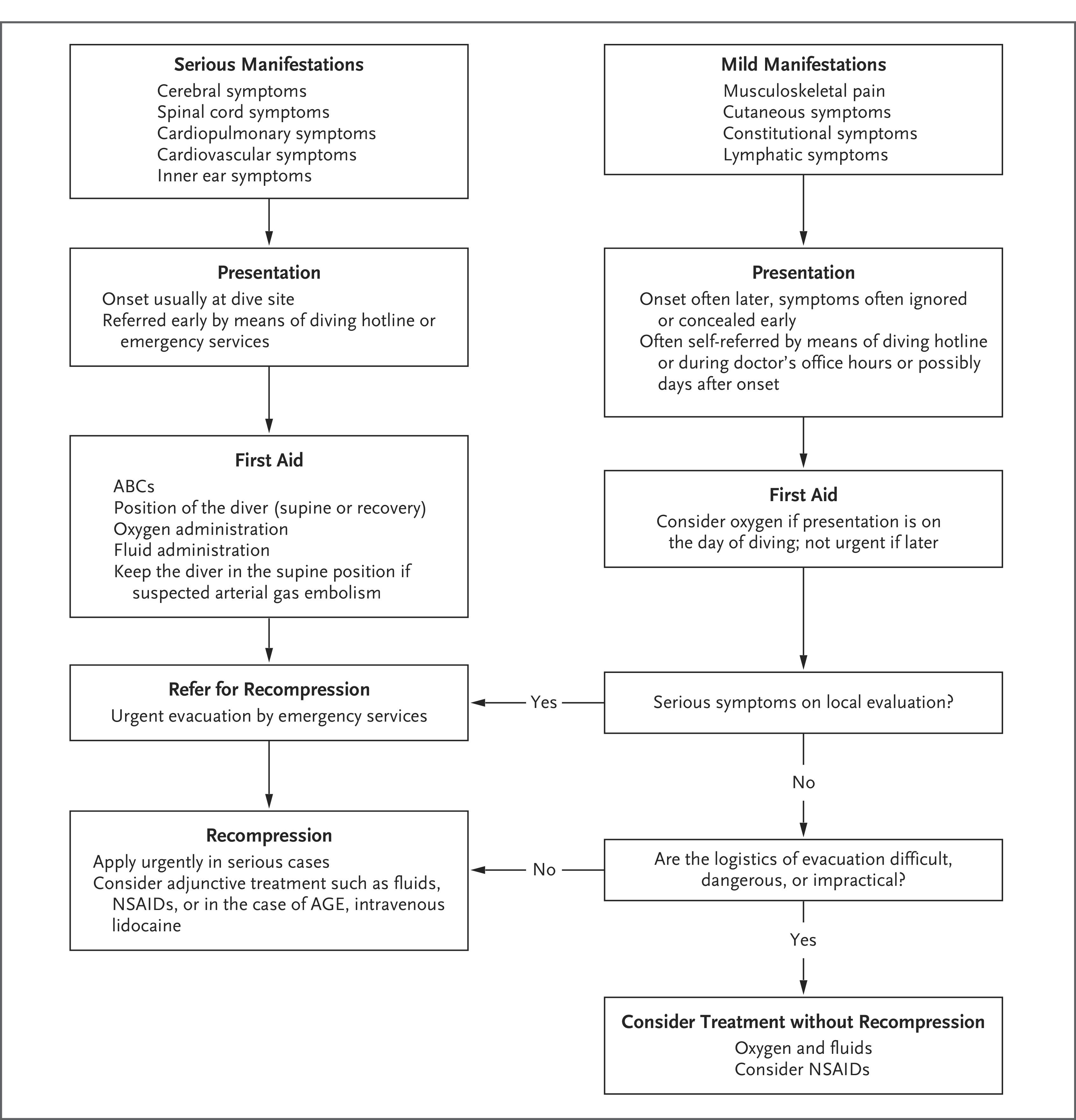
- Mild DCS: musculoskeletal pain, cutaneous symptoms (rash, itch), constitutional symptoms (fatigue, malaise), lymphatic swelling [1]
- Serious DCS: spinal cord involvement (weakness, sensory loss, bladder dysfunction), cerebral symptoms, inner ear DCS, cardiopulmonary symptoms ("the chokes"), cardiovascular collapse [1]
Symptom onset is typically within 1–6 hours of surfacing. Presentations are often nonspecific and easily misattributed to more familiar diagnoses. The cardinal rule:
Treatment Plan
First Aid (Pre-Hospital / ED)
- Position supine (not head-down — promotes cerebral edema) [1-2]
- 100% oxygen via nonrebreather or demand valve — continue even if symptoms resolve [1-2]
- IV fluids: isotonic, glucose-free crystalloids (e.g., normal saline, lactated Ringer's); target UOP >0.5–1 mL/kg/hr. Oral rehydration if conscious and mild [1]
- Contact diving medicine specialist: DAN Emergency Hotline or local hyperbaric facility [1][4]
Definitive Treatment
- Hyperbaric oxygen recompression using U.S. Navy Treatment Table 6: initial compression to 2.8 ATA breathing 100% O₂, total duration ~4 hours 45 minutes [1][5]
- Can be extended or repeated for incomplete response [1]
- Residual symptoms: daily recompressions at 2.0–2.8 ATA until resolution or plateau; most patients need only 2–3 sessions [2]
- Outcomes: 80–86% complete recovery with timely treatment; 97% complete relief when recompression is immediately available [2]
- Delayed recompression (>48 hours) still achieves 76% complete recovery — treatment should never be denied due to delay [10]
Adjunctive
- NSAIDs (e.g., tenoxicam/ibuprofen) may reduce recompression sessions needed [1][5]
- DVT prophylaxis with LMWH for immobile spinal cord DCS patients [1]
The following management algorithm from Mitchell et al. (NEJM 2022) illustrates the decision pathway for mild vs. serious DCS:
Disposition
- Admit / Transfer for recompression: all serious DCS (neurological, inner ear, cardiopulmonary, cardiovascular symptoms) — urgent transfer to nearest hyperbaric facility [1]
- Mild DCS: if symptoms are static or remitting, normal neurological exam, and recompression facility is remote/inaccessible, may be managed with surface O₂ and fluids with close observation — but this decision should involve a diving medicine physician [1][3]
- Post-recompression observation: mild cases — 2 hours within reach of chamber; severe cases — 6 hours; remain within 1 hour of chamber for 24 hours [2]
- Hospital admission: for severe or residual symptoms, hemodynamic instability, or need for ongoing recompression [2]
- Helicopter transport: low altitude (<300 m / 1,000 ft); fixed-wing: pressurized to 1 atm for severe cases [2]
Follow Up / Return Precautions
- Flying restrictions after DCS: [3]
- Complete symptom relief after recompression: no flying for at least 72 hours
- Residual symptoms: consult hyperbaric physician before any altitude exposure
- Symptom recurrence: worsening or new symptoms after treatment require immediate re-evaluation and possible repeat recompression [2]
- Return to diving: requires clearance by a diving medicine specialist; consider PFO screening (especially after neurological, cutaneous, or inner ear DCS) [1][8]
- PFO management: catheter-based PFO closure eliminated unprovoked DCS in long-term follow-up in the DIVE-PFO registry; conservative options include diving within conservative limits or cessation of diving [8][11]
- Patient counseling: educate on proper hydration, conservative dive profiles, slow ascent rates, safety stops, and avoidance of post-dive altitude exposure [3]
- Expected course: most mild cases resolve fully; spinal cord DCS may have long-term residual symptoms (numbness, bladder dysfunction, weakness) in a subset of patients [2]
References
1. Decompression Sickness and Arterial Gas Embolism. — Mitchell SJ, Bennett MH, Moon RE. The New England Journal of Medicine. 2022.
2. Decompression Illness. — Vann RD, Butler FK, Mitchell SJ, Moon RE. Lancet. 2011.
3. Scuba Diving: Decompression Illness and Other Dive-Related Injuries. — James Chimiak and Daniel A. Nord CDC Yellow Book. 2025.
4. Medical Problems Associated with Underwater Diving. — Melamed Y, Shupak A, Bitterman H. The New England Journal of Medicine. 1992.
5. Recompression and Adjunctive Therapy for Decompression Illness. — Bennett MH, Lehm JP, Mitchell SJ, Wasiak J. The Cochrane Database of Systematic Reviews. 2012.
6. Increased Risk of Decompression Sickness When Diving With a Right-to-Left Shunt: Results of a Prospective Single-Blinded Observational Study (The "Carotid Doppler" Study). — Germonpré P, Lafère P, Portier W, et al. Frontiers in Physiology. 2021.
7. Percutaneous Treatment of Patent Foramen Ovale and Atrial Septal Defects. — Tobis J, Shenoda M. Journal of the American College of Cardiology. 2012.
8. Screening and Risk Stratification Strategy Reduced Decompression Sickness Occurrence in Divers With Patent Foramen Ovale. — Honěk J, Šrámek M, Honěk T, et al. JACC. Cardiovascular Imaging. 2022.
9. Brain Under Pressure: Insights Into Diving-Related Lesions: A Descriptive Study. — Kouki N, Messelmani M, Moncef A, et al. Journal of the Neurological Sciences. 2025.
10. Delayed Recompression for Decompression Sickness: Retrospective Analysis. — Hadanny A, Fishlev G, Bechor Y, et al. PloS One. 2015.
11. Patent Foramen Ovale Closure Is Effective In Divers: Long-Term Results From the DIVE-PFO Registry. — Honěk J, Šrámek M, Honěk T, et al. Journal of the American College of Cardiology. 2020.
Figure
More from Deep Dive
Reviewed by Dr. Lucas Mastropaolo. SymptomDx is an educational tool for medical professionals — it does not replace clinical judgment. Verify all clinical data and drug dosages with authoritative sources.