Diverticulitis (Uncomplicated)
Uncomplicated diverticulitis refers to localized diverticular inflammation without abscess, perforation, fistula, obstruction, or bleeding. It accounts for approximately 85% of all diverticulitis cases and can be managed in the outpatient setting in the vast majority (~95%) of patients. [1-2]
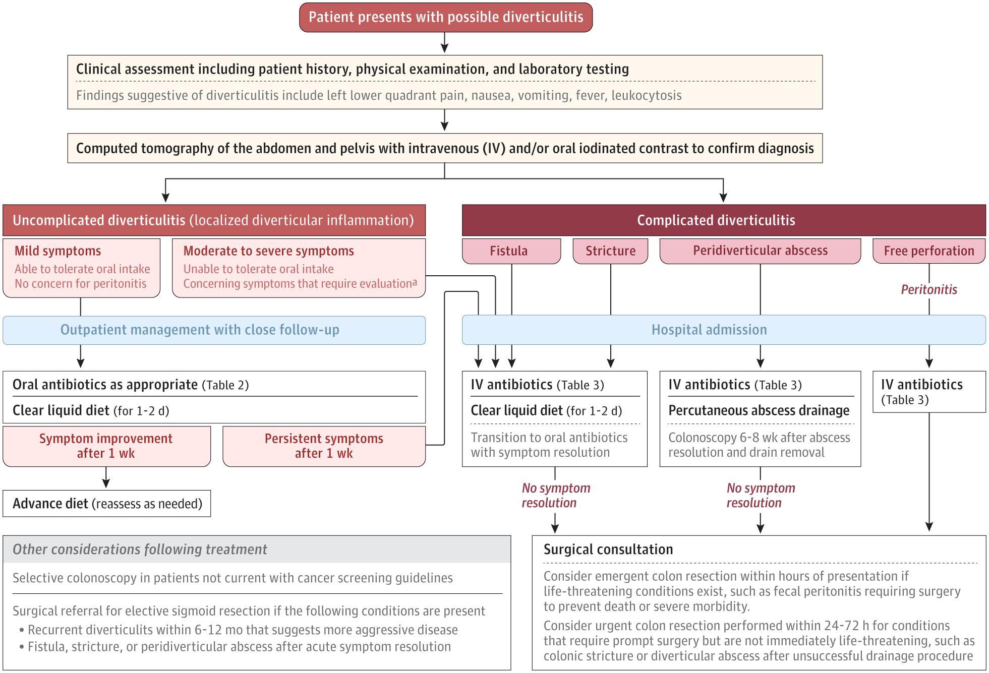
The following management algorithm from a 2025 JAMA review illustrates the clinical decision-making pathway:
History
- Location and quality of pain: Gradual-onset left lower quadrant pain (70% of cases); may be suprapubic or right-sided with right colonic diverticulitis or redundant sigmoid [1]
- Timing: Acute or subacute onset; characterize duration — symptoms >5 days before presentation predict progression to complicated disease [2]
- Associated symptoms: Nausea/vomiting (~30%), low-grade fever (~33%), change in bowel habits, anorexia, malaise [1][3]
- Important negatives: Rectal bleeding is not commonly seen in acute diverticulitis — its presence should prompt consideration of alternative diagnoses [3]
- Fistula screening: Ask about fecaluria, pneumaturia, pyuria (colovesical fistula) and stool per vagina (colovaginal fistula) [4]
- Prior episodes: Number, severity, imaging confirmation, and prior treatments
Alarm Features
- Diffuse abdominal guarding or rigidity → suspect perforation/peritonitis [1]
- Hemodynamic instability (tachycardia, hypotension) → complicated disease or sepsis [1]
- High fever >38.5°C (101.3°F) with persistent chills [1]
- Rising leukocytosis or WBC >15 × 10⁹/L [5]
- CRP >140 mg/L [2][5]
- Palpable abdominal mass (~20% of complicated cases) [1]
- Symptoms >5 days, pain score >7, vomiting — predictors of progression to complicated disease [2][5]
- CT findings: pericolic extraluminal air, fluid collection, or long inflamed segment (>86 mm) [5]
Medications
Medications that increase risk
- NSAIDs (regular use ≥2 times/week), opioids, corticosteroids — all increase risk of diverticulitis and complications [1][5]
- Immunosuppressive agents (chemotherapy, high-dose steroids, post-transplant medications) — increase risk of complicated disease [1]
Treatment — when antibiotics are NOT needed
- Immunocompetent patients with mild uncomplicated diverticulitis, no systemic inflammatory response, no vomiting, WBC <15 × 10⁹/L, CRP <140 mg/L → observation with pain control alone is appropriate (ACP, AGA, Cochrane) [2][5-6]
- Acetaminophen is the preferred analgesic [1]
Treatment — when antibiotics ARE indicated
If unable to tolerate PO: Ceftriaxone + metronidazole IV, or ampicillin-sulbactam IV [1]
Contraindicated: Avoid NSAIDs for pain control during acute episodes (use acetaminophen instead) [1][5]
Diet
- Acute phase: Clear liquid diet for 1–2 days after diagnosis, though evidence for this is limited; advance diet as symptoms improve [1][5]
- If unable to advance diet after 3–5 days → immediate follow-up [5]
- Long-term prevention: High-quality diet rich in fruits, vegetables, whole grains, and legumes; limit red meat and sweets [5][8]
- The old advice to avoid nuts, seeds, and popcorn has not been supported by evidence and is no longer recommended [5]
- Adequate hydration during acute illness
Review of Systems
- GI: Bowel habit changes (constipation or diarrhea), bloating, flatus, hematochezia (atypical — consider alternative dx), tenesmus
- GU: Dysuria, pneumaturia, fecaluria, pyuria (fistula), hematuria (kidney stone)
- GYN (if applicable): Vaginal discharge, abnormal bleeding, stool per vagina
- Constitutional: Fever, chills, rigors, weight loss, night sweats (malignancy)
- Systemic: Signs of sepsis — tachycardia, altered mental status, hypotension
Collateral History and Family History
- Prior imaging-confirmed episodes of diverticulitis and management
- Immunosuppressive medications or conditions (transplant, chemotherapy, HIV)
- Genetic factors: Variants in TNFSF15 gene; connective tissue disorders (Ehlers-Danlos, Marfan, polycystic kidney disease) increase risk [1]
- Family history of diverticular disease, colorectal cancer, or inflammatory bowel disease
- Social context: home support system, ability to tolerate PO, access to follow-up (critical for outpatient management decisions) [2]
Risk Factors
- Age >65 years (strongest demographic risk factor) [1]
- BMI ≥30 [1]
- Medications: NSAIDs, opioids, corticosteroids, immunosuppressants [1][9]
- Comorbidities: Hypertension, type 2 diabetes, CKD, cirrhosis, heart failure [1]
- Lifestyle: Smoking, physical inactivity, low-fiber diet, obesity [5][8]
- Connective tissue disorders: Polycystic kidney disease, Marfan syndrome, Ehlers-Danlos syndrome [1]
- Genetic predisposition: TNFSF15 gene variants [1]
Differential Diagnosis
- Colorectal cancer — cannot-miss; clinical diagnosis of diverticulitis is only accurate 40–65% of the time; colonoscopy recommended 6 weeks after resolution of complicated diverticulitis [3][5]
- Inflammatory bowel disease (Crohn disease, ulcerative colitis) [1]
- Appendicitis — especially with right-sided or suprapubic pain [1]
- Constipation / irritable bowel syndrome [4]
- Kidney stones / urinary tract infection [1][4]
- Bowel obstruction [1]
- Gynecologic pathology: Pelvic inflammatory disease, tubo-ovarian abscess, ovarian torsion, ectopic pregnancy [1]
- Ischemic colitis — particularly in elderly patients with vascular risk factors
Past Medical History
- Prior episodes of diverticulitis (number, severity, imaging confirmation)
- Prior abdominal/pelvic surgery
- History of colorectal cancer screening (last colonoscopy)
- Immunocompromising conditions (organ transplant, HIV, active malignancy, chronic steroid use)
- Chronic conditions affecting management: cirrhosis, CKD, heart failure, poorly controlled diabetes [1]
- Connective tissue disorders [1]
Physical Exam
- Vitals: Temperature (fever in ~33%), heart rate (tachycardia in ~27%), blood pressure (hypotension = red flag) [1]
- Abdomen: LLQ tenderness with localized guarding is the classic finding; assess for rebound, rigidity (peritonitis), distension (obstruction) [1]
- Palpable mass in LLQ → phlegmon or abscess (~20% of complicated cases) [1]
- Rectal exam: Tenderness may indicate pelvic abscess; assess for occult blood [1]
- Concerning findings: Diffuse peritonitis, hemodynamic instability, cool/clammy extremities → emergent surgical evaluation [1]
Lab Studies
- CBC with differential: Leukocytosis present in ~55% of febrile patients; WBC >15 × 10⁹/L is a predictor of progression [1][5]
- CRP: >140 mg/L predicts complicated disease and progression; CRP >150 mg/L discriminates uncomplicated from complicated diverticulitis [5][10]
- BMP: Assess renal function, electrolytes (especially if poor PO intake)
- Urinalysis: Rule out UTI; sterile pyuria may suggest colovesical fistula [4]
- Lactate: If concern for sepsis or ischemia
- Blood cultures: If febrile or signs of systemic infection [1]
- Pregnancy test: In women of reproductive age
- Procalcitonin and fecal calprotectin are being explored but not currently recommended for routine use by ASCRS [4]
Imaging
First-line: CT abdomen/pelvis with IV contrast — sensitivity 98–99%, specificity 99–100% [1]
- Findings: pericolic fat stranding, colonic wall thickening, diverticula; absence of abscess, free air, or extraluminal fluid confirms uncomplicated disease [1]
- CT in the ED can prevent unnecessary hospital admission by >50% [11]
Alternatives when CT is contraindicated
- Ultrasound (radiologist-performed): sensitivity 77–98%, specificity 80–99% [1]
- Point-of-care ultrasound: sensitivity 92.7%, specificity 90.9% in one multicenter study [1]
- MRI: sensitivity 86–94%, specificity 88–92%; preferred in pregnancy [1]
When imaging may be deferred
- Patients with a prior CT-confirmed diagnosis presenting with a typical mild recurrence may be managed clinically without repeat imaging (AGA) [5]
- However, err on the side of imaging if any predictors of complicated disease are present [2]
Avoid: Contrast enema (risk of perforation); plain radiographs have limited utility for diagnosis [1][5]
Special Tests
- Hinchey Classification: Grades severity of diverticulitis on CT (Ia = confined pericolic abscess; Ib = pelvic abscess; II = distant abscess; III = purulent peritonitis; IV = fecal peritonitis). Uncomplicated = Hinchey 0 [10]
- CRP >150 mg/L + free fluid on CT = significantly greater risk of mortality in one retrospective study [10]
- Colonoscopy: Avoid during acute episode (risk of perforation); consider 6 weeks after resolution in patients with complicated diverticulitis or those without recent high-quality colonoscopy — CRC risk is 6–8% in complicated diverticulitis [3][12]
ECG
- Not routinely indicated for uncomplicated diverticulitis
- Obtain if: elderly patient, hemodynamically unstable, concern for sepsis, or pre-procedural evaluation
- Rule out cardiac causes of referred abdominal pain in atypical presentations (especially elderly)
Assessment
Uncomplicated diverticulitis = localized inflammation without abscess, perforation, fistula, obstruction, or bleeding. [2] Approximately 85% of all diverticulitis cases. [1]
Key clinical pearl: Clinical diagnosis alone is only correct in 40–65% of patients — imaging confirmation is important, especially for first episodes. [3][5]
Severity stratification for antibiotic decision-making
- Low risk (no antibiotics needed): Immunocompetent, mild symptoms, no fever, WBC <15, CRP <140, no vomiting, symptoms <5 days, no high-risk CT features [2][5]
- Higher risk (antibiotics indicated): Age >80, immunocompromised, pregnant, significant comorbidities, WBC >15, CRP >140, vomiting, symptoms >5 days, pericolic air or fluid on CT [1][5]
Symptoms typically improve within 48–72 hours, with complete resolution in 1–2 weeks. [1] Recurrence rate is approximately 15% within 2 years. [1]
Treatment Plan
Initial stabilization
- IV access, fluid resuscitation if dehydrated or unable to tolerate PO
- Pain control with acetaminophen (avoid NSAIDs and opioids when possible) [1]
Outpatient management (majority of patients)
- Clear liquid diet × 1–2 days, advance as tolerated [1][5]
- Acetaminophen for pain
- ± Antibiotics based on risk stratification (see Medications section above)
- If antibiotics prescribed: 4–7 day course; re-evaluate within 7 days [1]
Inpatient management (when indicated)
- IV fluids, bowel rest
- IV antibiotics: ceftriaxone 1 g daily + metronidazole 500 mg q8h, or ampicillin-sulbactam [1][7]
- Transition to PO when tolerating diet (typically 48–72 hours)
- Repeat CT if no improvement after 3–5 days [1]
Long-term recurrence prevention
- High-fiber diet, maintain BMI 18–25, regular physical activity, smoking cessation [5][8]
- Avoid regular NSAID use (≥2×/week) except aspirin for cardiovascular prevention [5]
- Elective surgery is not routinely recommended based on number of episodes; individualize based on quality of life, severity, and patient preference [3][5]
Disposition
Discharge criteria (outpatient management)
- Immunocompetent, tolerating PO, mild symptoms, no systemic inflammatory response [2]
- Adequate home support and ability to follow up [2]
- No high-risk features (see Alarm Features)
Admission criteria
- High fever >38.5°C (101.3°F), sepsis or septic shock [1]
- Unable to tolerate oral intake [7]
- Complicated disease on CT [7]
- Immunosuppression [7]
- Serious comorbidities or medically frail [2]
- Inadequate home support or inability to follow up [7]
- Failure to improve with outpatient management [7]
- Intractable pain requiring parenteral analgesia [7]
Surgical consultation triggers
- Perforation with peritonitis → emergent laparotomy [1]
- Abscess ≥3 cm → percutaneous drainage [8]
- Failure to improve despite adequate medical therapy [3]
- Obstruction, fistula, or non-resolving abscess [3]
Follow Up / Return Precautions
Follow-up timing
- If treated with antibiotics: re-evaluate within 7 days for symptom resolution [1]
- If managed without antibiotics: follow up within 2–3 days if not improving, or sooner if worsening
- If unable to advance diet after 3–5 days → immediate reassessment [5]
- Colonoscopy 6 weeks after resolution — particularly important for complicated diverticulitis (6–8% CRC risk), first episode without recent colonoscopy, or atypical features [3][12]
Return precautions — instruct patients to return immediately for:
- Worsening or spreading abdominal pain
- New or worsening fever/chills
- Inability to tolerate any oral intake
- Bloody stools
- Dizziness, lightheadedness, or fainting
- Abdominal rigidity or distension
Expected recovery
- Symptoms improve within 48–72 hours; full resolution in 1–2 weeks [1]
- ~15% recurrence rate within 2 years [1]
- Risk of complicated disease is highest with the first presentation [5]
References
1. Diverticulitis. — Brown RF, Lopez K, Smith CB, Charles A. The Journal of the American Medical Association. 2025.
2. Diagnosis and Management of Acute Left-Sided Colonic Diverticulitis: A Clinical Guideline From the American College of Physicians. — Qaseem A, Etxeandia-Ikobaltzeta I, Lin JS, et al. Annals of Internal Medicine. 2022.
3. Colorectal Cancer Screening and Surveillance and Other Colon Conditions in the Older Adult. — Calderwood AH, Shaukat A. The American Journal of Gastroenterology. 2025.
4. Management of Diverticulitis: A Review. — Sacks OA, Hall J. JAMA Surgery. 2024.
5. AGA Clinical Practice Update on Medical Management of Colonic Diverticulitis: Expert Review. — Peery AF, Shaukat A, Strate LL. Gastroenterology. 2021.
6. Antibiotics for Uncomplicated Diverticulitis. — Dichman ML, Rosenstock SJ, Shabanzadeh DM. The Cochrane Database of Systematic Reviews. 2022.
7. Diverticulitis. — Young-Fadok TM. The New England Journal of Medicine. 2018.
8. Diverticular Disease: Rapid Evidence Review. — Bailey J, Dattani S, Jennings A. American Family Physician. 2022.
9. Revised Version Global Guidelines on Diverticular Disease of the Colon: The Fiesole Consensus Report. — Tursi A, Brandimarte G, Di Mario F, et al. Gut. 2025.
10. The American Society of Colon and Rectal Surgeons Clinical Practice Guidelines for the Treatment of Left-Sided Colonic Diverticulitis. — Hall J, Hardiman K, Lee S, et al. Diseases of the Colon and Rectum. 2020.
11. ACR Appropriateness Criteria® Left Lower Quadrant Pain: 2023 Update. — Weinstein S, Kim DH, Fowler KJ, et al. Journal of the American College of Radiology : JACR. 2023.
12. Evaluation and Management After Acute Left-Sided Colonic Diverticulitis : A Systematic Review. — Balk EM, Adam GP, Cao W, Mehta S, Shah N. Annals of Internal Medicine. 2022.
Figure
More from Deep Dive
Reviewed by Dr. Lucas Mastropaolo. SymptomDx is an educational tool for medical professionals — it does not replace clinical judgment. Verify all clinical data and drug dosages with authoritative sources.