Eastern Equine Encephalitis
Eastern equine encephalitis is a rare but devastating mosquito-borne alphavirus infection with the highest case fatality rate (~41%) of any arboviral encephalitis in the US and >50% of survivors suffering long-term neurologic sequelae. [1-2] There is no specific antiviral treatment and no licensed human vaccine; management is entirely supportive. [1]
History
- Exposure history: Outdoor activities (camping, gardening, agricultural work) in endemic forested wetland areas (Atlantic Coast, Gulf Coast, Great Lakes, New England) during June–October, peaking in late August [1]
- Incubation period: 4–10 days after mosquito bite [1]
- Prodrome: Fevers, chills, malaise, arthralgias, myalgias lasting days before neurologic onset (median 5 days of prodrome before CNS symptoms in older children/adults; abrupt onset in infants) [1]
- Neurologic symptoms: Headache, confusion, altered mental status, focal deficits (hemiparesis, dysarthria, facial droop), seizures, rapid progression to stupor or coma [1][3]
- Ask about: recent travel to endemic areas, mosquito exposure, outdoor occupation/hobbies, immunosuppressive medications or transplant history
Alarm Features
- Rapid neurologic deterioration — progression from prodrome to coma can occur within hours to days [1][4]
- Seizures or status epilepticus (clinical or nonconvulsive) [3]
- Inability to protect airway [1]
- Signs of elevated intracranial pressure (posturing, pupillary changes, Cushing triad) [1]
- Severe hyponatremia (Na ≤130 mmol/L) — associated with poor outcome [5-6]
- High CSF WBC count (>500/mm³) — predictor of poor prognosis [5]
Medications
- No approved antiviral therapy exists [1]
- Empiric coverage: Start IV acyclovir (for HSV encephalitis) and broad-spectrum antibiotics (for bacterial meningitis) until EEE is confirmed [3-4]
- Anticonvulsants: Levetiracetam commonly used; other standard AEDs as needed for seizure management [3]
- IVIG: Limited data suggest earlier administration may reduce long-term disability (average 5 days post-admission in one series, N=17); results remain inconclusive [1][7]
- High-dose IV methylprednisolone: Used in some cases for immunomodulation, but a meta-analysis of steroids for viral encephalitides showed no survival benefit [1][7]
- Avoid: Medications that lower seizure threshold; use caution with hypertonic solutions given risk of hyponatremia
Diet
- NPO if altered mental status or airway compromise anticipated
- Aggressive IV fluid resuscitation for dehydration from febrile illness, nausea, and vomiting [1]
- Monitor sodium closely — hyponatremia is common and prognostically significant; avoid hypotonic fluids [5-6]
- Enteral nutrition via NG/OG tube for prolonged ICU stays
Review of Systems
- Neurologic: Headache, confusion, personality changes, focal weakness, speech difficulty, vision changes, seizures
- Constitutional: Fever, chills, rigors, malaise, fatigue
- Musculoskeletal: Arthralgias, myalgias
- GI: Nausea, vomiting (common with meningeal irritation)
- Respiratory: Assess for aspiration risk, respiratory failure requiring intubation (50% in one series) [3]
- Ophthalmologic: Photophobia
Collateral History and Family History
- Collateral from family/witnesses regarding timeline of symptom onset, baseline mental status, and rapidity of decline
- Confirm geographic exposure — residence near or travel to deciduous forested wetlands, swamps, or marshes in endemic regions [1]
- Occupational exposure (agricultural workers, forestry, outdoor recreation)
- Immunosuppression history: Organ transplant recipients, patients on B-cell inhibiting monoclonal antibodies (e.g., rituximab) at higher risk for neuroinvasive disease [1]
- Family history is not a significant contributor; EEE is not hereditary
- One documented case of solid organ transplant transmission involving 3 recipients [1]
Risk Factors
- Age bimodal distribution: <5 years and >60 years at highest risk for neuroinvasive disease; case fatality highest in age ≥70 [1][8]
- Male sex: Predominance in reported cases (59–100% male in case series) [7][9]
- Geographic: Atlantic Coast, Gulf Coast, Great Lakes, and increasingly New England states (17/19 cases in 2024 from New England) [1]
- Seasonal: June–October, peak late August [1]
- Immunosuppression: Organ transplant, B-cell depleting therapies [1]
- Outdoor exposure: Living near or visiting forested wetlands/swamps; camping, gardening, agricultural work [1]
- Increased precipitation in preceding year may amplify mosquito vector populations [10]
Differential Diagnosis
- Herpes simplex encephalitis (HSV) — temporal lobe predominance on MRI (vs. basal ganglia/thalami in EEE); treat empirically with acyclovir until ruled out [6]
- West Nile virus encephalitis — similar arboviral presentation but lower mortality (~10%); may also involve basal ganglia but often shows anterior horn cell involvement [1][5]
- La Crosse encephalitis — primarily affects school-aged children; low mortality (~1%) [1][5]
- St. Louis encephalitis — flavivirus; similar geographic overlap [5]
- Bacterial meningitis — CSF with neutrophilic predominance early in EEE can mimic bacterial infection; low glucose favors bacterial etiology [1]
- Acute ischemic stroke — focal deficits (hemiparesis, dysarthria) may initially mimic stroke, as occurred in reported cases [4]
- Autoimmune encephalitis (anti-NMDA receptor, etc.) — subacute onset, psychiatric features
- Acute disseminated encephalomyelitis (ADEM) — post-infectious demyelination; may overlap clinically [4]
- Tuberculous meningitis — subacute, basilar meningeal enhancement, low glucose
Key distinguishing feature of EEE: Early, prominent basal ganglia and thalamic involvement on MRI differentiates EEE from HSV encephalitis, which typically spares these structures early. [6]
Past Medical History
- Prior arboviral infections or vaccinations (may cause cross-reactive serologies) [1]
- Immunosuppressive conditions or medications (transplant, rituximab, chemotherapy) [1]
- Seizure history (baseline vs. new-onset)
- Chronic neurologic conditions affecting baseline function
- Prior residence in or travel to endemic areas
Physical Exam
- Vitals: Fever (100% in one series), tachycardia; monitor for Cushing response (hypertension + bradycardia suggesting elevated ICP) [7]
- Neurologic:
- Mental status: GCS assessment; confusion → obtundation → coma
- Meningismus: Nuchal rigidity, Kernig/Brudzinski signs
- Focal deficits: Hemiparesis, facial droop, dysarthria, cranial nerve palsies [3-4]
- Seizure activity: Observe for subtle/nonconvulsive seizures
- Fundoscopic: Papilledema (elevated ICP)
- Skin: No characteristic rash (unlike some other arboviruses); look for mosquito bites
Lab Studies
- CSF analysis (critical):
- Serum: CBC, CMP (watch for hyponatremia ≤130 mmol/L as poor prognostic marker), blood cultures, coagulation studies [5-6]
- EEEV-specific IgM (serum and CSF) by ELISA or microsphere immunoassay — appears within first week of illness [1]
- Confirmatory: Plaque reduction neutralization test (PRNT) through public health laboratory [1]
- RT-PCR: Limited utility (viremia typically resolves by onset of neurologic symptoms); consider in immunocompromised patients who may not mount antibody response [1]
- Diagnosis is often delayed — median 24.5 days in one outbreak series due to initially negative serologies [9]
Imaging
- MRI brain with contrast (imaging modality of choice):
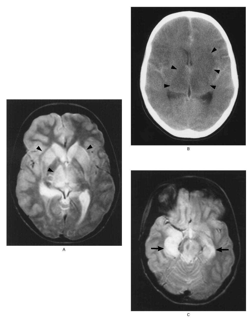
- T2/FLAIR hyperintensities in basal ganglia, thalami, and mesial temporal lobes — the hallmark pattern [1][6]
- May also involve brainstem (43% on MRI), cortical regions [6]
- Some cases show hyperintense ring encapsulation around basal ganglia/thalamus [3]
- Meningeal enhancement and periventricular white matter changes less common [6]
- CT head: Less sensitive; abnormal in ~66% of patients (vs. 100% on MRI in comatose patients) [6]
- May show focal basal ganglia lesions (56%) and diffuse cerebral swelling [6]
- Useful for rapid initial assessment to rule out hemorrhage or mass lesion
- Prognostic imaging features: Numerous and symmetrical abnormalities predict worse neurologic outcome [1]
The following figure from a landmark NEJM study demonstrates the characteristic basal ganglia and thalamic lesions on MRI and CT in a fatal case of EEE:
Special Tests
- EEG: δ and θ frequency slowing consistent with encephalopathy; epileptiform discharges; may reveal nonconvulsive status epilepticus (seen in all 4 patients in one series) [1][3]
- Continuous EEG monitoring recommended for encephalitic patients given high seizure burden
- Modified Rankin Scale (mRS): Used to track functional disability over time [7]
- Public health notification: Report suspected cases to state/local health department immediately to trigger mosquito surveillance and community prevention measures [1][8]
ECG
- No specific ECG findings associated with EEE
- Obtain baseline ECG to assess for arrhythmias related to electrolyte abnormalities (hyponatremia) or medication effects (QTc prolongation from antiemetics)
- Monitor telemetry in ICU setting given critical illness
Assessment
EEE is the most lethal arboviral encephalitis in the US. Among 193 cases reported to the CDC from 2005–2024, 99% were neuroinvasive and 41% died. [1] Death typically occurs within the first 3 weeks but can occur months later. [1] Among survivors, >50% develop long-term sequelae including cognitive impairment, aphasia, personality disorders, seizures, paralysis, and cranial nerve dysfunction. [1-2]
Severity stratification
- Mild/systemic illness only (fever, myalgias without CNS involvement) — generally self-limited, resolves in 1–2 weeks [1]
- Neuroinvasive disease (meningitis/encephalitis) — high mortality and morbidity; requires ICU-level care
Poor prognostic indicators: Hyponatremia (Na ≤130), high CSF WBC count (>500/mm³), symmetric/extensive MRI abnormalities, severely depressed EEG, immunosuppression, age extremes [1][5-6]
Treatment Plan
Initial stabilization
- ABCs — early intubation if GCS declining or inability to protect airway (50% required mechanical ventilation in one series) [3]
- IV access, continuous monitoring, ICU admission
Empiric therapy (until EEE confirmed)
- IV acyclovir 10 mg/kg q8h (cover HSV encephalitis)
- Broad-spectrum antibiotics (cover bacterial meningitis — e.g., vancomycin + ceftriaxone ± ampicillin if age >50 or immunocompromised)
- Dexamethasone if bacterial meningitis suspected
EEE-specific supportive care
- Seizure management: Levetiracetam first-line; continuous EEG monitoring; treat nonconvulsive status epilepticus aggressively [3]
- ICP management: Head of bed elevation, osmotic therapy if signs of elevated ICP; neurosurgical consultation for refractory cases [1]
- Fluid/electrolyte management: Avoid hypotonic fluids; correct and monitor hyponatremia closely [5]
- Antiemetics and analgesics for meningeal symptoms [1]
Immunomodulatory therapy (limited/inconclusive evidence)
- IVIG: May be considered early in hospitalization; shorter time to administration correlated with decreased long-term disability in one retrospective series (N=17) [1][7]
- High-dose IV methylprednisolone: Used in some cases; no proven survival benefit for viral encephalitides [1][7]
Disposition
Admission criteria (essentially all neuroinvasive cases)
- Any altered mental status, seizures, focal neurologic deficits, or meningismus → ICU admission [2]
- 98% of reported cases required hospitalization; average stay ~10 days among survivors [3][8]
Observation/floor admission
Discharge criteria
- Neurologically stable, seizure-free, able to protect airway, adequate oral intake
- Many survivors require discharge to rehabilitation facilities for long-term neurologic recovery [1][3]
Specialist consultation triggers
- Neurology (seizure management, EEG interpretation)
- Infectious disease (diagnostic workup, IVIG/steroid consideration)
- Neurosurgery (refractory elevated ICP)
- Critical care (ventilator management)
- Public health department notification (mandatory) [1][8]
Follow Up / Return Precautions
- Survivors require long-term neurologic follow-up: Cognitive rehabilitation, seizure monitoring, physical/occupational/speech therapy [1-2]
- Neurologic sequelae may include: cognitive/memory deficits, global aphasia, personality changes, seizure disorder, paralysis, cranial nerve dysfunction [1]
- One case demonstrated complete resolution of MRI abnormalities at follow-up, indicating some recovery potential [3]
- Return precautions: New or worsening seizures, declining mental status, new focal deficits, fever recurrence
- Prevention counseling: EPA-registered insect repellents (DEET, picaridin, IR3535), long-sleeved clothing, avoiding outdoor activity at dawn/dusk in endemic areas, eliminating standing water [1-2]
- No licensed human vaccine currently available; veterinary vaccines exist for horses [1][11]
- Counsel that humans are dead-end hosts — no person-to-person transmission risk [1]
References
1. Eastern Equine Encephalitis in the US. — Staples JE, Gould CV. The Journal of the American Medical Association. 2025.
2. What Is Eastern Equine Encephalitis?. — Roberts K. The Journal of the American Medical Association. 2025.
3. The Clinical and Radiographic Features of Eastern Equine Encephalitis: A Single-Center Retrospective Case Series. — Garcia-Dominguez MA, Moonis M, Kipkorir V, Srichawla BS. Medicine. 2024.
4. Eastern Equine Encephalitis Virus: A Case Report and Brief Literature Review of Current Therapeutic and Preventative Strategies. — Ruiz C, Gibson G, Rojas S, Friend K. Vector Borne and Zoonotic Diseases. 2024.
5. Viral Encephalitis: Familiar Infections and Emerging Pathogens. — Whitley RJ, Gnann JW. Lancet. 2002.
6. Clinical and Neuroradiographic Manifestations of Eastern Equine Encephalitis. — Deresiewicz RL, Thaler SJ, Hsu L, Zamani AA. The New England Journal of Medicine. 1997.
7. Eastern Equine Encephalitis and Use of IV Immunoglobulin Therapy and High-Dose Steroids. — Wilcox DR, Collens SI, Solomon IH, Mateen FJ, Mukerji SS. Neurology Neuroimmunology & Neuroinflammation. 2021.
8. Eastern Equine Encephalitis Virus in the United States, 2003-2016. — Lindsey NP, Staples JE, Fischer M. The American Journal of Tropical Medicine and Hygiene. 2018.
9. Clinical Characteristics of the 2019 Eastern Equine Encephalitis Outbreak in Michigan. — Ladzinski AT, Tai A, Rumschlag MT, et al. Open Forum Infectious Diseases. 2023.
10. Association of Human Eastern Equine Encephalitis With Precipitation Levels in Massachusetts. — Mermel LA. JAMA Network Open. 2020.
11. Notes From the Field: Increase in Eastern Equine Encephalitis Virus Activity - Vermont, 2023-2024. — Strelau KM, Pareles E, Kelso P, et al. MMWR. Morbidity and Mortality Weekly Report. 2026.
Figure
More from Deep Dive
Reviewed by Dr. Lucas Mastropaolo. SymptomDx is an educational tool for medical professionals — it does not replace clinical judgment. Verify all clinical data and drug dosages with authoritative sources.