Ectopic Pregnancy (Ruptured)
Ruptured ectopic pregnancy is a life-threatening surgical emergency requiring immediate resuscitation and operative intervention. It accounts for 2.7% of pregnancy-related deaths and is the leading cause of maternal mortality in the first trimester. [1-2] The following is a comprehensive clinical summary organized for emergency medicine and primary care workflows.
History
- Key HPI questions: Last menstrual period (LMP), sexual activity, contraceptive use, prior positive pregnancy test, IVF/ART use
- Classic triad: Amenorrhea + vaginal bleeding + abdominal/pelvic pain — though all three are present in a minority of cases
- Pain characterization: May be unilateral or bilateral, localized or generalized, dull/sharp/crampy; tubal rupture classically causes sudden, severe, generalized abdominal pain that may initially improve briefly after rupture [3]
- Shoulder pain (Kehr sign): Referred pain from diaphragmatic irritation by hemoperitoneum — a late and ominous sign [3]
- Vaginal bleeding: Typically light, intermittent spotting (from decidual sloughing), rarely exceeds menstrual flow; passage of tissue (decidual cast) does not rule out ectopic [3-4]
- Important negatives: Syncope/presyncope, dizziness, orthostatic symptoms, fever, dysuria, GI symptoms
Alarm Features
- Hemodynamic instability: Hypotension, tachycardia, diaphoresis, altered mental status [3-4]
- Peritoneal signs: Rebound tenderness, guarding, rigidity — suggest hemoperitoneum
- Shoulder pain: Indicates significant intraperitoneal hemorrhage irritating the diaphragm [3]
- Syncope or near-syncope in a reproductive-age female — always rule out ruptured ectopic
- Positive pregnancy test + shock = ruptured ectopic until proven otherwise
- Any patient with hemodynamic instability or peritoneal signs and a positive pregnancy test should have immediate OB/GYN surgical consultation [3][5]
Medications
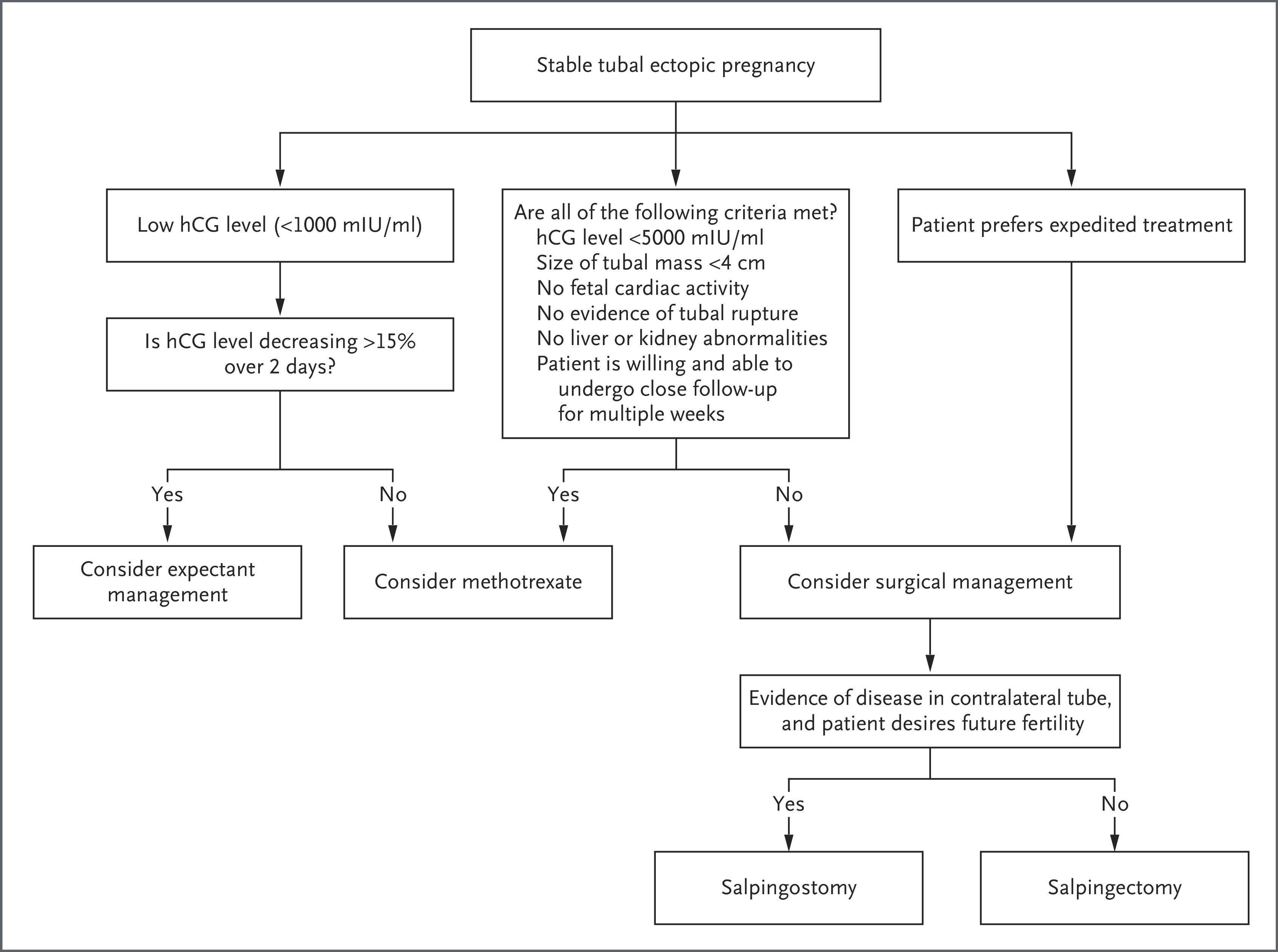
- Methotrexate is absolutely contraindicated in ruptured ectopic pregnancy — it is reserved only for hemodynamically stable, unruptured cases with β-hCG <5,000, no fetal cardiac activity, mass <4 cm, and reliable follow-up [5-6]
- Resuscitation medications: Crystalloid IV fluids, blood products (pRBCs, consider massive transfusion protocol), vasopressors if needed
- RhoGAM (anti-D immunoglobulin): Administer to all Rh-negative patients
- Analgesics: IV opioids for pain control; avoid NSAIDs if coagulopathy suspected
- Avoid delays for any medication that would postpone surgical intervention
Diet
- NPO status immediately upon suspicion of ruptured ectopic — surgical intervention is anticipated
- Not applicable for acute management; dietary counseling is relevant only in post-operative recovery
Review of Systems
- GYN: Vaginal bleeding, pelvic pain, dyspareunia, prior abnormal Pap or STI history
- GI: Nausea/vomiting (common in pregnancy and with hemoperitoneum), diarrhea, rectal pressure
- GU: Dysuria, urinary frequency (may mimic UTI)
- Neuro: Lightheadedness, syncope, visual changes (signs of hypovolemia)
- Constitutional: Fatigue, weakness, diaphoresis
Collateral History and Family History
- Collateral: Confirm LMP, pregnancy awareness, recent sexual partners, prior ED visits for pelvic pain/bleeding, IVF history
- Family history: Generally not a major contributor, though familial predisposition to endometriosis or tubal abnormalities may be relevant
- Social context: Access to follow-up care, transportation, social support — critical for any patient initially considered for non-surgical management of unruptured ectopic [6]
Risk Factors
- Prior ectopic pregnancy (strongest risk factor — 10× increased risk)
- History of pelvic inflammatory disease (PID) / chlamydia / gonorrhea [1][6]
- Prior tubal surgery (tubal ligation, salpingostomy, tubal reanastomosis)
- Current IUD use (does not increase absolute risk, but if pregnancy occurs with IUD in place, higher proportion are ectopic)
- Cigarette smoking (dose-dependent, impairs tubal motility) [1][6]
- Infertility and assisted reproductive technology (IVF/IUI)
- Age >35 years [7]
- Endometriosis, DES exposure
- Important: A significant number of patients with ectopic pregnancy have no identifiable risk factors [7]
Differential Diagnosis
- Ruptured hemorrhagic ovarian cyst — sudden pain, hemoperitoneum possible, but pregnancy test negative
- Ovarian torsion — acute unilateral pain, nausea/vomiting, may be pregnant or not
- Threatened/incomplete miscarriage — vaginal bleeding with open os, IUP on ultrasound
- Appendicitis — RLQ pain, fever, leukocytosis; always check pregnancy test
- Heterotopic pregnancy — coexisting IUP and ectopic (rare spontaneously ~1:30,000, but up to 1:100 with IVF)
- Ruptured splenic artery aneurysm — rare but catastrophic in pregnancy
- Placental abruption — later in pregnancy
- Distinguishing feature: Positive pregnancy test + no IUP on TVUS + free fluid = ruptured ectopic until proven otherwise [6][8]
Past Medical History
- Prior ectopic pregnancies and their management (medical vs. surgical)
- Prior pelvic/abdominal surgeries (adhesion risk)
- History of PID, STIs, endometriosis
- Infertility treatments
- Bleeding disorders or anticoagulant use
- Rh status
Physical Exam
- Vitals: Tachycardia and hypotension are late signs — may be initially compensated in young patients [3-4]
- Abdomen: Diffuse tenderness, rebound, guarding, rigidity, distension (hemoperitoneum)
- Pelvic exam:
- Cervical motion tenderness (chandelier sign) — highly suggestive
- Adnexal tenderness ± palpable adnexal mass
- Closed cervical os (open os suggests miscarriage)
- Vaginal bleeding — typically dark, scant
- Skin: Pallor, diaphoresis, delayed capillary refill
- Rectal: Fullness in cul-de-sac (blood pooling in pouch of Douglas)
Lab Studies
- Quantitative serum β-hCG: Confirms pregnancy; level does not reliably predict rupture — ruptured ectopic can occur at any β-hCG level, including very low levels [9]
- CBC: Baseline hemoglobin/hematocrit — lower preoperative Hgb associated with rupture; serial Hgb to track ongoing hemorrhage [10]
- Type and screen / crossmatch: Essential — anticipate need for transfusion
- Coagulation studies: PT/INR, fibrinogen if massive hemorrhage suspected (DIC risk)
- BMP: Renal function, electrolytes
- Blood gas (VBG/ABG): Lactate for perfusion assessment
- Rh status: For RhoGAM administration
- Urinalysis: Rule out UTI as confounder
Imaging
- First-line: Transvaginal ultrasound (TVUS) — single best diagnostic modality [11]
- Findings: Empty uterus + adnexal mass (tubal ring sign) ± free fluid
- Free fluid in the cul-de-sac and paracolic gutters strongly suggests hemoperitoneum from rupture [8]
- Echogenic free fluid (clotted blood) is more concerning than anechoic fluid
- Transabdominal ultrasound: Supplement to quantify free fluid in upper abdomen (Morison's pouch, splenorenal recess)
- Bedside FAST exam: Rapid assessment for free fluid in the unstable patient — do not delay surgery for formal imaging
- CT abdomen/pelvis: Not first-line but may identify hemoperitoneum if ectopic not initially suspected
- MRI: Rarely indicated acutely; may help with non-tubal ectopic locations
- When imaging is unnecessary: If the patient is hemodynamically unstable with a positive pregnancy test and clinical signs of rupture — proceed directly to OR [5]
Special Tests
- Bedside FAST/POCUS: Rapid identification of free fluid — critical in the unstable patient
- Culdocentesis: Historically used to confirm hemoperitoneum (aspiration of non-clotting blood from cul-de-sac); largely replaced by ultrasound but may still be useful in resource-limited settings
- Serial β-hCG trending: Not applicable in ruptured ectopic (used for pregnancy of unknown location); in ruptured cases, do not delay for serial values
- Discriminatory zone concept: β-hCG >3,500 mIU/mL with no IUP on TVUS is highly suggestive of ectopic, but rupture can occur well below this threshold [5-6]
ECG
- Indications: Obtain in patients with significant hemorrhage, tachycardia, or hemodynamic instability
- Expected findings: Sinus tachycardia
- Concerning patterns: ST changes or arrhythmias suggesting myocardial ischemia from hypovolemic shock
- Primarily used to assess cardiovascular status pre-operatively
Assessment
Ruptured ectopic pregnancy is a clinical diagnosis in the unstable patient — do not delay definitive treatment for confirmatory testing. The classic presentation is a reproductive-age female with abdominal pain, vaginal bleeding, amenorrhea, and hemodynamic instability with a positive pregnancy test. [3-4]
- Severity stratification:
- Compensated shock: Tachycardia, normal or borderline BP, mild peritoneal signs
- Decompensated shock: Hypotension, altered mental status, diffuse peritonitis
- Atypical presentations: Pain may be minimal or absent; β-hCG may be very low; high-altitude physiology can mask anemia; young patients may compensate until sudden cardiovascular collapse [9]
- Complications: Hemorrhagic shock, DIC, multiorgan failure, death, future infertility, recurrent ectopic pregnancy
Treatment Plan
Initial stabilization (simultaneous with diagnosis)
- Two large-bore IVs (16–18G), aggressive fluid resuscitation
- Activate massive transfusion protocol if hemodynamically unstable
- Type and crossmatch; transfuse uncrossmatched O-negative pRBCs if critical
- NPO, Foley catheter, continuous monitoring
Definitive treatment
- Emergent surgical intervention is mandatory for ruptured ectopic [5]
- Laparoscopic salpingectomy is the preferred approach when feasible [6][12]
- Laparotomy is indicated for massive hemorrhage, hemodynamic instability precluding laparoscopy, or dense adhesions [6]
- Salpingectomy is preferred over salpingostomy when the tube is ruptured [6]
- Salpingostomy may be considered if the contralateral tube is damaged/absent and future fertility is desired, though it carries higher risk of persistent trophoblast (requiring post-op β-hCG monitoring and possible methotrexate) [6]
Post-operative
- Serial β-hCG to confirm resolution to <5 mIU/mL (especially after salpingostomy)
- RhoGAM if Rh-negative
- Contraceptive counseling; delay conception ≥3 months if methotrexate was used [5]
- Mental health screening — pregnancy loss and ectopic pregnancy carry significant psychological burden [2]
The following figure from the 2025 NEJM review illustrates the treatment algorithm for stable tubal ectopic pregnancy (note: ruptured ectopic bypasses this algorithm and goes directly to surgery):
Disposition
- All ruptured ectopic pregnancies require admission — typically to the OR directly, then post-operative inpatient monitoring
- Admission criteria: Hemodynamic instability, need for transfusion, post-surgical monitoring, ongoing pain
- ICU admission: Consider for patients with massive hemorrhage, DIC, or persistent hemodynamic instability post-operatively
- Specialist consultation: OB/GYN — emergent, at time of diagnosis; do not delay for any reason [1][7]
- Transfer: If OB/GYN surgical capability is not available, initiate resuscitation and arrange emergent transfer
Follow Up / Return Precautions
- Post-operative follow-up: OB/GYN within 1–2 weeks; serial β-hCG weekly until undetectable
- Return precautions (counsel patient):
- Return immediately for increasing abdominal pain, heavy vaginal bleeding, fever, dizziness/syncope, or shoulder pain
- Wound care instructions for laparoscopic/laparotomy sites
- Expected recovery: Return to usual activities within ~2 weeks for laparoscopic approach [6]
- Future pregnancy counseling:
- Psychological support: Screen for grief, anxiety, PTSD — ectopic pregnancy loss is often underrecognized as a significant psychological event [2]
References
1. Ectopic Pregnancy: Diagnosis and Management. — Hendriks E, Rosenberg R, Prine L. American Family Physician. 2020.
2. Ectopic Pregnancy. — Chong KY, de Waard L, Oza M, et al. Nature Reviews. Disease Primers. 2024.
3. Does This Woman Have an Ectopic Pregnancy?The Rational Clinical Examination Systematic Review. — Crochet JR, Bastian LA, Chireau MV. The Journal of the American Medical Association. 2013.
4. Ectopic Pregnancy. — Barnhart KT. The New England Journal of Medicine. 2009.
5. ACOG Practice Bulletin No. 193: Tubal Ectopic Pregnancy. — Committee on Practice Bulletins—GynecologyThis Practice Bulletin was developed by the Committee on Practice Bulletins—Gynecology in collaboration with Kurt T Obstetrics and Gynecology. 2018.
6. Tubal Ectopic Pregnancy. — Schreiber CA, Sonalkar S. The New England Journal of Medicine. 2025.
7. Updates in Emergency Medicine: Ectopic Pregnancy. — Jeffers K, Koyfman A, Long B. The American Journal of Emergency Medicine. 2024.
8. ACR Appropriateness Criteria® First Trimester Vaginal Bleeding: 2025 Update. — Expert Panel on GYN and OB Imaging, Laifer-Narin SL, Fruauff A, et al. Journal of the American College of Radiology : JACR. 2025.
9. An Acute Pelvic Pain in High-Altitude Tourist: A Case Report of Ruptured Ectopic Pregnancy With Low Β-Human Chorionic Gonadotropin. — Yuan F, Cui ZC, Jiang ZZ, Liu XT. Medicine. 2025.
10. Risk Factors and Clinical Characteristics Associated With a Ruptured Ectopic Pregnancy: A 19-Year Retrospective Observational Study. — Li PC, Lin WY, Ding DC. Medicine. 2022.
11. ACR Appropriateness Criteria® Acute Pelvic Pain in the Reproductive Age Group: 2023 Update. — Brook OR, Dadour JR, Robbins JB, et al. Journal of the American College of Radiology : JACR. 2024.
12. The Diagnosis and Management of Extrauterine and Uterine Ectopic Pregnancy. — Farren J, Al Wattar BH, Jurkovic D. Human Reproduction Update. 2025.
13. Pregnancy Outcomes Following Medical Versus Surgical Treatment of Tubal Ectopic Pregnancy: A Population-Based Retrospective Cohort Study. — Rosen A, Palma L, Ordon M, et al. American Journal of Obstetrics and Gynecology. 2025.
Figure
More from Deep Dive
Reviewed by Dr. Lucas Mastropaolo. SymptomDx is an educational tool for medical professionals — it does not replace clinical judgment. Verify all clinical data and drug dosages with authoritative sources.