Electrical Burn Injury
Electrical burn injuries are a unique subset of burn trauma characterized by deep tissue destruction disproportionate to visible cutaneous injury, with potential for multisystem involvement including cardiac arrhythmias, rhabdomyolysis, compartment syndrome, and significant long-term neuropsychological sequelae. [1-3] All electrical burns warrant burn center referral per American Burn Association criteria. [4-5]
History
- Voltage: Low voltage (<1000 V, typically household 120–240 V) vs. high voltage (≥1000 V, industrial/power lines) — this is the single most important historical detail [3][6]
- Type of current: Alternating current (AC) vs. direct current (DC); AC causes tetanic muscle contraction and prolonged contact, DC causes a single forceful throw
- Duration of contact: Longer contact = greater tissue damage
- Pathway of current: Determine entry and exit wounds; vertical pathway (hand-to-foot) carries higher risk of cardiac and abdominal organ injury [7]
- Loss of consciousness or syncope: Present in ~69% of high-voltage injuries; key predictor of cardiac complications [3]
- Tetany: Inability to release the source (AC current)
- Associated trauma: Falls from height, blast injuries, secondary thermal burns
- Wet skin at time of contact: Decreases skin resistance, increases current flow [8]
- Workplace vs. domestic vs. lightning: Occupational injuries predominate in adults [2]
Alarm Features
- Cardiac arrest (VF, VT, asystole, PEA) — occurs in ~20% of high-voltage injuries [3]
- Loss of consciousness or syncope [1][9]
- High-voltage exposure (≥1000 V) — strongest predictor of arrhythmia (AUC 0.804) [6]
- Transthoracic current pathway (hand-to-hand or hand-to-foot) [10]
- Dark/cola-colored urine (myoglobinuria indicating rhabdomyolysis) [11]
- Tense, swollen extremity (compartment syndrome) — the 6 Ps: Pain, Pallor, Paresthesia, Paralysis, Pulselessness, Poikilothermia [12]
- Circumferential burns with vascular compromise
- Respiratory distress — associated with severe electrical injury [2]
- Visible deep tissue necrosis disproportionate to skin burns
Medications
- Fluid resuscitation: Crystalloid (LR or NS); goal urine output 1 mL/kg/h for high-voltage injuries (higher than standard burn goal of 0.5 mL/kg/h) [11]
- Sodium bicarbonate: Consider for urine alkalinization (target urine pH >6.5) in rhabdomyolysis, though evidence is limited [13]
- Mannitol: May be used as osmotic diuretic for rhabdomyolysis/compartment syndrome; limited evidence; monitor osmolal gap [13]
- Analgesics: Opioids for severe pain; acetaminophen/NSAIDs for mild injuries [14]
- Tetanus prophylaxis: Update as indicated for any open wound
- Antibiotics: Not routinely indicated unless signs of infection; consider antianaerobic coverage for deep necrotic wounds [15]
- Avoid: Succinylcholine in the subacute phase (hyperkalemia risk from rhabdomyolysis)
Diet
- Aggressive hydration is the cornerstone — patients may require up to 10 L/day in severe rhabdomyolysis [13]
- High-calorie, high-protein diet for wound healing in admitted patients with significant burns
- Monitor and correct electrolyte abnormalities (hypokalemia in 18%, hypocalcemia in 3.3%) [9]
- Adequate nutritional supply and early treatment of anemia expedite wound healing [15]
Review of Systems
- Cardiac: Palpitations, chest pain, syncope, dyspnea
- Neurological: Paresthesias (15%), weakness, confusion, memory problems, headache, seizures [9][16]
- Musculoskeletal: Myalgias, muscle weakness, joint pain (fractures/dislocations from tetanic contraction or falls)
- Renal/GU: Dark urine, decreased urine output
- Psychiatric: Anxiety, insomnia, flashbacks, nightmares — screen early [17-18]
- Ophthalmologic: Visual changes (cataracts can develop weeks to months post-injury)
- ENT: Hearing loss, tinnitus (especially lightning)
Collateral History and Family History
- Witnesses: Determine duration of contact, whether patient was thrown or had tetanic grip, any CPR performed
- Occupational details: Type of electrical source, voltage, safety equipment used
- Pre-existing cardiac history: Increases risk and monitoring requirements [19]
- Medication history: Beta-blockers or antiarrhythmics may mask or alter cardiac response
- Social context: Workplace safety compliance, potential for non-accidental injury in children (especially oral burns in toddlers)
Risk Factors
- High-voltage exposure (≥1000 V): Strongest predictor of arrhythmia and severe injury [3][6]
- Male sex: ~85–98% of electrical injuries [3][9]
- Occupational exposure: Electricians, linemen, construction workers [20]
- Age: Bimodal — toddlers (oral burns from biting cords) and working-age adults [2]
- Wet skin/water contact: Reduces skin resistance from ~100,000 Ω to ~1,000 Ω
- Vertical current pathway (upper-to-lower body): Predictor of myocardial damage [7]
- Greater TBSA burned: Only positively related factor to cardiac complications in one large series [21]
- Summer months: Peak incidence [2]
Differential Diagnosis
- Lightning strike: DC current, unique Lichtenberg figures (ferning pattern), different cardiac effects
- Thermal/flame burn: From arc flash or ignited clothing — may coexist
- Chemical burn: Especially in industrial settings
- Crush injury/rhabdomyolysis from other causes: Trauma, prolonged immobilization, drugs
- Non-accidental injury: Especially in pediatric patients with inconsistent history
- Acute coronary syndrome: If ECG changes or troponin elevation present
- Spinal cord injury: If neurological deficits present — may be from direct electrical injury or fall
Past Medical History
- Pre-existing cardiac disease: Arrhythmias, pacemakers/ICDs, coronary artery disease — lowers threshold for monitoring [19]
- Renal disease: Increases risk of AKI from rhabdomyolysis
- Epilepsy: May confound post-injury seizures
- Psychiatric history: Pre-existing conditions excluded from some outcome studies; baseline depression predicts worse long-term adjustment [22]
- Previous electrical injuries: Cumulative neurological effects possible
Physical Exam
- Entry and exit wounds: Charred, depressed, centrally necrotic lesions; exit wounds often larger and more destructive
- "Iceberg" phenomenon: Cutaneous burns dramatically underestimate deep tissue injury, especially in high-voltage injuries [23]
- Vital signs: Tachycardia, hypotension (hypovolemia from third-spacing), arrhythmias
- Vascular exam: Distal pulses (Doppler if needed), capillary refill — assess for compartment syndrome [24]
- Compartment assessment: Tense swollen extremities, pain with passive stretch
- Neurological exam: Motor/sensory deficits, cranial nerves, mental status
- Oral exam: In children — commissure burns from biting electrical cords (risk of delayed labial artery hemorrhage at 7–14 days)
- Musculoskeletal: Posterior shoulder dislocations, vertebral compression fractures from tetanic contraction
- Secondary survey: Full trauma assessment — falls, blast injuries
Lab Studies
- CK (creatine kinase): Key marker for rhabdomyolysis; CK >850 U/L is an independent risk factor for arrhythmia; serial monitoring recommended [6]
- Troponin: Elevated in ~5.7% of cases; CK >850 and troponin >250 ng/mL predict arrhythmia [6][9]
- BMP/CMP: Potassium (hyperkalemia from rhabdomyolysis), calcium, creatinine, BUN for renal function
- Lactate: >2.1 mmol/L is an independent predictor of arrhythmia [6]
- Urinalysis: Myoglobinuria (dipstick positive for blood without RBCs on microscopy)
- CBC: Baseline
- Coagulation studies: DIC can occur with severe injuries [25]
- ABG/VBG: Assess acid-base status in severe injuries
- CK-MB: Less useful than troponin for cardiac risk stratification [10]
An algorithmic approach to lab testing can reduce unnecessary workup without increasing mortality or readmission rates. [26]
Imaging
- CT head: If loss of consciousness, altered mental status, or fall from height
- CT C-spine/trauma CT: Per trauma protocol if associated fall or blast
- X-rays: Shoulder (posterior dislocation), spine (compression fractures from tetanic contraction)
- CT angiography: If vascular injury suspected
- Echocardiography: If troponin elevated, wall motion abnormalities suspected, or hemodynamic instability [21]
- MRI: For delayed neurological deficits or spinal cord injury evaluation
- Imaging of the burn wound itself is generally not useful acutely — clinical assessment guides management
Special Tests
- Compartment pressure measurement: When clinical exam is equivocal; >30 mmHg indicates need for fasciotomy [12]
- Doppler ultrasound: Assess distal perfusion in circumferential or extremity burns [24]
- McMahon score: Risk stratification tool for rhabdomyolysis-associated AKI [27]
- Nerve conduction studies/EMG: For delayed peripheral neuropathy evaluation [16]
- Neuropsychological testing: For cognitive complaints in follow-up [17][20]
ECG
- Obtain 12-lead ECG on all patients with electrical injury [1][9]
- Common findings: Sinus tachycardia (4.4%), sinus bradycardia (10.4%), ST-T wave changes (3%), conduction abnormalities (8.1%) [9-10]
- Dangerous patterns: VF, VT, PEA — typically occur immediately at time of injury, not delayed [9-10]
- Normal initial ECG: Strong negative predictor of severe electrical injury; a normal ECG within 1 hour post-injury correlates with low risk of severe complications [2]
- Delayed arrhythmias: Malignant delayed arrhythmias were not observed in multiple large series; however, monitoring is still recommended for high-risk patients [9-10]
- Monitoring indications: High voltage, LOC/syncope, abnormal initial ECG, transthoracic pathway, cardiac history — monitor for at least 24 hours [1][19]
Assessment
Severity stratification
- Low-risk: Low voltage (<1000 V), no LOC, normal ECG, no significant burns, asymptomatic → may be candidates for ED discharge [1][9][28]
- Moderate-risk: Low voltage with LOC, abnormal ECG, or significant burns → cardiac monitoring, lab workup, observation
- High-risk: High voltage (≥1000 V), cardiac arrest, rhabdomyolysis, compartment syndrome, large TBSA → ICU admission, burn center transfer [3][6]
Key clinical pearls
- Surface burns dramatically underestimate the extent of deep tissue injury ("iceberg effect") [23]
- Cardiac arrhythmias develop in ~21% of patients overall; high voltage is the strongest predictor [6]
- Rhabdomyolysis occurs in ~12–20% of cases; compartment syndrome in ~16% of high-voltage injuries [17]
- Amputation rate: ~24% for high-voltage vs. ~6% for low-voltage injuries [3]
Treatment Plan
Initial stabilization
- ABCs per ATLS; treat as a trauma patient with potential multisystem injury
- Secure airway early if facial/neck burns or inhalation injury [29]
- Aggressive IV crystalloid resuscitation: Standard Parkland formula underestimates needs in electrical injury; target urine output 1 mL/kg/h (vs. 0.5 mL/kg/h for thermal burns) [11][30]
- If myoglobinuria present: increase fluids to achieve UOP 200–300 mL/h; consider sodium bicarbonate for urine alkalinization [13][31]
Surgical management
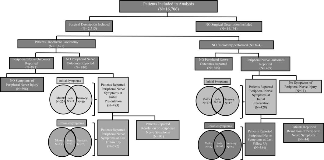
- Fasciotomy: Emergent for compartment syndrome — more commonly needed than escharotomy in electrical burns. Fasciotomy may also provide long-term protection against chronic peripheral nerve dysfunction. [23-24][32]
- Escharotomy: For circumferential burns with vascular compromise; performed in ~40% of electrical burn patients in one series [33]
- Serial debridement: Delayed 24–48 hours to allow tissue demarcation unless compartment syndrome present [23]
- Amputation: Required in ~13.5–24% of high-voltage injuries [3][33]
The following figure illustrates the role of fasciotomy in protecting against long-term peripheral nerve dysfunction in electrical burn patients:
Cardiac management
Renal protection
Disposition
Discharge from ED (low-risk criteria — all must be met): [1][9-10][28]
- Low-voltage injury (<1000 V)
- No loss of consciousness
- Normal 12-lead ECG
- No significant burns or trauma
- Asymptomatic
- Normal labs (if obtained)
Admit for monitoring (≥24 hours)
- High-voltage injury
- Abnormal ECG
- LOC or syncope
- Significant burns
- Elevated CK, troponin, or lactate [6]
- Transthoracic current pathway
- Pre-existing cardiac disease [19]
Burn center transfer (per ABA criteria): [4-5]
- All electrical burns warrant burn center referral
- Especially: high-voltage injuries, significant TBSA, injuries to hands/feet/face/genitalia, compartment syndrome, rhabdomyolysis
ICU admission
- Cardiac arrest survivors
- Active rhabdomyolysis with AKI
- Compartment syndrome requiring fasciotomy
- Hemodynamic instability
- Large TBSA burns requiring aggressive resuscitation
Follow Up / Return Precautions
Follow-up timing
- Burn clinic within 24–48 hours of discharge for wound reassessment
- Ophthalmology referral within 1–2 weeks (cataracts can develop weeks to years post-injury)
- Neuropsychological screening at follow-up — neurological symptoms (81.6%) and psychological symptoms (71%) are the most common long-term sequelae [20]
Long-term sequelae to monitor: [16-17][37-38]
- Neurological: Paresthesias, neuropathy, chronic pain, seizures, memory problems — may develop up to 5 years post-injury [16]
- Psychiatric: PTSD (33%), anxiety (62%), depression (25%), insomnia (68%) — occur regardless of voltage; baseline depression predicts worse long-term adjustment [17][22]
- Functional: 19% of work-related injuries unable to return to any employment; 61% return to preinjury work [17]
- Aesthetic sequelae: 43.9% at 2-year follow-up [2]
Return precautions (patient counseling)
- Return immediately for: chest pain, palpitations, syncope, dark urine, increasing limb swelling/pain/numbness, fever, wound changes
- Oral commissure burns in children: risk of delayed labial artery hemorrhage at 7–14 days post-injury — parents must be counseled
- Expected recovery: minor low-voltage injuries typically resolve within days; high-voltage injuries may require months of rehabilitation with potential for permanent disability
References
1. Electrical Cardiac Injuries: Current Concepts and Management. — Waldmann V, Narayanan K, Combes N, et al. European Heart Journal. 2018.
2. Outcomes of Electrical Injuries in the Emergency Department: Epidemiology, Severity Predictors, and Chronic Sequelae. — Karray R, Chakroun-Walha O, Mechri F, et al. European Journal of Trauma and Emergency Surgery : Official Publication of the European Trauma Society. 2025.
3. Electrical Injuries and Outcomes: A Retrospective Review. — Khor D, AlQasas T, Galet C, et al. Burns : Journal of the International Society for Burn Injuries. 2023.
4. Adherence to Burn Center Referral Criteria for Pediatric Burns. — Gus E, To T, Fish J, Diong C, Saunders N. JAMA Network Open. 2026.
5. Wilderness Medical Society Clinical Practice Guideline on Care of Burns in the Wilderness. — Bitter CC, Storkan M, Overmiller A, et al. Wilderness & Environmental Medicine. 2025.
6. Prevalence and Risk Factors of Developing Cardiac Arrhythmia in Patients Presenting to the Emergency Department With Electrical Injuries. — Yazıcı R, Bulut B, Genç M, et al. PloS One. 2025.
7. Clinical Predictors of Myocardial Damage After High Voltage Electrical Injury. — Chandra NC, Siu CO, Munster AM. Critical Care Medicine. 1990.
8. Cardiac Monitoring of Children With Household Electrical Injuries. — Bailey B, Gaudreault P, Thivierge RL, Turgeon JP. Annals of Emergency Medicine. 1995.
9. Arrhythmias and Laboratory Abnormalities After an Electrical Accident: A Single-Center, Retrospective Study of 333 Cases. — Seyfrydova M, Rokyta R, Rajdl D, Huml M. Clinical Research in Cardiology : Official Journal of the German Cardiac Society. 2023.
10. Risk of Cardiac Arrhythmias After Electrical Accident: A Single-Center Study of 480 Patients. — Pilecky D, Vamos M, Bogyi P, et al. Clinical Research in Cardiology : Official Journal of the German Cardiac Society. 2019.
11. Volume Resuscitation in Patients With High-Voltage Electrical Injuries. — Culnan DM, Farner K, Bitz GH, et al. Annals of Plastic Surgery. 2018.
12. Releasing Burn-Induced Compartment Syndrome by Enzymatic Escharotomy-Debridement: A Case Study. — Mataro I, Lanza A, Di Franco S, et al. Journal of Burn Care & Research : Official Publication of the American Burn Association. 2020.
13. Rhabdomyolysis and Acute Kidney Injury. — Bosch X, Poch E, Grau JM. The New England Journal of Medicine. 2009.
14. 2024 American Heart Association and American Red Cross Guidelines for First Aid. — Hewett Brumberg EK, Douma MJ, Alibertis K, et al. Circulation. 2024.
15. Experience of the Treatment of Severe Electric Burns on Special Parts of the Body. — Xu X, Zhu W, Wu Y. Annals of the New York Academy of Sciences. 1999.
16. Neurological Symptoms and Disorders Following Electrical Injury: A Register-Based Matched Cohort Study. — Nielsen KJ, Carstensen O, Kærgaard A, Vestergaard JM, Biering K. PloS One. 2022.
17. Acute and Long-Term Clinical, Neuropsychological and Return-to-Work Sequelae Following Electrical Injury: A Retrospective Cohort Study. — Radulovic N, Mason SA, Rehou S, Godleski M, Jeschke MG. BMJ Open. 2019.
18. Mental Disorders Following Electrical Injuries-a Register-Based, Matched Cohort Study. — Biering K, Vestergaard JM, Kærgaard A, Carstensen O, Nielsen KJ. PloS One. 2021.
19. Experience With Guidelines for Cardiac Monitoring After Electrical Injury in Children. — Bailey B, Gaudreault P, Thivierge RL. The American Journal of Emergency Medicine. 2000.
20. Long-Term Sequelae of Low-Voltage Electrical Injury. — Singerman J, Gomez M, Fish JS. Journal of Burn Care & Research : Official Publication of the American Burn Association. 2008.
21. Retrospective Study of Prognosis and Relating Factors of Cardiac Complications Associated With Electrical Injuries at a Single Centre in Korea. — Choi JH, Han D, Kang SH, et al. BMJ Open. 2019.
22. Psychiatric Outcome Over a Decade After Electrical Injury: Depression as a Predictor of Long-Term Adjustment. — Hahn-Ketter A, Aase DM, Paxton J, et al. Journal of Burn Care & Research : Official Publication of the American Burn Association. 2014.
23. Electrical Injuries of the Hand and Upper Extremity. — Lee DH, Desai MJ, Gauger EM. The Journal of the American Academy of Orthopaedic Surgeons. 2019.
24. Escharotomy in Burns. — Pegg SP. Annals of the Academy of Medicine, Singapore. 1992.
25. Electric Injury, Part II: Specific Injuries. — Fish RM. The Journal of Emergency Medicine. 2000.
26. Institutional Experience Using a Treatment Algorithm for Electrical Injury. — Nisar S, Keyloun JW, Kolachana S, et al. Journal of Burn Care & Research : Official Publication of the American Burn Association. 2021.
27. Updates on Rhabdomyolysis: A Clinically Oriented Narrative Review. — Richert Q, Miller N, Cameron S, et al. Chest. 2026.
28. Cardiac Monitoring in Patients With Electrical Injuries. An Analysis of 268 Patients at the Charité Hospital. — Searle J, Slagman A, Maaß W, Möckel M. Deutsches Arzteblatt International. 2013.
29. Prehospital and Emergency Management. — Williams JM, Ingle CL, Schauer SG, Maddry JK. The Surgical Clinics of North America. 2023.
30. Burns in Children: Standard and New Treatments. — Jeschke MG, Herndon DN. Lancet. 2014.
31. An Evidence-Based Narrative Review of the Emergency Department Evaluation and Management of Rhabdomyolysis. — Long B, Koyfman A, Gottlieb M. The American Journal of Emergency Medicine. 2019.
32. The Roles of Fasciotomy and Peripheral Nerve Decompression in Electric Burn Patients: A Systematic Review and Meta‐Analysis. — Blears EE, Ballou J, Weitzner A, Caffrey J, Dellon AL. Microsurgery. 2025.
33. Predictors for Limb Amputation and Reconstructive Management in Electrical Injuries. — Pedrazzi N, Klein H, Gentzsch T, et al. Burns : Journal of the International Society for Burn Injuries. 2023.
34. Part 10: Adult and Pediatric Special Circumstances of Resuscitation: 2025 American Heart Association Guidelines for Cardiopulmonary Resuscitation and Emergency Cardiovascular Care. — Cao D, Arens AM, Chow SL, et al. Circulation. 2025.
35. Early Fluid Resuscitation in Patients With Rhabdomyolysis. — Better OS, Abassi ZA. Nature Reviews. Nephrology. 2011.
36. Continuous Renal Replacement Therapy (CRRT) for Rhabdomyolysis. — Zeng X, Zhang L, Wu T, Fu P. The Cochrane Database of Systematic Reviews. 2014.
37. Electrical Injury: Chronic Pain, Somatosensory Dysfunction, Post Traumatic Stress and Movement Disorders. — Lun EWY, Tan AC, Andrews CJ, Champion GD. Injury. 2022.
38. Long-Term Consequences of Electrical Injury Without Initial Signs of Severity: The AFTER-ELEC Study. — Chauveau N, Renard A, Gasperini G, Cazes N. The American Journal of Emergency Medicine. 2021.
Figure
More from Deep Dive
Reviewed by Dr. Lucas Mastropaolo. SymptomDx is an educational tool for medical professionals — it does not replace clinical judgment. Verify all clinical data and drug dosages with authoritative sources.