Empyema
Empyema — pus within the pleural space — is most commonly a complication of pneumonia (parapneumonic effusion progressing through exudative → fibrinopurulent → organized stages). Overall mortality is approximately 20%, rising to >30% in patients over 65 and 47% in hospital-acquired cases. [1-2] Incidence is rising, and delays in drainage are associated with substantially higher mortality. [2]
History
- Duration and progression of respiratory symptoms: cough, fever, dyspnea, pleuritic chest pain, sputum production
- Preceding pneumonia or recent respiratory illness — parapneumonic effusions occur in 20–57% of pneumonia cases, progressing to empyema in 5–10% [1-2]
- Timing of antibiotic initiation relative to symptom onset
- Recent thoracic surgery, trauma, or procedures (risk for hospital-acquired empyema) [3-4]
- Aspiration risk factors: alcohol use, altered consciousness, dysphagia, poor dentition (anaerobic organisms)
- Important: elderly patients often present atypically — with anemia, fatigue, and failure to thrive rather than classic cough/fever/chest pain [2]
Alarm Features
- Sepsis or septic shock: tachycardia, hypotension, altered mental status, high lactate
- Rapidly enlarging effusion or tension physiology (mediastinal shift, tracheal deviation)
- Failure to improve on appropriate antibiotics within 48–72 hours
- Frank purulence on thoracentesis
- Hospital-acquired infection (mortality 47% vs. 17% for community-acquired) [2]
- RAPID score 5–7 (≥30% 12-week mortality) — warrants more aggressive initial therapy [2]
Medications
- Community-acquired empyema: [5]
- Ceftriaxone + metronidazole, OR
- Ampicillin/sulbactam (covers anaerobes without additional agent)
- Clindamycin is an alternative to metronidazole for anaerobic coverage
- Hospital-acquired/post-procedural empyema: [5]
- Vancomycin + cefepime + metronidazole, OR
- Vancomycin + piperacillin/tazobactam
- Meropenem + vancomycin if ESBL-producing organisms suspected
- Continue anaerobic coverage even with negative anaerobic cultures (difficult to culture) [5]
- Aminoglycosides are NOT recommended — inactivated in acidic empyema fluid and have poor pleural penetration [5-6]
- No role for intrapleural antibiotic administration [3][5]
- Do not delay antibiotics for pleural analysis unless the patient is clinically stable with an indolent infection [7]
- Intrapleural fibrinolytics: tPA 10 mg + DNase 5 mg (twice daily × 3 days = 6 doses) for loculated/complicated collections [2][6]
Diet
- Nutritional optimization is critical — all patients should receive adequate nutrition [2]
- Albumin <2.7 g/dL is a component of the RAPID prognostic score and predicts poor outcomes [2]
- Ensure adequate caloric and protein intake during prolonged hospitalization
- No specific dietary triggers; focus on preventing malnutrition in the setting of catabolic sepsis
Review of Systems
- Pulmonary: cough, dyspnea, pleuritic chest pain, hemoptysis, sputum character
- Constitutional: fever, chills, rigors, night sweats, weight loss, fatigue
- GI: dysphagia, vomiting (aspiration risk), abdominal pain (subdiaphragmatic abscess)
- Musculoskeletal: chest wall tenderness
- Hematologic: anemia symptoms (especially in elderly with atypical presentation)
- Screen for immunocompromise: HIV risk factors, steroid use, diabetes, malignancy
Collateral History and Family History
- Confirm antibiotic use prior to presentation (may alter pleural fluid characteristics — can convert neutrophilic to lymphocyte-predominant fluid) [6]
- Recent hospitalizations, surgeries, or invasive procedures
- Functional status and surgical candidacy (critical for treatment planning)
- Substance use: alcohol (aspiration risk), IV drug use (endocarditis → septic emboli)
- No significant hereditary predisposition, but immunodeficiency states increase risk
Risk Factors
- Pneumonia (most common antecedent) [2]
- Age >65 years [1-2]
- Diabetes mellitus, chronic liver disease, chronic kidney disease
- Immunosuppression (HIV, chemotherapy, chronic steroids)
- Alcohol use disorder and aspiration risk
- Poor dentition (anaerobic flora)
- Recent thoracic surgery or trauma (32.8% cumulative incidence in trauma patients) [3]
- Prolonged ICU stay (up to 62% of ICU pneumonia patients develop parapneumonic effusions) [7]
- GERD, esophageal disease
Differential Diagnosis
- Complicated parapneumonic effusion (CPPE) — the distinction from empyema is the presence of frank pus; management overlaps significantly [4]
- Lung abscess — may coexist; look for air-fluid level within parenchyma
- Malignant pleural effusion — lymphocyte-predominant, cytology positive in ~60%
- Tuberculous pleurisy — lymphocyte-predominant, elevated ADA (>40 U/L), consider in endemic areas [8]
- Chylothorax — milky fluid, elevated triglycerides
- Hemothorax — bloody fluid, hematocrit >50% of peripheral blood
- Subdiaphragmatic abscess with sympathetic effusion
- Esophageal rupture (Boerhaave syndrome) — low pH, high amylase, salivary origin
Past Medical History
- Prior pneumonia episodes or pleural infections
- Previous thoracic surgery or chest tube placement
- History of lung resection (risk for bronchopleural fistula and post-pneumonectomy empyema) [6]
- Chronic lung disease (COPD, bronchiectasis)
- Malignancy (especially lung, breast, lymphoma)
- Immunosuppressive conditions or medications
Physical Exam
- Vitals: fever, tachycardia, tachypnea, hypoxia; hypotension suggests sepsis
- Inspection: splinting, decreased chest wall movement on affected side
- Percussion: stony dullness over the effusion
- Auscultation: decreased or absent breath sounds; bronchial breathing at the upper border of the effusion; decreased tactile fremitus
- Palpation: tracheal deviation away from the affected side (large effusion)
- Signs of sepsis: altered mental status, mottled skin, delayed capillary refill
- Assess for subcutaneous emphysema (bronchopleural fistula)
- Examine dentition (source of anaerobic organisms)
Lab Studies
- Pleural fluid analysis (cornerstone of diagnosis): [5-6]
- pH <7.2 → most important predictor of need for drainage
- Glucose <40 mg/dL
- LDH >1000 IU/L
- Gram stain and culture (inoculate aerobic AND anaerobic blood culture bottles at bedside — increases yield) [5]
- Cell count with differential (neutrophil predominant)
- Protein, LDH for Light's criteria (exudate confirmation)
- Frank pus = empyema by definition — no need to check pH [7]
- Serum labs: CBC, BMP (renal function for RAPID score), lactate, blood cultures, albumin (<2.7 g/dL = poor prognosis), CRP, procalcitonin
- Serum CRP >200 mg/L combined with pleural glucose <60 mg/dL has 98% specificity for complicated effusion [6]
- Culture sensitivity is only ~56%; bedside inoculation of blood culture bottles improves yield [7]
Imaging
- Point-of-care ultrasound (POCUS) — first-line and superior to CT for detecting septations and loculations [6-7]
- Chest X-ray: costophrenic angle blunting; lateral decubitus film to assess free-flowing vs. loculated fluid
- Contrast-enhanced CT (venous/pleural phase): indicated when sepsis persists >48 hours after drainage [6]
- "Split pleura" sign — enhancement of both visceral and parietal pleura
- Thickened parietal pleura, increased extrapleural fat attenuation
- Gas bubbles within the effusion (anaerobic organisms)
- Evaluates for malpositioned chest tubes, lung abscess, subdiaphragmatic abscess, bronchopleural fistula
- MRI: no established role in adults; may be considered in pediatrics as radiation-free alternative [6]
Special Tests
- RAPID Score (Renal function, Age, Purulence, Infection source, Dietary/albumin) — scores 0–7; score 5–7 = high risk (≥30% 12-week mortality) [2]
- Light's Criteria — confirms exudative effusion (pleural/serum protein >0.5, pleural/serum LDH >0.6, or pleural LDH > two-thirds upper limit of normal) [7]
- Bedside thoracentesis — diagnostic and therapeutic; use ultrasound guidance
- Adenosine deaminase (ADA) — if TB is suspected (sensitivity ≥79%, specificity ≥83%) [8]
- Pleural fluid CRP >100 mg/L has comparable discriminative value to pH and glucose for CPPE [6]
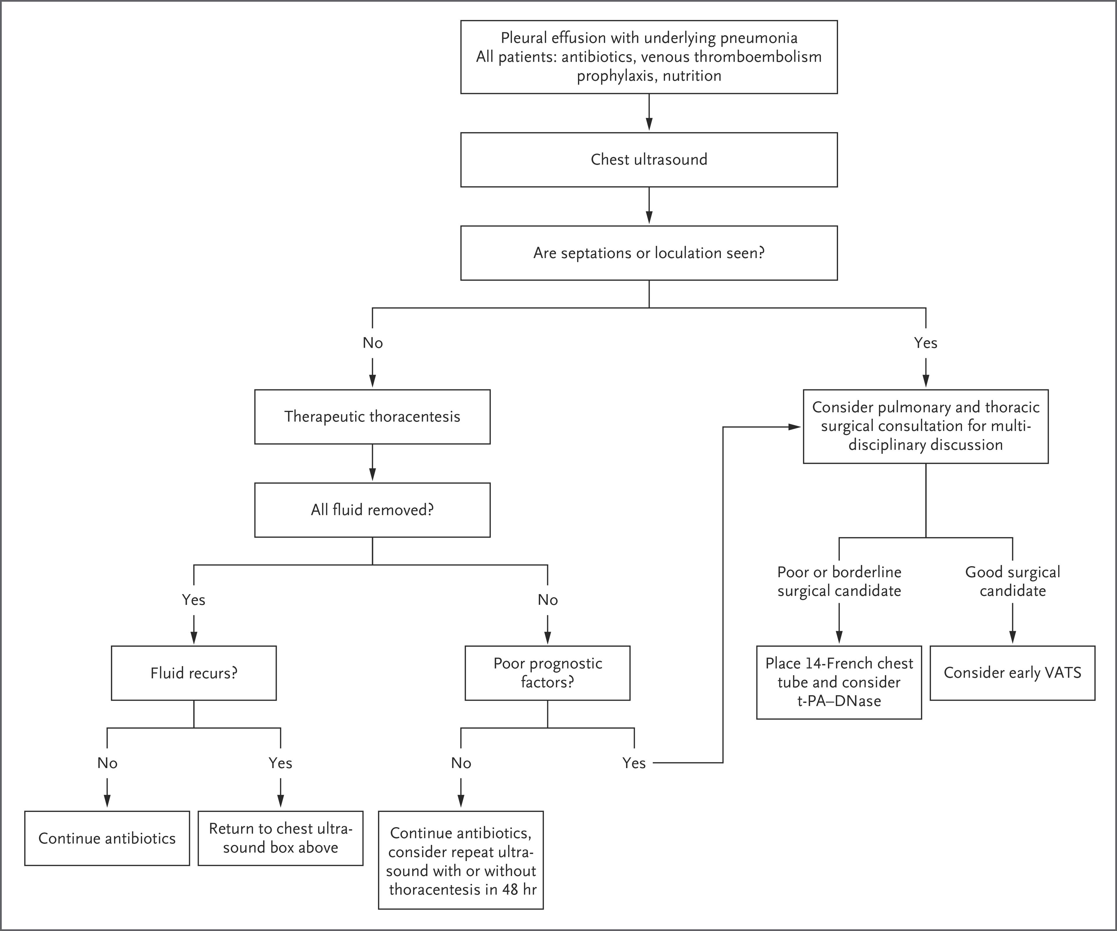
The following figure from the NEJM illustrates a practical management algorithm for parapneumonic effusions and empyema:
ECG
- Not a primary diagnostic tool for empyema
- Obtain to evaluate for tachycardia, atrial fibrillation (common in sepsis), or signs of right heart strain
- Rule out pericarditis if chest pain is diffuse or positional (diffuse ST elevation, PR depression)
- Large left-sided effusions may cause low voltage or electrical alternans
Assessment
Empyema progresses through three stages: [4]
- Stage I (Exudative): free-flowing sterile exudate; may resolve with antibiotics alone
- Stage II (Fibrinopurulent): bacterial invasion, fibrin deposition, septations/loculations, pH <7.2 — requires drainage
- Stage III (Organized): fibroblast proliferation, pleural peel ("rind") formation — often requires surgical decortication
Key prognostic factors: hospital-acquired source, advanced age, renal impairment, hypoalbuminemia, and delayed drainage all predict worse outcomes. [2] Mortality approaches 20% overall and is substantially higher with treatment delays. [2]
Treatment Plan
All patients: [2]
- Appropriate IV antibiotics (do not delay)
- DVT prophylaxis
- Adequate nutrition
- Peripheral blood cultures
Drainage strategy (stepwise escalation): [2][6][9]
- Small-bore chest tube (14-French pigtail catheter preferred; avoid <12-French due to higher failure rate) [2]
- Intrapleural tPA + DNase if inadequate drainage: tPA 10 mg + DNase 5 mg, administered sequentially or concurrently, twice daily × 3 days (6 doses total) [2][6][9]
- Associated with 60% reduction in pleural fluid on imaging
- Does NOT reduce mortality but reduces surgical referral and hospital stay [2]
- VATS decortication — if medical management fails or patient is a good surgical candidate [6]
- Open thoracotomy — reserved for VATS failure or late-stage disease; higher morbidity and mortality (3.7% vs. 2.8% for VATS) [6]
Antibiotic duration: typically 2–4 weeks; guided by clinical response, inflammatory markers, and imaging
Disposition
- All confirmed empyema cases require hospital admission — this is not an outpatient diagnosis [2]
- ICU admission: sepsis/septic shock, hemodynamic instability, respiratory failure requiring mechanical ventilation
- Surgical consultation triggers: [6]
- Failure to improve after 48–72 hours of antibiotics + drainage
- Persistent sepsis at day 3 post-chest tube
- Organized/loculated collection not amenable to tube drainage
- Failed tPA/DNase therapy
- Discharge criteria: afebrile, improving inflammatory markers, chest tube removed (output minimal), tolerating oral antibiotics, adequate nutrition
- Mean hospital stay: ~12 days with tPA/DNase; ~5–7 days with early VATS [2]
Follow Up / Return Precautions
- Follow-up imaging (chest X-ray or ultrasound) at 4–6 weeks post-discharge to assess for residual collection, trapped lung, or pleural thickening [3]
- Pulmonary function monitoring after discharge, especially post-surgical patients [3]
- Transition to oral antibiotics guided by culture sensitivities; complete full antibiotic course
- Return precautions: recurrent fever, worsening dyspnea, chest pain, purulent sputum, hemoptysis
- Consider repeat imaging sooner if symptoms recur
- Nutritional rehabilitation — ensure albumin recovery
- Patients with RAPID score 5–7 warrant closer follow-up given ≥30% 12-week mortality risk [2]
- Long-term: most patients recover well with early treatment; residual pleural thickening is common but usually does not impair function significantly
References
1. Intra-Pleural Fibrinolytic Therapy Versus Placebo, or a Different Fibrinolytic Agent, in the Treatment of Adult Parapneumonic Effusions and Empyema. — Altmann ES, Crossingham I, Wilson S, Davies HR. The Cochrane Database of Systematic Reviews. 2019.
2. Pleural Disease. — Feller-Kopman D, Light R. The New England Journal of Medicine. 2018.
3. Thoracic Trauma WSES-AAST Guidelines. — Coccolini F, Cremonini C, Moore EE, et al. World Journal of Emergency Surgery : WJES. 2025.
4. Surgical Versus Non-Surgical Management for Pleural Empyema. — Redden MD, Chin TY, van Driel ML. The Cochrane Database of Systematic Reviews. 2017.
5. The American Association for Thoracic Surgery Consensus Guidelines for the Management of Empyema. — Shen KR, Bribriesco A, Crabtree T, et al. The Journal of Thoracic and Cardiovascular Surgery. 2017.
6. ERS/ESTS Statement on the Management of Pleural Infection in Adults. — Bedawi EO, Ricciardi S, Hassan M, et al. The European Respiratory Journal. 2023.
7. Pleural Effusion: Diagnostic Approach in Adults. — Shen-Wagner J, Gamble C, MacGilvray P. American Family Physician. 2023.
8. Guide to Utilization of the Microbiology Laboratory for Diagnosis of Infectious Diseases: 2024 Update by the Infectious Diseases Society of America (IDSA) and the American Society for Microbiology (ASM). — Miller JM, Binnicker MJ, Campbell S, et al. Clinical Infectious Diseases : An Official Publication of the Infectious Diseases Society of America. 2024.
9. Comprehensive Review of Chest Tube Management: A Review. — Anderson D, Chen SA, Godoy LA, Brown LM, Cooke DT. JAMA Surgery. 2022.
Figure
More from Deep Dive
Reviewed by Dr. Lucas Mastropaolo. SymptomDx is an educational tool for medical professionals — it does not replace clinical judgment. Verify all clinical data and drug dosages with authoritative sources.