Endocarditis
Infective endocarditis (IE) is an infection of the cardiac endothelium — most commonly involving heart valves — with in-hospital mortality of approximately 15–30%. It presents acutely (high fever, sepsis, rapid valve destruction) or subacutely (weeks of malaise, low-grade fevers, weight loss). [1-3] Diagnosis relies on the 2023 Duke-ISCVID Criteria, integrating clinical, microbiologic, and imaging findings. [2][4]
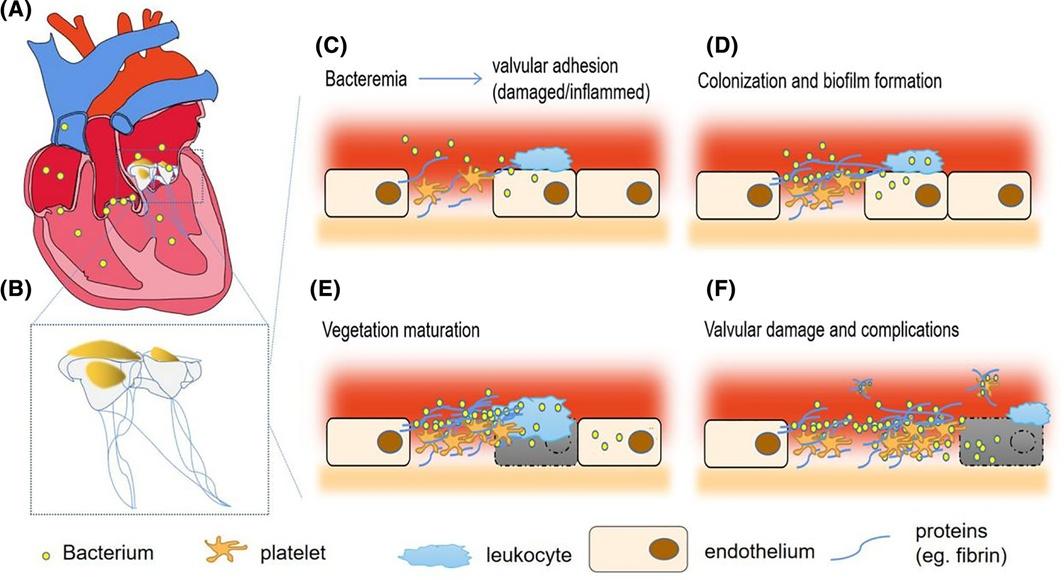
The following figure illustrates the pathogenesis from initial bacteremia through vegetation formation and embolic complications:
History
- Onset and tempo: Acute (hours–days) vs. subacute (weeks–months); sudden high fever with rigors suggests S. aureus, while insidious fatigue/malaise suggests viridans streptococci or coagulase-negative staphylococci [1][3]
- Symptom characterization: Fever (present in ~78% of cases), chills, night sweats, dyspnea, back pain, arthralgias, weight loss [1-2]
- Embolic symptoms: New focal neurologic deficits (stroke), pleuritic chest pain (septic pulmonary emboli), flank pain (splenic/renal infarct), abdominal pain [6]
- Cardiac symptoms: Progressive dyspnea, orthopnea, lower extremity edema (heart failure from acute valvular regurgitation) [2]
- Important negatives: Absence of fever does not exclude IE, especially in elderly, immunocompromised, or subacute presentations [6]
- Key questions: Recent dental procedures? IV drug use? Recent hospitalization or intravascular catheter? Prior valve surgery or prosthetic valve? History of IE? Recent skin/soft tissue infection?
Alarm Features
- Acute heart failure — new dyspnea, pulmonary edema, cardiogenic shock from acute severe valvular regurgitation [7-8]
- Persistent bacteremia (>5–7 days on appropriate antibiotics) — suggests uncontrolled infection, abscess, or need for surgery [7]
- Neurologic events — stroke, intracranial hemorrhage, mycotic aneurysm rupture, brain abscess [6][9]
- Septic shock (occurs in ~7% of cases) [2]
- New conduction abnormalities (heart block) — suggests perivalvular abscess, especially with aortic valve IE [7-8]
- Large vegetations >10 mm with embolic events — high risk for recurrent embolization [7]
- Paravalvular abscess, fistula, or prosthetic valve dehiscence [8-9]
Medications
Empiric therapy (before culture results)
- Native valve: Vancomycin + ceftriaxone [1][6][10]
- Prosthetic valve: Vancomycin + cefepime or piperacillin-tazobactam [6][10]
- If oral/GI source suspected: ampicillin-sulbactam may replace ceftriaxone [6]
- If Pseudomonas risk: cefepime or piperacillin-tazobactam [6]
Definitive therapy (organism-directed)
- MSSA: Nafcillin/oxacillin or cefazolin (4–6 weeks); beta-lactams preferred over vancomycin for higher cure rates [1][6][11]
- MRSA: Vancomycin (30 mg/kg/day divided q8–12h) or daptomycin (6 weeks) [6][11]
- Viridans streptococci: Ceftriaxone 2g IV daily or penicillin G (4 weeks) [6][11]
- Enterococcus: Ampicillin + ceftriaxone (preferred) or ampicillin + gentamicin (≥6 weeks regardless of valve type); avoid gentamicin in elderly/CKD patients [1][6]
Oral step-down: The POET trial supports transition to oral antibiotics in the continuation phase for stable patients with left-sided IE who no longer require surgery [2][10]
Cautions
- Aminoglycosides: nephrotoxicity/ototoxicity — limit duration to ≤2 weeks; avoid in elderly/renal impairment [6]
- Rifampin: reserve for prosthetic valve IE; do not use as monotherapy [1][10]
- Avoid empiric antibiotics before blood cultures are drawn whenever possible [1][8]
Diet
- No specific dietary triggers or restrictions for IE itself
- Adequate nutrition is critical during prolonged hospitalization; consider nutritional support for cachectic patients
- Ensure adequate hydration, particularly with nephrotoxic antibiotics (vancomycin, aminoglycosides)
- Patients with heart failure from IE should follow sodium and fluid restriction as appropriate
Review of Systems
- Constitutional: Fever, chills, rigors, night sweats, weight loss, fatigue, malaise [1][6]
- Cardiovascular: Dyspnea, orthopnea, PND, palpitations, chest pain, syncope [2]
- Neurologic: Headache, focal weakness, vision changes, confusion, seizures [6]
- Musculoskeletal: Back pain, arthralgias (may suggest spondylodiscitis or septic arthritis) [1]
- Skin: New rash, painful fingertip lesions, painless palmar/plantar lesions [1][6]
- Renal: Hematuria, decreased urine output (glomerulonephritis, renal infarct) [9]
- Pulmonary: Cough, pleuritic chest pain, hemoptysis (right-sided IE with septic pulmonary emboli) [12]
- Abdominal: Left upper quadrant pain (splenic infarct/abscess) [6]
Collateral History and Family History
- Collateral: IV drug use history (type of drug, injection practices, water source), recent dental work, recent hospitalization or procedures, indwelling catheters, prior antibiotic use [3][11]
- Social context: Homelessness (risk for Bartonella quintana), farm animal exposure (risk for Coxiella burnetii/Q fever) [1]
- Family history: Generally not a major contributor; however, congenital heart disease (e.g., bicuspid aortic valve) may be familial and is a predisposing condition [2-3]
- Prior cardiac history: Previous IE (strong risk factor for recurrence), known valvular disease, prior valve surgery [2][9]
Risk Factors
Cardiac
- Prior IE, prosthetic heart valve, prior valve repair [2][9]
- Valvular heart disease (degenerative, rheumatic, congenital — especially bicuspid aortic valve) [1][3]
- Cardiac implantable electronic devices (CIEDs), ventricular assist devices [2]
- Hypertrophic obstructive cardiomyopathy [9]
Non-cardiac
- IV drug use — most common risk factor in younger patients; predominantly right-sided (tricuspid) IE [11-12]
- Age >60 years, male sex [2-3]
- Hemodialysis, central venous catheters, indwelling intravascular devices [1][3]
- Poor dentition, recent dental procedures [1-2]
- Immunosuppression, diabetes, chronic liver disease, neoplastic disease [1][3]
- Recent hospitalization (nosocomial IE accounts for up to one-third of US cases) [6]
Differential Diagnosis
- Bacteremia without endocarditis — most common mimic; distinguish by echocardiographic findings and persistence of bacteremia [1][12]
- Nonbacterial thrombotic endocarditis (marantic endocarditis) — sterile vegetations in malignancy or hypercoagulable states; culture-negative [3]
- Libman-Sacks endocarditis — associated with SLE/antiphospholipid syndrome; sterile vegetations [3]
- Atrial myxoma — intracardiac mass mimicking vegetation on echo; can cause embolic phenomena and constitutional symptoms
- Rheumatic fever — fever, murmur, arthralgias; distinguished by Jones criteria, negative blood cultures
- Systemic vasculitis (e.g., ANCA-associated) — can mimic immunologic phenomena of IE
- Occult malignancy — fever of unknown origin, weight loss, elevated inflammatory markers
- Other causes of FUO — lymphoma, connective tissue disease, deep-seated abscess
Past Medical History
- Prior IE — strongest predictor of recurrence [2][9]
- Prosthetic valve or valve repair — prosthetic valve IE has distinct microbiology (early: coagulase-negative staphylococci; late: similar to native valve) [4][6]
- Congenital heart disease — especially unrepaired or palliated lesions [9]
- Rheumatic heart disease — most common predisposing condition in developing countries [1]
- Chronic kidney disease/hemodialysis — increased risk and impacts antibiotic selection [1][3]
- Diabetes, chronic liver disease, immunosuppression [1]
- History of IV drug use — even remote history is relevant [11]
Physical Exam
Vital signs
Cardiac
- New murmur (48%) or worsening of known murmur (20%) — most common sign [6]
- New aortic regurgitation murmur is particularly concerning
- Signs of heart failure: JVD, pulmonary crackles, peripheral edema [2]
Skin/extremities
- Janeway lesions — painless erythematous macules/papules on palms and soles (~3.5%) [2][6]
- Osler nodes — painful, tender nodules on fingers/toes (~2%) [2][6]
- Splinter hemorrhages — linear subungual hyperpigmentation [6]
- Petechiae — conjunctival, oral mucosa, skin [1]
Eyes
- Roth spots — hemorrhagic retinal lesions with pale centers (~1.4%); perform fundoscopy [2]
- Conjunctival hemorrhages [1]
Abdominal
- Splenomegaly (more common in subacute IE) [2]
- LUQ tenderness (splenic infarct/abscess)
Neurologic
Musculoskeletal
Lab Studies
Essential
- Blood cultures — at least 2–3 sets from separate venipuncture sites before antibiotics; first and last drawn ≥1 hour apart [1][8][12]
- CBC with differential (leukocytosis, anemia of chronic disease)
- CRP, ESR (elevated in >90%)
- BMP/CMP (renal function for antibiotic dosing, electrolytes)
- Urinalysis (hematuria from glomerulonephritis or renal infarct)
- Procalcitonin (may help distinguish IE from non-infectious causes)
If culture-negative
- Serologies: Coxiella burnetii phase I IgG (>1:800 is a major Duke criterion), Bartonella IgG/IgM [1][4][6]
- PCR: Bartonella, C. burnetii, Tropheryma whipplei [2][4][6]
- Amplicon/metagenomic sequencing if available [2][4]
Monitoring
- Repeat blood cultures q48h until clearance documented [1]
- Vancomycin trough levels (target AUC/MIC-based dosing)
- Renal function (especially with vancomycin, aminoglycosides) [6]
- Rheumatoid factor, complement levels (immunologic phenomena) [9]
Imaging
First-line
Second-line / when TTE is negative or equivocal
- Transesophageal echocardiography (TEE) — sensitivity ~90–96% for both native and prosthetic valves; recommended for all prosthetic valves, possible IE by Duke criteria, or suspected complicated IE [6][11]
- If TEE is negative but suspicion remains high, repeat in 5–7 days [1]
Advanced imaging (2023 Duke-ISCVID criteria now include these):
- 18F-FDG PET/CT — particularly useful for prosthetic valve IE, CIEDs, and detecting metastatic infection (splenic abscess, spondylodiscitis); may be more sensitive than TEE for prosthetic valves [2][4][6]
- Cardiac CT — identifies abscess, pseudoaneurysm, fistula; useful for surgical planning; less sensitive for small (<4 mm) vegetations [6]
- Brain MRI — for neurologic symptoms; detects silent cerebral emboli in up to 80% of left-sided IE
- CT abdomen/pelvis — for splenic/renal infarcts, abscesses [6]
- Chest CT — for septic pulmonary emboli in right-sided IE
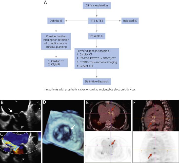
The following figure illustrates an integrated imaging algorithm for suspected IE:
Special Tests
Diagnostic scoring
- 2023 Duke-ISCVID Criteria — definite IE requires 2 major criteria, or 1 major + 3 minor, or 5 minor criteria; more sensitive than modified Duke criteria (84.2% vs 74.9%) without significant loss of specificity [2][4]
- Intraoperative inspection is now a new major criterion [4]
Point-of-care
- Bedside TTE (limited but can identify large vegetations in the ED)
- Point-of-care lactate (sepsis assessment)
Specialty tests
- Histopathology of resected valve tissue or embolized vegetation (definitive diagnosis) [8]
- In situ hybridization on valve tissue [2][4]
ECG
- New conduction abnormalities — PR prolongation, new bundle branch block, or complete heart block strongly suggest perivalvular/aortic root abscess, especially in aortic valve IE [7-8]
- Obtain baseline ECG and monitor serially
- ST changes may indicate coronary embolization (rare)
- Tachycardia (sepsis, heart failure)
- Low voltage or electrical alternans if pericardial effusion develops
Assessment
Severity stratification
- Acute IE — rapid onset, high fever, sepsis, S. aureus predominant; high mortality (~25–40% for S. aureus IE) [1][3]
- Subacute IE — indolent course, viridans streptococci or enterococci; lower mortality but delayed diagnosis common [1][12]
- Complicated IE — heart failure, abscess, embolic events, persistent bacteremia; requires urgent surgical evaluation [7-8]
Typical vs. atypical presentations
- Classic triad of fever + murmur + positive blood cultures is present in a minority [12]
- Elderly/immunocompromised patients may be afebrile with nonspecific symptoms [6]
- Right-sided IE (IVDU) may lack peripheral embolic phenomena but presents with septic pulmonary emboli (cough, pleuritic pain, bilateral nodular infiltrates) [12]
Complications
- Heart failure (most common indication for surgery, ~27% of cases) [2]
- Systemic embolization (~20–50%, highest in first 2 weeks) [5]
- Stroke (~15–20% of left-sided IE) [6]
- Mycotic aneurysm, perivalvular abscess, intracardiac fistula [7][9]
- Glomerulonephritis, renal failure [9]
Treatment Plan
Initial stabilization
- ABCs, hemodynamic resuscitation, vasopressors if septic shock
- Draw blood cultures immediately, then start empiric IV antibiotics [2][6]
- Source control: remove infected intravascular catheters, evaluate for abscess drainage [6]
Empiric antibiotic regimens (see Medications section above) [6][10]
Definitive therapy: Organism-directed based on culture and susceptibility results; minimum 4 weeks for NVE, 6 weeks for PVE (enterococcal IE: ≥6 weeks regardless) [6]
Oral step-down: Three RCTs support transition to oral therapy in the continuation phase for stable patients with left-sided NVE or PVE who are clinically improving, afebrile, with negative blood cultures, and no longer require surgery [2][10]
Surgical indications (early surgery during initial hospitalization):
- Heart failure from valve dysfunction [7-8]
- Uncontrolled infection: abscess, fistula, enlarging vegetation, persistent bacteremia >5–7 days [7-8]
- Prevention of embolism: recurrent emboli with persistent vegetations, or vegetations >10 mm after embolic event [7]
- Fungal or highly resistant organisms [7]
- Prosthetic valve dehiscence [7-8]
Timing of surgery
- Emergency (within 24 hours): refractory pulmonary edema, cardiogenic shock [7]
- Urgent (within days): most other surgical indications [7]
- Stroke is NOT a contraindication to surgery unless there is extensive neurologic damage or intracranial hemorrhage [8]
Multidisciplinary team: Infectious disease, cardiology, cardiothoracic surgery, neurology (if neurologic complications) — early involvement is associated with improved survival [6][8][13]
Disposition
Admission criteria
- All patients with suspected or confirmed IE require hospitalization for IV antibiotics, monitoring, and workup [2][6]
- ICU admission for hemodynamic instability, septic shock, acute heart failure, or neurologic emergencies
Transfer criteria
Observation
Discharge criteria
- Clinically stable, afebrile, blood cultures negative, no surgical indication, and appropriate oral step-down regimen available (per POET trial criteria) — may complete therapy as outpatient with OPAT or oral antibiotics [2][10]
- Ensure reliable follow-up and compliance
Follow Up / Return Precautions
Follow-up timing
- Repeat echocardiography at completion of therapy to establish new baseline [8]
- Infectious disease follow-up within 1–2 weeks of discharge
- Cardiology follow-up for valve function monitoring
- Dental evaluation for source control [3]
Return precautions — instruct patients to seek immediate care for:
- Recurrent fever, chills, or rigors
- New neurologic symptoms (weakness, speech difficulty, severe headache)
- Worsening dyspnea, chest pain, or lower extremity swelling
- Skin lesions on palms/soles or painful fingertip nodules
- Hematuria or decreased urine output
Expected recovery
- Clinical improvement typically within 5–7 days of appropriate antibiotics [2]
- Total treatment duration: 4–6 weeks minimum
- Recurrence rate: ~2–6% within 1 year; higher with ongoing IV drug use [11]
Long-term considerations
- Antibiotic prophylaxis for future dental procedures in patients with prosthetic valves, prior IE, certain congenital heart diseases, or cardiac transplant with valvulopathy per AHA guidelines [8][12]
- Substance use disorder treatment and harm reduction for patients with drug use-associated IE [11]
- Colonoscopy if S. gallolyticus (formerly S. bovis) is isolated — associated with colorectal neoplasia [1]
References
1. Native-Valve Infective Endocarditis. — Chambers HF, Bayer AS. The New England Journal of Medicine. 2020.
2. Infective Endocarditis. — Li M, Kim JB, Sastry BKS, Chen M. Lancet. 2024.
3. Management Considerations in Infective Endocarditis: A Review. — Wang A, Gaca JG, Chu VH. The Journal of the American Medical Association. 2018.
4. The 2023 Duke-International Society for Cardiovascular Infectious Diseases Criteria for Infective Endocarditis: Updating the Modified Duke Criteria. — Fowler VG, Durack DT, Selton-Suty C, et al. Clinical Infectious Diseases : An Official Publication of the Infectious Diseases Society of America. 2023.
5. Infective endocarditis complicated by embolic events: Pathogenesis and predictors. — Hu W, Wang X, Su G. Clinical Cardiology. 2021.
6. Infective Endocarditis: Diagnosis and Treatment. — Nohria R, Romaine A, Garcia-Sampson G. American Family Physician. 2026.
7. Challenges in Infective Endocarditis. — Cahill TJ, Baddour LM, Habib G, et al. Journal of the American College of Cardiology. 2017.
8. 2020 ACC/AHA Guideline for the Management of Patients With Valvular Heart Disease: A Report of the American College of Cardiology/American Heart Association Joint Committee on Clinical Practice Guidelines. — Otto CM, Nishimura RA, Bonow RO, et al. Journal of the American College of Cardiology. 2021.
9. Recent Insights Into Native Valve Infective Endocarditis: JACC Focus Seminar 4/4. — Dayer MJ, Quintero-Martinez JA, Thornhill MH, et al. Journal of the American College of Cardiology. 2024.
10. Guidelines for Diagnosis and Management of Infective Endocarditis in Adults: A WikiGuidelines Group Consensus Statement. — McDonald EG, Aggrey G, Aslan AT, et al. JAMA Network Open. 2023.
11. Diagnosis and Management of Infective Endocarditis in People Who Inject Drugs: JACC State-of-the-Art Review. — Yucel E, Bearnot B, Paras ML, et al. Journal of the American College of Cardiology. 2022.
12. Infective Endocarditis in Adults: Diagnosis, Antimicrobial Therapy, and Management of Complications: A Scientific Statement for Healthcare Professionals From the American Heart Association. — Baddour LM, Wilson WR, Bayer AS, et al. Circulation. 2015.
13. Staphylococcus Aureus Infective Endocarditis: JACC Patient Pathways. — Grapsa J, Blauth C, Chandrashekhar YS, et al. Journal of the American College of Cardiology. 2022.
Figures
More from Deep Dive
Reviewed by Dr. Lucas Mastropaolo. SymptomDx is an educational tool for medical professionals — it does not replace clinical judgment. Verify all clinical data and drug dosages with authoritative sources.