Erysipelas
Erysipelas is an acute, non-purulent infection of the superficial dermis and lymphatics, predominantly caused by beta-hemolytic streptococci (primarily Group A Streptococcus), presenting as a sharply demarcated, bright-red, raised plaque with systemic symptoms. [1-2] It most commonly affects the lower extremities (~76–85%) and the face (~17%). [3-4]
History
- Onset and progression: Acute onset of a well-demarcated, painful, warm, red area — often preceded by constitutional symptoms (fever, chills, malaise) before the erythema appears. In one study, 91.4% of erysipelas patients reported constitutional symptoms at or before erythema onset. [5]
- Entry portal: Ask about recent skin breaks — wounds, insect bites, tinea pedis, leg ulcers, eczema, surgical sites, IV drug use. [6-7]
- Timing: Rapid spread over hours to days; tongue-like extensions are characteristic. [1]
- Prior episodes: Recurrence rate is 8–20% annually; up to 40% lifetime recurrence. Ask about number and location of prior episodes. [8-9]
- Associated symptoms: Fever, rigors, malaise, nausea. Systemic symptoms may precede skin findings by hours. [9]
- Important negatives: Purulent drainage (suggests abscess/cellulitis), pain out of proportion (necrotizing fasciitis), crepitus, skin necrosis, numbness.
Alarm Features
- Pain out of proportion to exam findings → necrotizing fasciitis [2][10]
- Rapidly spreading erythema with dusky/violaceous discoloration, bullae, skin necrosis, or crepitus [10-11]
- Hemodynamic instability — hypotension, tachycardia, altered mental status (SIRS/sepsis) [9]
- Cutaneous numbness or anesthesia over the affected area
- Failure to improve within 48–72 hours of appropriate antibiotics [5]
- Immunocompromised host (chemotherapy, neutropenia, HIV) with rapidly progressive infection [9]
Medications
- First-line treatment: Penicillin — erysipelas responds reliably (98.3% within 2 days). Penicillin V 500 mg PO QID or amoxicillin 500 mg PO TID for outpatient; IV penicillin G for inpatient. [1][3][5][9]
- Alternatives (penicillin allergy): Cephalexin 500 mg PO QID, clindamycin 300–450 mg PO TID, or macrolides (azithromycin, erythromycin). [9][12]
- Duration: 5 days per IDSA, extended if no improvement. [9][13]
- IV options: Penicillin G 2–4 million units IV q4–6h; cefazolin 1–2 g IV q8h. [9]
- MRSA coverage is generally unnecessary for classic non-purulent erysipelas. Add MRSA coverage only if purulent drainage, penetrating trauma, IVDU, MRSA colonization, or SIRS. [9]
- Recurrence prophylaxis: Penicillin V 250 mg PO BID for ≥2 episodes/year; reduces recurrence by ~69% while on therapy (NNT = 6). [14-15]
- Medication contributors: Immunosuppressants (corticosteroids, biologics, chemotherapy) increase susceptibility.
Diet
- No specific dietary triggers for erysipelas.
- Hydration is important during febrile illness.
- Long-term: Weight management is relevant, as obesity is an independent risk factor for both primary and recurrent erysipelas. [4][6][16]
Review of Systems
- Constitutional: Fever, chills, rigors, malaise, fatigue (present in majority) [3][5]
- Skin: Erythema, warmth, swelling, pain, vesicles/bullae, lymphangitic streaking
- Musculoskeletal: Joint pain/swelling near affected area (rule out septic arthritis, gout)
- Vascular: Unilateral leg swelling (consider DVT — prevalence ~2–3% in lower extremity erysipelas) [17-18]
- Endocrine: Diabetes symptoms (polyuria, polydipsia) — diabetes is a key comorbidity [4][16]
Collateral History and Family History
- Prior episodes of erysipelas/cellulitis — location, frequency, treatments used
- Surgical history: Lymph node dissection (axillary for breast cancer, inguinal), saphenous vein harvest, radical pelvic surgery — all predispose to lymphedema and recurrence [7][19]
- Social context: Homelessness, IVDU, living conditions [9]
- Family history is generally not a major contributor, though familial lymphedema syndromes (e.g., Milroy disease) may predispose.
Risk Factors
- Disruption of cutaneous barrier — the strongest risk factor (OR 23.8): leg ulcers, wounds, tinea pedis/toe-web intertrigo, eczema, psoriasis [6]
- Lymphedema (OR 71.2 in one case-control study) [6]
- Venous insufficiency (OR 2.9) [6]
- Leg edema from any cause (OR 2.5) [6]
- Obesity (OR 2.0) [4][6][16]
- Diabetes mellitus [4][16][20]
- Prior episode of erysipelas/cellulitis [9][14]
- Prior surgery with lymphatic disruption [7][16]
- Immunosuppression [14]
- Tinea pedis — population attributable risk of 61% for leg erysipelas [6]
Differential Diagnosis
- Cellulitis — deeper, poorly demarcated, darker red, less likely to have early constitutional symptoms [1-2][5]
- Necrotizing fasciitis — pain out of proportion, dusky skin, crepitus, rapid progression, systemic toxicity; requires emergent surgical exploration [2][10]
- Deep vein thrombosis — unilateral leg swelling, erythema, calf tenderness; use Wells score and duplex ultrasound [17][21]
- Stasis dermatitis — bilateral, chronic, associated with hemosiderin deposition and varicosities; a common cellulitis mimic [2][21]
- Contact dermatitis — pruritic, well-demarcated, often bilateral; exposure history [21]
- Gout/septic arthritis — periarticular erythema and swelling; joint aspiration differentiates [21]
- Abscess — fluctuant collection; point-of-care ultrasound differentiates [2]
- Erythema migrans (Lyme) — expanding annular erythema with central clearing; tick exposure history
- Herpes zoster — dermatomal, vesicular, painful
Past Medical History
- Prior erysipelas/cellulitis episodes (strongest predictor of recurrence) [9][14]
- Lymphedema (primary or secondary) [6][19]
- Venous insufficiency, chronic leg edema, varicose veins [6][20]
- Diabetes mellitus [4][16]
- Obesity [4][6]
- Cancer (especially breast cancer with axillary dissection) [4][7]
- Immunosuppressive conditions or medications [14]
- Chronic skin conditions (eczema, psoriasis, tinea pedis) [6][14]
- Saphenous vein harvest for CABG [7]
Physical Exam
- Vital signs: Fever (present in 25–77% depending on setting), tachycardia; hypotension and altered mental status suggest severe infection [2-3][9]
- Skin: Sharply demarcated, raised, bright-red, warm, tender plaque with tongue-like extensions; peau d'orange appearance from lymphatic involvement [1-2][9]
- Vesicles/bullae and petechiae/ecchymoses may develop in severe cases [9]
- Lymphangitis: Linear erythematous streaking proximal to the lesion [2]
- Regional lymphadenopathy: Tender, enlarged nodes draining the affected area [9]
- Interdigital toe spaces: Examine for fissuring, maceration, tinea pedis — a critical and often overlooked entry portal [6][9]
- Concerning findings suggesting necrotizing fasciitis: Dusky/violaceous discoloration, skin necrosis, crepitus, cutaneous anesthesia, pain out of proportion [2][10]
- Mark the borders of erythema with a skin marker to track progression.
Lab Studies
- Routine labs are not required for uncomplicated cases. [9]
- When obtained: Leukocytosis and elevated CRP are typical; CRP ≥ 3.27 mg/dL distinguishes erysipelas from limited cellulitis with 75% sensitivity and 73% specificity. [22]
- Elevated ESR found in ~60% of cases. [3]
- Blood cultures: Positive in ≤5% of cases; obtain only if systemic toxicity (high fever, hypotension), immunocompromised, malignancy, or unusual exposures. [9]
- Wound/aspirate cultures: Not routinely recommended; consider in immunocompromised patients, animal bites, immersion injuries. [9]
- To rule out dangerous conditions:
- WBC > 15,400/μL or Na < 135 mmol/L → raises concern for necrotizing fasciitis [10]
- Lactate, CK, coagulation studies if necrotizing infection suspected
- BMP for renal function (antibiotic dosing)
Imaging
- Imaging is generally unnecessary for uncomplicated erysipelas. [9]
- Point-of-care ultrasound: Useful to rule out abscess (anechoic/hypoechoic fluid collection) or DVT. [2][21]
- CT with contrast: If concern for necrotizing fasciitis — may show fascial thickening, gas tracking, or fluid along fascial planes. However, imaging should not delay surgical exploration if clinical suspicion is high. [10]
- MRI: Most sensitive for deep soft tissue infection and necrotizing fasciitis, but rarely needed acutely.
- Duplex ultrasound: Consider if clinical concern for DVT (unilateral leg swelling disproportionate to erythema). [17-18]
Special Tests
- LRINEC Score (Laboratory Risk Indicator for Necrotizing Fasciitis): Based on WBC, hemoglobin, sodium, glucose, creatinine, and CRP. Score ≥ 6 has PPV 92% and NPV 96% for necrotizing fasciitis vs. cellulitis in the index study. Use as an adjunct — clinical judgment remains paramount. [10]
- Wells Score for DVT: Apply if DVT is in the differential; pooled DVT incidence in erysipelas/cellulitis is low (~2–3% for proximal DVT), comparable to low-risk Wells patients. [17-18]
- Skin marking: Outline the erythema border with a marker and timestamp — a simple but critical bedside tool to track progression or response to therapy.
- Bedside ultrasound: Cobblestoning of subcutaneous tissue supports soft tissue infection; fluid collection suggests abscess.
ECG
- ECG is not routinely indicated for erysipelas.
- Obtain if sepsis is suspected (tachycardia, hypotension) or in patients with cardiac comorbidities.
- Consider ECG in patients with atrial fibrillation or other cardiac conditions that may affect management (e.g., anticoagulation decisions). [4]
Assessment
Erysipelas is a clinical diagnosis based on the characteristic sharply demarcated, raised, bright-red erythema with tongue-like extensions and early systemic symptoms (fever/chills preceding or concurrent with skin findings). [1-2][5] It is distinguished from cellulitis by its more superficial involvement, brighter erythema, sharper borders, and more prominent early constitutional symptoms. [1][5][22]
Severity stratification per IDSA: [9]
- Mild: No systemic signs of infection → outpatient oral antibiotics
- Moderate: Systemic signs (fever, tachycardia, leukocytosis) → systemic antibiotics, consider observation/admission
- Severe: SIRS criteria, hemodynamic instability, immunocompromised, or concern for deeper infection → hospitalization, IV antibiotics, surgical consultation if necrotizing infection suspected
Complications: Abscess formation, bacteremia (<5%), lymphatic damage leading to chronic lymphedema, recurrence (8–20%/year), and rarely progression to necrotizing fasciitis. [8-9]
Treatment Plan
Initial stabilization
- IV fluid resuscitation if septic; hemodynamic support as needed.
- Elevation of the affected extremity to reduce edema. [9]
Antibiotic therapy
Adjunctive measures
- Treat the entry portal — antifungal therapy for tinea pedis (topical terbinafine or clotrimazole), wound care for ulcers. [6][9]
- Compression stockings and lymphedema management for venous insufficiency/edema. [14]
- Weight management counseling for obese patients. [4][6]
- Recurrence prophylaxis (≥2 episodes/year): Penicillin V 250 mg PO BID; duration is indefinite as recurrence resumes after discontinuation. [9][14]
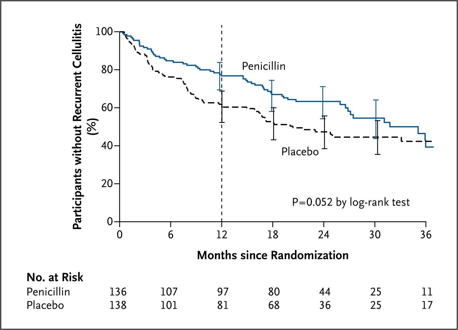
The PATCH II trial demonstrated that penicillin prophylaxis significantly reduced recurrence during the treatment period (HR 0.51), but the benefit was lost after cessation of prophylaxis. [14][23]
Disposition
- Discharge (outpatient): No SIRS, hemodynamically stable, able to tolerate oral antibiotics, reliable follow-up, no concern for deeper infection. [9]
- Observation/admission:
- Surgical consultation: Suspected necrotizing fasciitis, abscess requiring I&D, or rapidly progressive infection despite IV antibiotics. [10]
Follow Up / Return Precautions
- Follow-up: Reassess in 48–72 hours to confirm clinical improvement (reduction in erythema, pain, fever). Note: erythema may initially worsen slightly after starting antibiotics due to bacterial lysis releasing inflammatory mediators — this does not necessarily indicate treatment failure. [9]
- Return precautions — instruct patients to return immediately for:
- Worsening redness, swelling, or pain despite antibiotics
- New skin discoloration (purple/black), blistering, or skin breakdown
- High fever, rigors, or feeling significantly worse
- Numbness over the affected area
- Red streaking spreading up the limb
- Patient counseling:
- Complete the full antibiotic course
- Keep the affected limb elevated as much as possible
- Treat and prevent tinea pedis (keep feet dry, use antifungal powder)
- Moisturize skin to prevent cracking
- Manage edema with compression if applicable
- Expected recovery: Clinical improvement typically within 24–48 hours of appropriate antibiotics. Full resolution of erythema may take 1–2 weeks. Residual skin changes (hyperpigmentation, mild edema) may persist. [3][5]
References
1. Frequent Bacterial Skin and Soft Tissue Infections: Diagnostic Signs and Treatment. — Sunderkötter C, Becker K. Journal Der Deutschen Dermatologischen Gesellschaft = Journal of the German Society of Dermatology : JDDG. 2015.
2. Cellulitis: A Review. — Raff AB, Kroshinsky D. The Journal of the American Medical Association. 2016.
3. Analysis of Epidemiology, Clinical Features and Management of Erysipelas. — Krasagakis K, Valachis A, Maniatakis P, et al. International Journal of Dermatology. 2010.
4. Primary and Recurrent Erysipelas-Epidemiological Patterns in a Single-Centre Retrospective Analysis. — Matych M, Ciosek A, Miler K, et al. Journal of Clinical Medicine. 2025.
5. Constitutional Symptoms and Response to Penicillin G in Erysipelas and Cellulitis - A Monocentric, Retrospective, Explorative Study. — Schieffers H, Sunderkötter C. Journal Der Deutschen Dermatologischen Gesellschaft = Journal of the German Society of Dermatology : JDDG. 2026.
6. Risk Factors for Erysipelas of the Leg (Cellulitis): Case-Control Study. — Dupuy A, Benchikhi H, Roujeau JC, et al. BMJ. 1999.
7. Streptococcal Infections of Skin and Soft Tissues. — Bisno AL, Stevens DL. The New England Journal of Medicine. 1996.
8. Intracellular Streptococcal Uptake and Persistence: A Potential Cause of Erysipelas Recurrence. — Jendoubi F, Rohde M, Prinz JC. Frontiers in Medicine. 2019.
9. Practice Guidelines for the Diagnosis and Management of Skin and Soft Tissue Infections: 2014 Update by the Infectious Diseases Society of America. — Stevens DL, Bisno AL, Chambers HF, et al. Clinical Infectious Diseases : An Official Publication of the Infectious Diseases Society of America. 2014.
10. Necrotizing Soft Tissue Infections: A Review. — McDermott J, Kao LS, Keeley JA, et al. JAMA Surgery. 2024.
11. Erysipelas and Necrotizing Fasciitis. — Hammar H, Wanger L. The British Journal of Dermatology. 1977.
12. Assessment of Antibiotic Treatment of Cellulitis and Erysipelas: A Systematic Review and Meta-analysis. — Brindle R, Williams OM, Barton E, Featherstone P. JAMA Dermatology. 2019.
13. Appropriate Use of Short-Course Antibiotics in Common Infections: Best Practice Advice From the American College of Physicians. — Lee RA, Centor RM, Humphrey LL, et al. Annals of Internal Medicine. 2021.
14. Interventions for the Prevention of Recurrent Erysipelas and Cellulitis. — Dalal A, Eskin-Schwartz M, Mimouni D, et al. The Cochrane Database of Systematic Reviews. 2017.
15. Antibiotic Prophylaxis for Preventing Recurrent Cellulitis: A Systematic Review and Meta-Analysis. — Oh CC, Ko HC, Lee HY, et al. The Journal of Infection. 2014.
16. Risk Factors of Recurrent Erysipelas in Adult Chinese Patients: A Prospective Cohort Study. — Li A, Wang N, Ge L, Xin H, Li W. BMC Infectious Diseases. 2021.
17. Risk of Deep Vein Thrombosis in Patients With Cellulitis and Erysipelas: A Systematic Review and Meta-Analysis. — Gunderson CG, Chang JJ. Thrombosis Research. 2013.
18. Incidence of Deep Vein Thrombosis in Erysipelas or Cellulitis of the Lower Extremities. — Mortazavi M, Samiee MM, Spencer FA. International Journal of Dermatology. 2013.
19. Recurrent Erysipelas--Risk Factors and Clinical Presentation. — Inghammar M, Rasmussen M, Linder A. BMC Infectious Diseases. 2014.
20. Epidemiology and Comorbidity of Erysipelas in Primary Care. — Bartholomeeusen S, Vandenbroucke J, Truyers C, Buntinx F. Dermatology. 2007.
21. Clinical Mimics: An Emergency Medicine-Focused Review of Cellulitis Mimics. — Blumberg G, Long B, Koyfman A. The Journal of Emergency Medicine. 2017.
22. Diagnostic Value of Laboratory Parameters for the Discrimination Between Erysipelas and Limited Cellulitis. — Drerup C, Eveslage M, Sunderkoetter C, Ehrchen J. Journal Der Deutschen Dermatologischen Gesellschaft = Journal of the German Society of Dermatology : JDDG. 2020.
23. Penicillin to Prevent Recurrent Leg Cellulitis. — Thomas KS, Crook AM, Nunn AJ, et al. The New England Journal of Medicine. 2013.
Figure
More from Deep Dive
Reviewed by Dr. Lucas Mastropaolo. SymptomDx is an educational tool for medical professionals — it does not replace clinical judgment. Verify all clinical data and drug dosages with authoritative sources.