First-Degree AV Block
First-degree AV block (more accurately termed first-degree AV delay) is defined as a PR interval >200 ms with 1:1 AV conduction preserved — no P waves are dropped. [1-2] It is generally considered a benign conduction abnormality, though emerging data associate it with increased risks of atrial fibrillation, heart failure, and mortality, particularly in older adults and those with structural heart disease. [3-5]
History
- Onset and duration of any symptoms: fatigue, exercise intolerance, lightheadedness, dyspnea on exertion, near-syncope, or syncope
- Temporal correlation of symptoms with bradycardia or pauses
- Medication history — specifically beta-blockers (including ophthalmic), non-dihydropyridine calcium channel blockers (verapamil, diltiazem), digoxin, antiarrhythmics (amiodarone, flecainide, sotalol, propafenone), lithium, donepezil, SSRIs, opioids, and fingolimod [1][6]
- Recent tick exposure, erythema migrans rash, travel to Lyme-endemic areas [1][7]
- History of cardiac surgery, catheter ablation, or TAVR [1]
- Athletic training level (high vagal tone is a common benign cause in young athletes) [8-9]
- Symptoms of hypothyroidism, sarcoidosis, or autoimmune disease
Alarm Features
- PR interval ≥300 ms — can cause "pseudo-pacemaker syndrome" with hemodynamic compromise (loss of AV synchrony, decreased cardiac output, elevated pulmonary capillary wedge pressure) [1][10]
- Concurrent wide QRS or bundle branch block — suggests infranodal disease with risk of progression [1-2]
- Syncope or presyncope temporally correlated with bradycardia
- New first-degree AV block in the setting of acute MI (especially inferior) [2]
- New AV block with tick exposure — Lyme carditis can progress from first-degree to complete heart block within minutes to hours [7]
- First-degree AV block in patients with neuromuscular disease (myotonic dystrophy, Kearns-Sayre, Emery-Dreifuss) — unpredictable progression to high-grade block and sudden death [1-2]
- First-degree AV block in lamin A/C cardiomyopathy — associated with ventricular arrhythmias and sudden cardiac death [1-2]
Medications
- Common causative agents: [1][6]
- Beta-blockers (including ophthalmic timolol)
- Non-dihydropyridine CCBs (verapamil, diltiazem)
- Digoxin
- Antiarrhythmics: amiodarone, dronedarone, flecainide, propafenone, sotalol, procainamide
- Others: lithium, donepezil, phenytoin, SSRIs/TCAs, fingolimod, ivabradine, clonidine
- Key pearl: Therapeutic doses of AV-nodal blocking agents are rarely the sole cause of new AV block — most patients presenting with new AV block on these medications ultimately require permanent pacing [1-2]
- First-degree AV block is not an absolute contraindication to AV-nodal blocking drugs, but the PR interval should be monitored to ensure no progression [6]
- For digoxin toxicity: anti-digoxin Fab antibody fragments for severe cases [1]
- Avoid combining multiple AV-nodal blocking agents when possible [6]
Diet
- No specific dietary triggers for first-degree AV block
- In the setting of hyperkalemia-related conduction delay, restrict potassium intake and correct the electrolyte abnormality urgently [1]
- Ensure adequate hydration, particularly in patients with vagally mediated bradycardia
Review of Systems
- Cardiovascular: palpitations, chest pain, dyspnea on exertion, orthopnea, lower extremity edema, syncope/presyncope
- Neurologic: muscle weakness, myotonia, ptosis (neuromuscular disease screening)
- Infectious: fever, rash (erythema migrans), arthralgias, recent tick bite
- Endocrine: cold intolerance, weight gain, constipation, dry skin (hypothyroidism)
- Pulmonary: cough, dyspnea (sarcoidosis)
- Rheumatologic: joint pain, skin changes (SLE, scleroderma, RA) [1]
Collateral History and Family History
- Family history of sudden cardiac death, cardiomyopathy, or conduction disease
- Family history of neuromuscular disease (myotonic dystrophy, Emery-Dreifuss, limb-girdle dystrophy) [2][10]
- Family history of lamin A/C mutations — autosomal dominant with high risk of progressive AV block and ventricular arrhythmias [1-2]
- Maternal history of SLE — associated with congenital AV block [1]
- Social history: athletic conditioning level, cannabis use [1]
Risk Factors
- Age — prevalence follows a J-shaped curve: common in young athletes (vagal tone), then increases from the 5th–6th decade onward, peaking in the 9th–10th decade due to degenerative fibrosis [9]
- Chronic hypertension and diabetes mellitus — associated with degenerative conduction system disease [2]
- Structural heart disease — ischemic or nonischemic cardiomyopathy [2]
- AV-nodal blocking medications [1][6]
- High vagal tone — athletes, sleep, obstructive sleep apnea [1-2]
- Infiltrative/inflammatory disease — sarcoidosis, amyloidosis, myocarditis [1]
- Post-cardiac surgery or TAVR [1]
Differential Diagnosis
- Medication effect — most common iatrogenic cause; review all AV-nodal blocking agents [1]
- High vagal tone — benign in young, athletic patients; resolves with exercise [8]
- Lyme carditis — cannot-miss diagnosis in endemic areas; can rapidly progress to complete heart block [7]
- Acute inferior MI — AV block due to right coronary artery supply to AV node; usually transient [2]
- Cardiac sarcoidosis — may present with AV block as initial manifestation [2]
- Hypothyroidism — reversible cause, though treatment often does not eliminate need for pacing if otherwise indicated [1-2]
- Hyperkalemia — reversible with correction [1]
- Infiltrative disease — amyloidosis (AL or ATTR), especially with low voltage on ECG [2]
- Neuromuscular disease — myotonic dystrophy, Kearns-Sayre syndrome [2][10]
- Degenerative fibrosis (Lev's/Lenegre's disease) — most common cause in elderly [1-2]
Past Medical History
- Prior episodes of AV block or documented PR prolongation
- History of myocardial infarction (especially inferior)
- Congenital heart disease (e.g., congenitally corrected transposition of the great arteries) [8]
- Prior cardiac surgery or catheter ablation [1]
- Known cardiomyopathy, valvular disease
- Neuromuscular disorders
- Autoimmune disease (SLE, sarcoidosis)
- Prior Lyme disease
Physical Exam
- Vital signs: Heart rate (may be normal), blood pressure (assess for hemodynamic compromise)
- Cardiac auscultation: Soft S1 (due to prolonged PR allowing partial mitral valve closure before ventricular systole), cannon A waves in JVP if atrial contraction occurs against a closed tricuspid valve, murmurs suggesting structural disease
- Signs of heart failure: JVD, peripheral edema, pulmonary crackles
- Skin: Erythema migrans (Lyme), malar rash (SLE), skin thickening (scleroderma)
- Neurologic: Muscle weakness, myotonia, ptosis (neuromuscular disease)
- Thyroid exam: Goiter, signs of hypothyroidism
Lab Studies
- Recommended based on clinical suspicion: [1]
- BMP/CMP: Potassium (hyperkalemia), calcium, magnesium, renal function
- TSH: Rule out hypothyroidism
- Digoxin level: If on digoxin therapy
- Lyme serologies (ELISA + Western blot): In endemic areas or with suggestive history [1]
- Troponin: If acute coronary syndrome suspected
- ESR/CRP, ACE level: If sarcoidosis suspected
- ANA: If autoimmune etiology suspected
- No systematic studies have validated routine lab panels specifically for isolated first-degree AV block [1]
Imaging
- Echocardiogram: Indicated if structural heart disease is suspected (abnormal exam, abnormal ECG beyond PR prolongation, symptoms of heart failure) [8]
- Cardiac MRI with gadolinium: If sarcoidosis, myocarditis, or lamin A/C cardiomyopathy suspected — can identify myocardial fibrosis and inflammation [2]
- Chest X-ray: If heart failure or sarcoidosis suspected
Special Tests
- Ambulatory ECG monitoring (Holter or event monitor): Reasonable in patients with symptoms of unclear etiology and first-degree AV block to establish symptom-rhythm correlation (Class IIa) [2]
- Exercise treadmill test: Reasonable for exertional symptoms — improvement in AV conduction with exercise suggests AV nodal block (benign); worsening suggests infranodal disease (concerning) [2][8]
- Electrophysiology study (EPS): Rarely needed; may be considered if infranodal block is suspected (wide QRS, exercise-induced worsening) to measure HV interval [2][8]
- Carotid sinus massage/pharmacologic challenge: May be considered in selected cases to determine level of block [2]
ECG
- Defining feature: PR interval >200 ms with every P wave followed by a QRS complex (1:1 conduction) [1-2][10]
- QRS is typically narrow — a wide QRS raises concern for concomitant infranodal disease [2]
- Look for:
- PR ≥300 ms — "marked" first-degree AV block with hemodynamic implications [1][10]
- Bundle branch block pattern — RBBB, LBBB, or bifascicular block suggesting infranodal conduction disease [1]
- Alternating bundle branch block — high risk for sudden complete heart block [1]
- ST changes suggesting ischemia
- Low voltage suggesting infiltrative disease
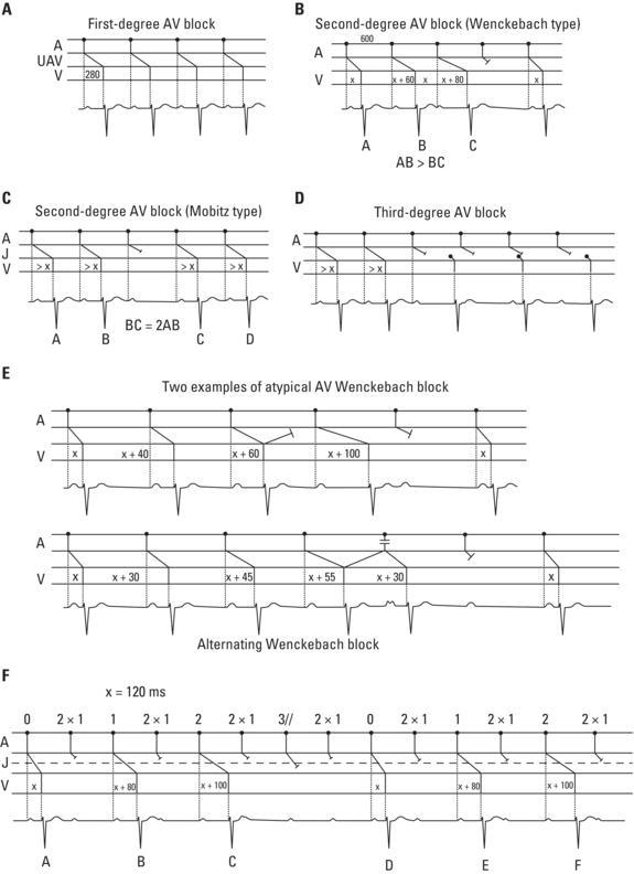
- The following figure illustrates the ECG patterns of different degrees of AV block:
Assessment
- First-degree AV block is typically benign and does not progress suddenly to complete heart block when the block is at the AV nodal level [1-2]
- However, a prolonged PR interval is not entirely benign — meta-analysis data show associations with a 1.45× increased risk of AF, 1.39× increased risk of heart failure, and 1.24× increased risk of mortality. The Framingham Heart Study demonstrated a 2-fold adjusted risk of AF and 3-fold adjusted risk of pacemaker implantation in individuals with first-degree AV block [3-4]
- In patients with stable CAD, first-degree AV block is associated with a 2.3× increased risk of HF hospitalization and 1.6× increased risk of mortality [5]
- In patients ≥85 years, first-degree AV block as a single ECG finding was the strongest predictor of mortality [9]
- Severity stratification:
Treatment Plan
- Asymptomatic, isolated first-degree AV block: No treatment required. Permanent pacing is not indicated (Class III — Harm per ACC/AHA/HRS) [2][10]
- Identify and treat reversible causes:
- Symptomatic first-degree AV block (PR ≥300 ms with pseudo-pacemaker syndrome — fatigue, exertional intolerance, hemodynamic compromise):
- Neuromuscular disease with any degree of AV block:
- Lamin A/C cardiomyopathy with first-degree AV block:
- pacing + defibrillator capability[1-2]
- Acute hemodynamic instability (rare with isolated first-degree AV block):
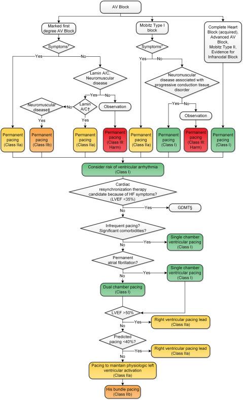
The following algorithm from the 2018 ACC/AHA/HRS guidelines outlines the management approach for chronic AV block:
Disposition
- Discharge: The vast majority of patients with isolated, asymptomatic first-degree AV block with PR <300 ms and narrow QRS can be safely discharged with outpatient follow-up [2]
- Observation/Admission criteria:
- New first-degree AV block with suspected Lyme carditis (risk of rapid progression) [7]
- Symptomatic bradycardia or hemodynamic compromise
- PR ≥300 ms with symptoms of pseudo-pacemaker syndrome
- Concurrent high-degree AV block, wide QRS, or alternating bundle branch block
- Acute MI with new conduction abnormality
- Drug toxicity (digoxin, beta-blocker, CCB overdose) requiring monitoring
- Cardiology consultation: For symptomatic patients, marked PR prolongation, wide QRS/bundle branch block, suspected neuromuscular or infiltrative disease, or consideration of pacemaker implantation
Follow Up / Return Precautions
- Outpatient follow-up with PCP or cardiology within 2–4 weeks for new findings; routine annual ECG monitoring is reasonable for stable, asymptomatic patients
- Return precautions — instruct patients to return for:
- Syncope or near-syncope
- New or worsening dizziness, lightheadedness
- Exertional intolerance or unexplained fatigue
- Palpitations or irregular heartbeat
- Chest pain or dyspnea
- Expected course: Isolated first-degree AV block is typically stable and does not progress suddenly to higher-degree block when at the AV nodal level. Progression to more advanced heart block is infrequent [1-2][9]
- Long-term awareness: Although benign in isolation, first-degree AV block is associated with increased long-term risk of AF, heart failure, and mortality — particularly in patients with structural heart disease, CAD, or advanced age. This may warrant periodic reassessment of cardiac status [3-5]
References
1. 2018 ACC/AHA/HRS Guideline on The Evaluation and Management Of Patients With Bradycardia and Cardiac Conduction Delay: A Report of the American College of Cardiology/American Heart Association Task Force on Clinical Practice Guidelines and the Heart Rhythm Society. — Kusumoto FM, Schoenfeld MH, Barrett C, et al. Journal of the American College of Cardiology. 2019.
2. 2018 ACC/AHA/HRS Guideline on The evaluation and Management Of patients With Bradycardia and Cardiac conduction Delay: A Report of the American College of Cardiology/American Heart Association Task Force on Clinical Practice Guidelines and the Heart Rhythm Society. — Writing Committee Members, Kusumoto FM, Schoenfeld MH, et al. Heart Rhythm. 2019.
3. Prolonged PR Interval, First-Degree Heart Block and Adverse Cardiovascular Outcomes: A Systematic Review and Meta-Analysis. — Kwok CS, Rashid M, Beynon R, et al. Heart. 2016.
4. Long-Term Outcomes in Individuals With Prolonged PR Interval or First-Degree Atrioventricular Block. — Cheng S, Keyes MJ, Larson MG, et al. The Journal of the American Medical Association. 2009.
5. First-Degree Atrioventricular Block Is Associated With Heart Failure and Death in Persons With Stable Coronary Artery Disease: Data From the Heart and Soul Study. — Crisel RK, Farzaneh-Far R, Na B, Whooley MA. European Heart Journal. 2011.
6. Drug-Induced Arrhythmias: A Scientific Statement From the American Heart Association. — Tisdale JE, Chung MK, Campbell KB, et al. Circulation. 2020.
7. Diagnosis and Treatment of Lyme Carditis: JACC Review Topic of the Week. — Yeung C, Baranchuk A. Journal of the American College of Cardiology. 2019.
8. Eligibility and Disqualification Recommendations for Competitive Athletes With Cardiovascular Abnormalities: Task Force 9: Arrhythmias and Conduction Defects: A Scientific Statement From the American Heart Association and American College of Cardiology. — Zipes DP, Link MS, Ackerman MJ, et al. Circulation. 2015.
9. Arrhythmias in Patients ≥80 Years of Age: Pathophysiology, Management, and Outcomes. — Curtis AB, Karki R, Hattoum A, Sharma UC. Journal of the American College of Cardiology. 2018.
10. 2012 ACCF/AHA/HRS Focused Update Incorporated Into the ACCF/AHA/HRS 2008 Guidelines for Device-Based Therapy of Cardiac Rhythm Abnormalities: A Report of the American College of Cardiology Foundation/American Heart Association Task Force on Practice Guidelines and the Heart Rhythm Society. — Epstein AE, DiMarco JP, Ellenbogen KA, et al. Journal of the American College of Cardiology. 2013.
11. Mechanisms, Classification, and Clinical Aspects of Arrhythmias. — Antoni Bayés De Luna, Miquel Fiol‐Sala, Antoni Bayés‐Genís, et al. Clinical Electrocardiography. 2021.
12. Cardiac Pacemakers: Function, Troubleshooting, and Management: Part 1 of a 2-Part Series. — Mulpuru SK, Madhavan M, McLeod CJ, Cha YM, Friedman PA. Journal of the American College of Cardiology. 2017.
13. 2018 ACC/AHA/HRS Guideline on The Evaluation and Management of Patients With Bradycardia and Cardiac Conduction Delay: Executive Summary: A Report of the American College of Cardiology/American Heart Association Task Force on Clinical Practice Guidelines, and the Heart Rhythm Society. — Kusumoto FM, Schoenfeld MH, Barrett C, et al. Journal of the American College of Cardiology. 2019.
Figures
More from Deep Dive
Reviewed by Dr. Lucas Mastropaolo. SymptomDx is an educational tool for medical professionals — it does not replace clinical judgment. Verify all clinical data and drug dosages with authoritative sources.