Gastroenteritis (Viral)
Viral gastroenteritis is a self-limited syndrome of rapid-onset nausea, vomiting, watery (non-bloody, non-mucoid) diarrhea, and cramping abdominal pain caused most commonly by norovirus, rotavirus, sapovirus, astrovirus, and enteric adenovirus. [1] It is the most common cause of acute nausea and vomiting and accounts for the majority of acute diarrheal illness in developed countries. [2-3] The cornerstone of management is oral rehydration and early refeeding; antibiotics are not indicated. [1][3]
History
- Onset and timeline: Abrupt onset typically 24–48 hours after exposure; preformed toxin food poisoning has a shorter incubation (2–7 hours) [4]
- Symptom characterization: Watery, non-bloody, non-mucoid diarrhea; nausea and vomiting (often predominant); diffuse crampy abdominal pain; malaise [1]
- Volume assessment: Quantify oral intake, urine output, number of emesis/diarrheal episodes, last void
- Exposures: Sick contacts, daycare/institutional setting, cruise ship, recent food (shellfish, salad bars, buffets), contaminated water [5]
- Important negatives: Absence of bloody stool, high fever, severe focal abdominal pain, and tenesmus makes viral etiology more likely [3-4]
- Medications: Recent antibiotics (C. difficile risk), PPIs, chemotherapy, laxatives [4]
- Travel history: International travel raises concern for bacterial/parasitic causes [3]
Alarm Features
- Bloody or mucoid stools → suggests inflammatory/invasive bacterial pathogen [3]
- High fever ≥38.5°C (101.3°F) → suggests bacterial etiology or systemic infection [4]
- Signs of severe dehydration: altered mental status, syncope, oliguria, sunken eyes, poor skin turgor [4][6]
- Severe abdominal pain or peritoneal signs (especially age >50) → workup for surgical abdomen [4]
- Symptoms >7 days → consider non-viral or parasitic etiology [3]
- Immunocompromised patient → risk of severe, prolonged, or opportunistic infection (CMV, Cryptosporidium) [1]
- Extremes of age (infants <3 months, elderly ≥65) → higher risk of dehydration and complications [3][7]
Medications
- Oral rehydration solution (ORS): First-line therapy; low-osmolar solutions (200–250 mOsm/L) preferred [1][3]
- Ondansetron: Safe and effective for recalcitrant vomiting to facilitate oral rehydration; may be used in patients >4 years; may increase stool volume [1][7]
- Loperamide (adults only): 4 mg initial dose, then 2 mg after each unformed stool (max 8 mg/day for ≤48 hours); reduces stool frequency in acute watery diarrhea [4][8]
- Bismuth subsalicylate: Mildly effective antisecretory; safe alternative when loperamide is contraindicated [3][8]
- Loperamide + simethicone: Combination provides faster relief of diarrhea and gas-related discomfort [3]
Contraindicated/Avoid
- Loperamide in bloody/inflammatory diarrhea (risk of toxic megacolon), febrile dysentery, and children <18 years per IDSA guidelines [7]
- Loperamide contraindicated in children <2 years (respiratory depression risk) [9]
- Bismuth subsalicylate in children <12 years (Reye syndrome risk) [9]
- Antibiotics are not indicated for viral gastroenteritis [5]
Diet
- Early refeeding (within 4–6 hours of rehydration) shortens illness duration and improves nutritional outcomes [1][3]
- Age-appropriate regular diet is recommended; no evidence supports the traditional BRAT diet over a normal diet [3]
- Avoid: High-sugar beverages (juice, soda), caffeine, alcohol, and high-fat foods acutely, as these can worsen osmotic diarrhea
- Lactose-containing foods may transiently worsen diarrhea due to transient lactase deficiency but routine avoidance is not universally recommended
- Hydration: Frequent small sips of ORS; commercial formulations (Pedialyte 250 mOsm/L) or homemade solutions [1]
Review of Systems
- GI: Nausea, vomiting, diarrhea frequency/character, abdominal pain location/quality, blood in stool, tenesmus
- Constitutional: Fever, malaise, myalgias, weight loss
- Neurologic: Dizziness, lightheadedness, syncope, confusion (dehydration markers)
- Urinary: Decreased urine output, dark urine
- Cardiopulmonary: Palpitations, chest pain (electrolyte-related), dyspnea
- Skin: Rash (consider viral exanthem, reactive arthritis)
Collateral History and Family History
- Sick contacts: Household members, coworkers, classmates with similar symptoms — secondary attack rates are 15% for norovirus and 28% for rotavirus within households [10]
- Institutional exposure: Nursing home, daycare, cruise ship, hospital — norovirus outbreaks are common in congregate settings [5]
- Food exposure: Shared meals, restaurant visits, raw shellfish consumption
- Family history: Generally not contributory for acute viral gastroenteritis; consider IBD if recurrent episodes
- Immunocompromised household members at risk for prolonged shedding and severe disease
Risk Factors
- Age extremes: Children <5 years (highest incidence) and adults ≥65 years [1][11]
- Congregate living: Cruise ships, long-term care facilities, dormitories, military barracks [5]
- Foodborne exposure: Raw/undercooked shellfish, contaminated salad greens, food prepared by infected handlers [1]
- Contaminated water or inadequately treated drinking water [1]
- Seasonality: Peak in winter months in temperate climates ("winter vomiting disease") [1]
- Immunosuppression: HIV/AIDS, transplant recipients, chemotherapy — risk of prolonged/severe illness [1]
- Lack of rotavirus vaccination in children [7]
- Fomite exposure: Norovirus persists on environmental surfaces and can cause recurrent outbreaks [5]
Differential Diagnosis
Cannot-miss diagnoses
- Appendicitis / surgical abdomen — focal RLQ pain, peritoneal signs, fever
- Mesenteric ischemia — severe pain out of proportion to exam, age >50, atrial fibrillation
- Bowel obstruction — distension, obstipation, bilious vomiting
- Diabetic ketoacidosis — nausea/vomiting with hyperglycemia, Kussmaul breathing
- Hemolytic uremic syndrome — bloody diarrhea followed by AKI, thrombocytopenia (post-STEC)
Important alternative diagnoses
- Bacterial gastroenteritis (Salmonella, Campylobacter, Shigella, STEC) — fever, bloody/mucoid stool, more severe abdominal pain [3]
- C. difficile — recent antibiotics or hospitalization [12]
- Preformed toxin food poisoning (S. aureus, B. cereus) — onset <6 hours after suspect meal [1]
- Parasitic infection (Giardia, Cryptosporidium) — prolonged watery diarrhea, travel history [1]
- Medication-induced diarrhea — antibiotics, PPIs, metformin, colchicine, SSRIs
- Early IBD flare — recurrent episodes, weight loss, extraintestinal manifestations [3]
- Hepatitis A/E — nausea, vomiting, jaundice, transaminase elevation [1]
Past Medical History
- Prior episodes of gastroenteritis and their frequency
- Immunocompromising conditions: HIV, transplant, chemotherapy, biologics
- Chronic kidney disease — higher risk from dehydration and electrolyte derangements
- Cardiac disease — less tolerance for volume depletion and electrolyte shifts
- Diabetes — risk of DKA with dehydration; medication adjustments may be needed (hold metformin, SGLT2 inhibitors)
- IBD — distinguish flare from superimposed infection
- Prior abdominal surgery — adhesive obstruction can mimic gastroenteritis
- Rotavirus vaccination status in children [7]
Physical Exam
Vital signs
- Tachycardia, orthostatic hypotension → dehydration
- Fever — low-grade is common; high/persistent fever suggests non-viral cause [1]
Focused exam
- General: Ill-appearing, listless (severe); alert and well-appearing (mild)
- Mucous membranes: Dry or tacky → dehydration
- Eyes: Sunken (especially children) [6]
- Skin turgor: Decreased; capillary refill >2 seconds in children
- Abdomen: Diffuse mild tenderness without rebound or guarding; hyperactive bowel sounds. Focal tenderness, rigidity, or peritoneal signs → surgical evaluation
- Rectal exam: Assess for occult blood if inflammatory cause suspected [4]
- Neurologic: Altered sensorium suggests severe dehydration or alternative diagnosis
Dehydration assessment scales: The Clinical Dehydration Scale (CDS) in children uses general appearance, eyes, mucous membranes, and tears to grade severity. [6][13]
Lab Studies
Most cases require no laboratory testing. [1][12]
When indicated (moderate-severe illness, high-risk patients):
- BMP/CMP: Electrolytes (hyponatremia, hypokalemia), BUN/Cr (pre-renal azotemia, AKI), glucose, bicarbonate [1][3]
- Expected findings: Metabolic alkalosis (predominant vomiting) or hyperchloremic non-anion gap metabolic acidosis (predominant diarrhea); elevated BUN:Cr ratio; lactic acidosis if hypovolemic [1][14]
- CBC: Leukocytosis may suggest bacterial etiology; thrombocytopenia raises concern for HUS [7]
- Lactate: If concern for sepsis or significant hypovolemia
Stool testing — reserve for
- Bloody, mucoid, or febrile diarrhea
- Immunocompromised patients
- Symptoms ≥7 days
- Suspected outbreak (food handlers, institutional settings)
- Infants <3 months [1][3][12]
- Multiplex PCR (e.g., BioFire FilmArray GI Panel) is preferred over culture when testing is indicated [3][15]
Imaging
- Imaging is generally unnecessary for uncomplicated viral gastroenteritis
- Abdominal X-ray or CT abdomen/pelvis if concern for obstruction, perforation, appendicitis, or ischemia
- CT with IV contrast is the study of choice when surgical pathology is suspected
- Ultrasound may be useful in pediatric patients to evaluate for appendicitis or intussusception
Special Tests
- Clinical Dehydration Scale (CDS): Validated 4-item tool for children (general appearance, eyes, mucous membranes, tears) — scores 0 (none), 1–4 (some), 5–8 (moderate-severe) [13]
- Point-of-care ultrasound (POCUS): IVC collapsibility for volume assessment in adults
- Stool multiplex PCR panels: Detect 22 pathogens (bacterial, viral, parasitic) with results in ~60 minutes; useful for outbreak investigation and infection control [16]
- Rapid norovirus PCR (e.g., Xpert Norovirus): Sensitivity 92–100%, specificity 93–100%, results in 90 minutes [1]
ECG
- Not routinely indicated for uncomplicated viral gastroenteritis
- Obtain ECG when:
- Severe dehydration with electrolyte abnormalities suspected
- Elderly patients or those with cardiac comorbidities
- Palpitations, chest pain, or syncope
- Findings to recognize:
Assessment
Viral gastroenteritis presents as acute-onset watery, non-bloody diarrhea with nausea/vomiting, typically self-limited within 1–3 days. [1] Norovirus is the most common cause in all age groups in the post-rotavirus vaccine era, responsible for ~18% of global acute gastroenteritis cases. [1][5] Globally, rotavirus remains the leading cause of diarrheal deaths, followed by norovirus. [18]
Severity stratification
- Mild: Tolerating oral fluids, no dehydration signs, <6 stools/day
- Moderate: Some dehydration, frequent vomiting limiting oral intake, 6–10 stools/day
- Severe: Signs of significant dehydration or shock, inability to tolerate oral fluids, altered mental status
Complications: Dehydration (most common), electrolyte disturbances (hyponatremia in 16% of hospitalized children with moderate-severe dehydration), AKI, metabolic acidosis, and rarely seizures in children. [6][19]
Treatment Plan
Initial stabilization
Rehydration strategy by severity: [1]
- Minimal/no dehydration: Ad lib oral fluids; ORS as tolerated; half-strength apple juice is acceptable in children with mild illness [13]
- Mild-moderate dehydration: ORS 50–100 mL/kg over 3–4 hours; frequent small sips; reassess frequently
- Severe dehydration/shock: IV isotonic crystalloid (NS or LR) bolus 20 mL/kg; transition to oral rehydration once stabilized [6]
Pharmacotherapy (adults)
- Ondansetron 4–8 mg PO/IV/ODT for vomiting (facilitates oral rehydration)
- Loperamide 4 mg initially, then 2 mg after each unformed stool (max 8 mg/day × 48 hours) for watery diarrhea only [4]
- Bismuth subsalicylate 525 mg PO q30–60 min (max 8 doses/24 hours) as alternative [8]
Pharmacotherapy (children)
- Ondansetron ODT 0.15 mg/kg (max 4 mg) × 1 dose to facilitate oral rehydration in children >4 years [1][7]
- Avoid loperamide in children <18 years per IDSA [7]
- Zinc supplementation recommended in low-income settings [20]
Refeeding: Resume age-appropriate regular diet within 4–6 hours of rehydration. [1][3]
Disposition
Discharge criteria
- Tolerating oral fluids without recurrent vomiting
- Adequate urine output
- No signs of significant dehydration
- Reliable follow-up and ability to return if worsening
- Caregiver able to monitor (pediatric patients)
Admission criteria
- Severe dehydration unresponsive to oral/IV rehydration in ED
- Inability to tolerate oral fluids despite antiemetics
- Hemodynamic instability or signs of shock
- Significant electrolyte abnormalities requiring correction
- Infants <3 months with dehydration
- Immunocompromised with severe symptoms
- Concern for alternative surgical diagnosis
Observation: Consider for patients with moderate dehydration requiring IV fluids who may improve with a few hours of rehydration and reassessment. [13]
Specialist consultation
- Surgery if peritoneal signs or concern for surgical abdomen
- GI if prolonged symptoms (>7 days), suspected IBD, or immunocompromised with refractory disease
- Infectious disease for immunocompromised patients with severe or prolonged illness
Follow Up / Return Precautions
Follow-up timing
- Most patients recover within 1–3 days [1]
- PCP follow-up in 2–3 days if symptoms persist; sooner for high-risk patients (elderly, infants, immunocompromised)
Return precautions — instruct patients to return immediately for:
- Inability to keep down any fluids for >12–24 hours
- Bloody or black stools
- High fever (>101.3°F / 38.5°C)
- Severe abdominal pain or distension
- Decreased urination, no tears (children), dizziness/lightheadedness
- Confusion or lethargy
- Symptoms worsening after initial improvement or lasting >7 days
Patient counseling
- Hand hygiene is the most effective prevention measure; alcohol-based sanitizers have limited efficacy against norovirus (soap and water preferred) [5]
- Contagion period: Patients remain infectious for ≥48 hours after symptom resolution; food handlers should not return to work until 48 hours symptom-free [5]
- Environmental decontamination: Norovirus persists on surfaces; clean with bleach-based solutions [5]
- Rotavirus vaccination for eligible infants [7]
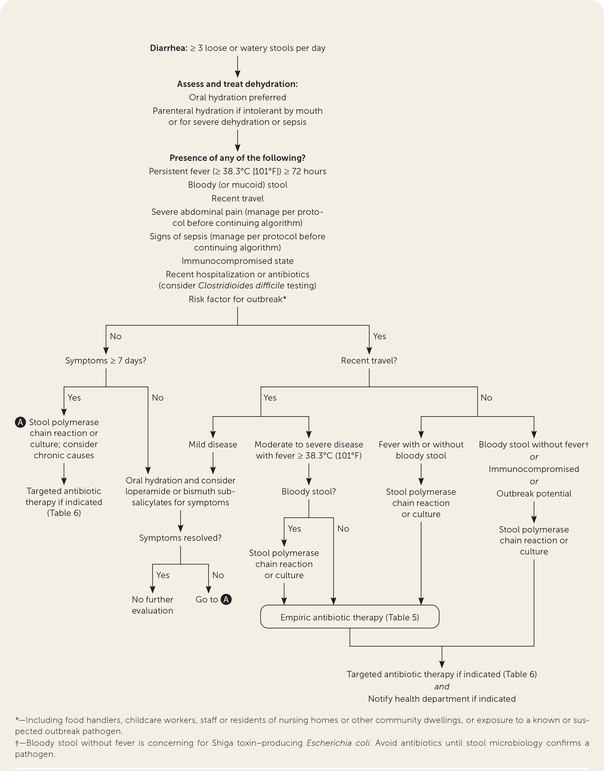
The following algorithm from the AAFP provides a practical framework for evaluating and managing acute diarrhea in the ED and primary care settings:
References
1. Viral Gastroenteritis. — Flynn TG, Olortegui MP, Kosek MN. Lancet. 2024.
2. Evaluation and Treatment of Nausea and Vomiting in Adults. — Johns T, Lawrence E. American Family Physician. 2024.
3. Acute Diarrhea in Adults. — Meisenheimer ES, Epstein C, Thiel D. American Family Physician. 2022.
4. Acute Infectious Diarrhea in Immunocompetent Adults. — DuPont HL. The New England Journal of Medicine. 2014.
5. Norovirus. — Sara A. Mirza and Jan Vinjé CDC Yellow Book. 2025.
6. Balanced Crystalloid Solutions Versus 0.9% Saline for Treating Acute Diarrhoea and Severe Dehydration in Children. — Florez ID, Sierra J, Pérez-Gaxiola G. The Cochrane Database of Systematic Reviews. 2023.
7. 2017 Infectious Diseases Society of America Clinical Practice Guidelines for the Diagnosis and Management of Infectious Diarrhea. — Shane AL, Mody RK, Crump JA, et al. Clinical Infectious Diseases : An Official Publication of the Infectious Diseases Society of America. 2017.
8. ACG Clinical Guideline: Diagnosis, Treatment, and Prevention of Acute Diarrheal Infections in Adults. — Riddle MS, DuPont HL, Connor BA. The American Journal of Gastroenterology. 2016.
9. Traveling Safely with Infants and Children. — Michelle Weinberg and Patrick W. Hickey CDC Yellow Book. 2025.
10. Community Burden and Transmission of Acute Gastroenteritis Caused by Norovirus and Rotavirus in the Netherlands (RotaFam): A Prospective Household-Based Cohort Study. — Quee FA, de Hoog MLA, Schuurman R, Bruijning-Verhagen P. The Lancet. Infectious Diseases. 2020.
11. Norovirus and Other Viral Causes of Medically Attended Acute Gastroenteritis Across the Age Spectrum: Results From the Medically Attended Acute Gastroenteritis Study in the United States. — Burke RM, Mattison CP, Marsh Z, et al. Clinical Infectious Diseases : An Official Publication of the Infectious Diseases Society of America. 2021.
12. Guide to Utilization of the Microbiology Laboratory for Diagnosis of Infectious Diseases: 2024 Update by the Infectious Diseases Society of America (IDSA) and the American Society for Microbiology (ASM). — Miller JM, Binnicker MJ, Campbell S, et al. Clinical Infectious Diseases : An Official Publication of the Infectious Diseases Society of America. 2024.
13. Gastroenteritis in Children. — Hartman S, Brown E, Loomis E, Russell HA. American Family Physician. 2019.
14. Integration of Acid–Base and Electrolyte Disorders. — Seifter JL. The New England Journal of Medicine. 2014.
15. FilmArray GI Panel in the Assessment of Acute Gastroenteritis. — Bryce C. American Family Physician. 2024.
16. Clinical Impact of Syndromic Molecular Point-of-Care Testing for Gastrointestinal Pathogens in Adults Hospitalised With Suspected Gastroenteritis (GastroPOC): A Pragmatic, Open-Label, Randomised Controlled Trial. — Brendish NJ, Beard KR, Malachira AK, et al. The Lancet. Infectious Diseases. 2023.
17. Electrocardiographic Manifestations: Electrolyte Abnormalities. — Diercks DB, Shumaik GM, Harrigan RA, Brady WJ, Chan TC. The Journal of Emergency Medicine. 2004.
18. Global, Regional, and National Age-Sex-Specific Burden of Diarrhoeal Diseases, Their Risk Factors, and Aetiologies, 1990-2021, for 204 Countries and Territories: A Systematic Analysis for the Global Burden of Disease Study 2021. — GBD 2021 Diarrhoeal Diseases Collaborators. The Lancet. Infectious Diseases. 2025.
19. The Incidence and Factors Associated With Dysnatremia in Children With Acute Gastritis/Gastroenteritis. — Chaiyapak T, Sommai K, Banluetanyalak P, et al. Pediatrics International : Official Journal of the Japan Pediatric Society. 2024.
20. Comparison of Recommendations in Clinical Practice Guidelines for Acute Gastroenteritis in Children. — Lo Vecchio A, Dias JA, Berkley JA, et al. Journal of Pediatric Gastroenterology and Nutrition. 2016.
Figure
More from Deep Dive
Reviewed by Dr. Lucas Mastropaolo. SymptomDx is an educational tool for medical professionals — it does not replace clinical judgment. Verify all clinical data and drug dosages with authoritative sources.