Hypertrophic Cardiomyopathy (HOCM)
Hypertrophic cardiomyopathy is the most common inherited cardiac disease (prevalence ~1 in 200–500), characterized by left ventricular hypertrophy (typically asymmetric septal thickening) in the absence of another loading condition, with dynamic left ventricular outflow tract obstruction (LVOTO) present in ~50–75% of patients. [1-3] It is an autosomal dominant disorder caused by sarcomeric gene mutations and is a leading cause of sudden cardiac death (SCD) in young people. [1][4]
History
- Exertional symptoms: dyspnea on exertion, exercise intolerance, fatigue — the hallmark complaints [3][5]
- Chest pain: anginal-type, often exertional; due to microvascular ischemia and increased myocardial oxygen demand
- Syncope/presyncope: particularly with exertion or postural changes — a red flag for SCD risk [4][6]
- Palpitations: may indicate atrial fibrillation (present in >50% over time) or ventricular arrhythmias [2-3]
- Timing and triggers: symptoms worsen with dehydration, Valsalva, standing, post-prandial states, exercise, and heat — all reduce preload and worsen LVOTO [6]
- Severity/progression: assess NYHA functional class; use cardiopulmonary exercise testing if uncertain [6]
- Important negatives: many patients are asymptomatic and diagnosed incidentally via murmur, abnormal ECG, or family screening [3][5]
Alarm Features
- Syncope (especially exertional or recurrent unexplained) — major SCD risk factor [4][6]
- Cardiac arrest or sustained ventricular tachycardia — secondary prevention ICD indication [6-7]
- Family history of HCM-related sudden death in a close relative [4][6]
- Acute hypotension in obstructive HCM — a medical urgency requiring immediate volume resuscitation and vasoconstrictors (phenylephrine); avoid inotropes [8]
- New-onset heart failure symptoms (orthopnea, PND, edema) — may indicate progression to end-stage/burned-out HCM with LVEF <50% [2]
- Rapid atrial fibrillation — poorly tolerated due to loss of atrial kick and shortened diastolic filling
Medications
First-line (symptomatic obstructive HCM)
- Beta-blockers (e.g., metoprolol succinate) — first-line; titrate to resting heart rate suppression [8]
- Non-dihydropyridine calcium channel blockers (verapamil, diltiazem) — alternative if beta-blockers not tolerated; do not combine with beta-blockers (risk of excessive bradycardia/hypotension) [3][6]
Second-line / escalation
- Disopyramide — potent negative inotrope; must be combined with an AV nodal blocker (beta-blocker or CCB) to prevent AF with rapid conduction [3][8]
- Mavacamten (Camzyos) — first-in-class cardiac myosin inhibitor; FDA-approved for symptomatic NYHA II–III obstructive HCM; requires REMS program with serial LVEF monitoring (do not initiate if LVEF <55%; interrupt if LVEF <50%) [8-10]
- Aficamten (Myqorzo) — next-in-class cardiac myosin inhibitor; FDA-approved for symptomatic obstructive HCM [9][11]
Contraindicated / avoid in obstructive HCM
- Vasodilators (nitroglycerin, nifedipine, amlodipine, ACE inhibitors, ARBs)
- High-dose diuretics
- Positive inotropes (dobutamine, dopamine, milrinone)
- Digoxin
- Stimulant medications (ADHD stimulants)
- β-agonists [6][8]
Drug interactions with mavacamten: Contraindicated with strong CYP2C19 inhibitors and moderate-to-strong CYP2C19/CYP3A4 inducers [10]
Diet
- Maintain adequate hydration — dehydration reduces preload and worsens LVOTO [6][12]
- Avoid excessive alcohol — can cause vasodilation and dehydration; also increases AF risk [6]
- Avoid excessive caffeine [6]
- Maintain healthy body weight [6]
- No specific dietary restrictions beyond standard cardiovascular health recommendations
Review of Systems
- Cardiovascular: chest pain, dyspnea, palpitations, syncope/presyncope, orthopnea, lower extremity edema
- Neurological: lightheadedness, dizziness, near-syncope (especially positional or exertional)
- Pulmonary: exertional dyspnea, paroxysmal nocturnal dyspnea
- Constitutional: exercise intolerance, fatigue
- Musculoskeletal/systemic: screen for features of phenocopies (e.g., Fabry disease — neuropathic pain, angiokeratomas; Noonan syndrome — dysmorphic features; Danon disease — skeletal myopathy) [8]
Collateral History and Family History
- Three-generation family history is recommended as part of initial evaluation (Class I recommendation) [8]
- Ask specifically about: relatives with HCM, unexplained sudden death (especially <50 years), heart failure, ICD placement, cardiac transplantation [5][8]
- Family history of SCD in a close relative is a major risk factor for ICD consideration [4][6]
- Genetic counseling by a counselor versed in cardiac genetics is highly recommended before genetic testing [3]
- Genetic variants found in only 30–60% of patients; 8+ genes implicated [3]
- All first-degree relatives require screening with ECG and echocardiography [7-8]
Risk Factors
- Autosomal dominant inheritance — sarcomeric gene mutations (MYH7, MYBPC3 most common) [1-2]
- Family history of HCM or sudden cardiac death
- Young age — higher SCD risk in younger patients [4][8]
- Massive LVH (wall thickness ≥30 mm) [4][6]
- LV apical aneurysm with regional scarring [4][6]
- Extensive late gadolinium enhancement on CMR (>15–20% of LV mass) [12-13]
- LVEF <50% (end-stage progression) [4][12]
- NSVT on ambulatory monitoring [6]
- Unexplained syncope [4][6]
- Abnormal blood pressure response to exercise [7]
Differential Diagnosis
- Athlete's heart — physiologic LVH; typically concentric, wall thickness rarely >13 mm, normal diastolic function, regression with deconditioning [8]
- Hypertensive heart disease — concentric LVH; wall thickness rarely >20 mm; history of uncontrolled hypertension [12]
- Aortic stenosis — fixed obstruction (not dynamic); crescendo-decrescendo murmur radiating to carotids; does NOT increase with Valsalva
- Cardiac amyloidosis — infiltrative; biventricular thickening, diastolic dysfunction, low voltage on ECG despite thick walls; consider in older patients [8][12]
- Fabry disease — X-linked; concentric LVH, neuropathic pain, renal dysfunction, angiokeratomas; check alpha-galactosidase A [8]
- Glycogen storage diseases (Pompe, Danon) — consider in young patients with massive hypertrophy [8]
- RASopathies (Noonan syndrome) — in infants/children with dysmorphic features [8]
- Mitochondrial cardiomyopathy — multisystem involvement
Past Medical History
- Prior episodes of syncope, presyncope, or cardiac arrest
- Known arrhythmias (AF, VT, NSVT)
- Prior septal reduction therapy (myectomy or alcohol septal ablation)
- ICD placement history
- Heart failure history and NYHA class trajectory
- Comorbidities that affect management: hypertension (complicates medication choices), coronary artery disease, renal disease
- Pregnancy history (HCM generally well-tolerated but requires specialized management) [14]
- Surgical history relevant to anesthesia planning (avoid general anesthesia pitfalls — see below)
Physical Exam
Vital signs
- Blood pressure may be normal; hypotension is a danger sign [8]
- Heart rate: may be normal or tachycardic
Key findings
- Harsh crescendo-decrescendo systolic murmur at left lower sternal border/apex [8][15]
- Increases with Valsalva, standing, dehydration (decreased preload)
- Decreases with squatting, passive leg raise (increased preload/afterload)
- Murmur of mitral regurgitation at apex — posteriorly directed jet from SAM [8]
- Prominent apical impulse (sustained, laterally displaced) [8]
- Abnormal carotid pulse — brisk upstroke with bisferiens quality ("spike and dome") [8]
- S4 gallop (due to decreased compliance) [8]
- Patients without LVOTO may have a normal exam [8]
Dynamic maneuvers (critical for ED/clinic)
- Valsalva → increases murmur (decreases preload)
- Squat-to-stand → increases murmur (decreases preload)
- Squatting → decreases murmur (increases preload and afterload)
Lab Studies
- BNP / NT-proBNP — elevated in symptomatic HCM; useful for monitoring and prognostication [16]
- Troponin — may be chronically mildly elevated due to myocardial ischemia/fibrosis; acute elevation raises concern for ACS or myocardial injury [16]
- Basic metabolic panel — assess renal function (relevant for medication dosing and contrast use)
- Thyroid function — exclude thyrotoxicosis as cause of symptoms
- CBC — rule out anemia exacerbating symptoms
- Genetic testing — recommended after genetic counseling; identifies sarcomeric mutations in 30–60% [3][8]
- Alpha-galactosidase A — if Fabry disease suspected [8]
- Serum/urine protein electrophoresis — if amyloidosis suspected
Imaging
First-line
- Transthoracic echocardiography (TTE) — diagnostic cornerstone [3][8][12]
- LV wall thickness ≥15 mm (≥13 mm with family history or positive genotype) [1][3]
- Assess LVOT gradient at rest and with provocative maneuvers
- SAM of the mitral valve, mitral regurgitation, diastolic dysfunction
- Exercise stress echocardiography if resting gradient <50 mmHg — to unmask latent obstruction [8][17]
Gold standard / advanced
- Cardiac MRI (CMR) — superior for wall thickness measurement, apical variants, and late gadolinium enhancement (LGE) quantification for fibrosis/SCD risk stratification [12-13]
When imaging is unnecessary
Special Tests
- Ambulatory ECG monitoring (24–48 hr Holter or ≥2-week wireless patch) — detect NSVT and paroxysmal AF; part of SCD risk stratification; repeat every 1–3 years [5]
- Cardiopulmonary exercise testing (CPET) — assess functional capacity, peak VO₂, blood pressure response to exercise [6][17]
- Exercise stress echocardiography — provoke latent LVOT obstruction when resting gradient <50 mmHg [8][17]
- Genetic testing — after pretest genetic counseling; identifies pathogenic sarcomeric variants [3][8]
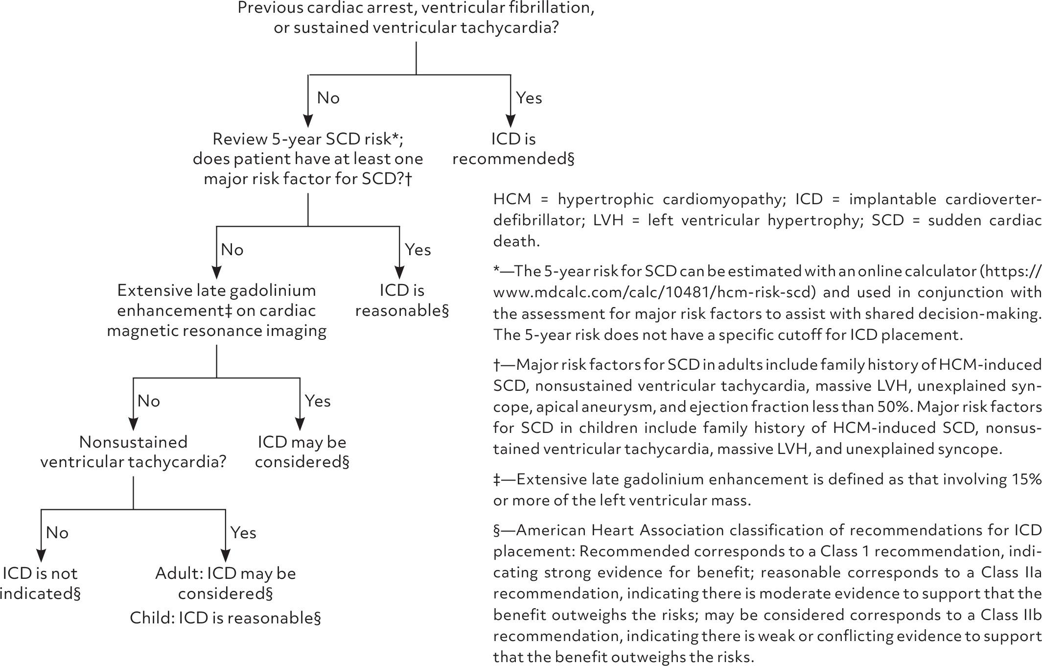
- HCM Risk-SCD Calculator (ESC model) — estimates 5-year SCD risk using age, wall thickness, LA diameter, LVOT gradient, family history of SCD, NSVT, and syncope; useful for shared decision-making but should not be the sole criterion for ICD decisions [6][12-13]
The AHA/ACC 2024 guidelines recommend the ICD assessment algorithm shown below:
ECG
ECG is abnormal in up to 95% of HCM patients and is the most sensitive routine screening test. [1][12][15]
Common findings
- LVH voltage criteria (large R waves in lateral leads, deep S waves in V1–V2)
- Prominent Q waves — typically in inferior (II, III, aVF) and lateral leads (I, aVL, V5–V6); represent septal hypertrophy, not infarction [15][18]
- ST-segment depression and T-wave inversions — repolarization abnormalities [15]
- Giant T-wave inversions in precordial leads — suggestive of apical HCM variant [15]
- Left axis deviation [15][18]
- P-wave abnormalities (left atrial enlargement) [15]
- Atrial fibrillation — found in >50% over time on ambulatory monitoring [3]
- NSVT on Holter — SCD risk marker [6]
Dangerous patterns
- Sustained VT or VF — immediate ICD indication [6-7]
- Rapid AF — poorly tolerated; urgent rate control needed
- A normal ECG does not exclude HCM (5–10% have normal ECG) [12][15]
Assessment
- HCM is classified as obstructive (resting or provocable LVOT gradient ≥30 mmHg; hemodynamically significant ≥50 mmHg) or nonobstructive [2][12]
- Most patients have a favorable prognosis with modern management, compatible with normal longevity [5]
- Annual SCD risk is approximately 0.5–1% overall; substantially lower in patients without risk markers [4][13]
- Atypical presentations: apical HCM (giant T-wave inversions, apical aneurysm risk), midventricular obstruction, end-stage/burned-out HCM (LVEF <50%, LV dilation) [2][4]
- Complications: SCD, AF with stroke risk, progressive heart failure, infective endocarditis (rare), end-stage HCM requiring transplant [2][4]
Treatment Plan
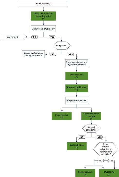
The 2020 AHA/ACC guideline management algorithm for symptomatic obstructive HCM is shown below:
Initial stabilization (ED setting)
- Acute hypotension/hemodynamic compromise: IV fluids (volume resuscitation), IV phenylephrine (pure alpha-agonist to increase afterload), IV beta-blocker if tachycardic; avoid dobutamine, milrinone, vasodilators [8-9]
- Rapid AF: rate control with IV beta-blocker or IV diltiazem; avoid digoxin; anticoagulation is strongly recommended for all HCM patients with AF regardless of CHA₂DS₂-VASc score [2]
- VT/VF/cardiac arrest: standard ACLS; defibrillation; avoid isoproterenol
Chronic pharmacological management (obstructive HCM)
- Step 1: Beta-blocker (titrate to symptom relief and heart rate suppression) [8]
- Step 2: Verapamil or diltiazem (if beta-blocker intolerant/ineffective) [8]
- Step 3 (refractory symptoms): Mavacamten (5 mg daily starting dose, max 15 mg; requires REMS, serial LVEF monitoring) or aficamten or disopyramide (with AV nodal blocker) [3][8-10]
- Avoid vasodilators, high-dose diuretics, positive inotropes [6][8]
Septal reduction therapy (refractory to medical therapy)
- Surgical myectomy — gold standard; preferred when concomitant cardiac surgery needed [19-20]
- Alcohol septal ablation — alternative for non-surgical candidates [19-20]
- Should be performed at experienced, high-volume HCM centers [3][8]
SCD prevention
- Secondary prevention ICD: all survivors of cardiac arrest or sustained VT [6-7]
- Primary prevention ICD: consider with ≥1 major risk factor (massive LVH ≥30 mm, family history of SCD, unexplained syncope, NSVT, extensive LGE, apical aneurysm, LVEF <50%) [4][6][8]
- SCD risk is low in patients >60 years; ICD generally discouraged in this age group unless specific high-risk features [4][8]
Disposition
Admit if
- Syncope (especially exertional or unexplained) — requires telemetry monitoring and SCD risk assessment
- Acute hemodynamic compromise / hypotension
- New-onset rapid atrial fibrillation with hemodynamic instability
- Sustained VT, VF, or cardiac arrest
- Acute heart failure symptoms
- New diagnosis with concerning features (massive LVH, NSVT on initial monitoring)
Observation
- New-onset AF, hemodynamically stable, requiring rate control optimization
- Presyncope with unclear etiology pending workup
Discharge criteria
- Hemodynamically stable, asymptomatic or at baseline symptoms
- Known HCM with stable presentation and no new risk factors
- Adequate follow-up arranged with cardiology/HCM center
Specialist consultation triggers
- All newly diagnosed HCM patients should be referred to a comprehensive HCM center [3][5]
- Cardiology/electrophysiology for SCD risk stratification and ICD discussion
- Cardiac surgery for septal myectomy evaluation
- Genetics for counseling and testing [3][8]
Follow-Up / Return Precautions
Follow-up timing
- Annual clinical evaluation with echocardiogram and 12-lead ECG [5]
- Ambulatory ECG monitoring every 1–3 years (or sooner if arrhythmia symptoms) [5]
- Cardiac MRI every 3–5 years (or sooner if clinical concern for progression) [5]
- SCD risk reassessment every 1–3 years or with any change in clinical profile [5][8]
- First-degree relative screening: ECG + echo at diagnosis; repeat every 1–2 years (children) or 3–5 years (adults) [8]
Return precautions — instruct patients to seek immediate care for:
- Syncope or near-syncope, especially with exertion
- Sustained palpitations or racing heartbeat
- New or worsening chest pain
- Progressive dyspnea or exercise intolerance
- Lightheadedness or dizziness with position changes
Patient counseling
- HCM is a treatable disease compatible with normal longevity in most patients [5]
- Maintain adequate hydration; avoid dehydration, excessive alcohol, and excessive caffeine [6]
- Exercise: newer data support an individualized approach rather than blanket restriction; shared decision-making with cardiology is essential [2][12]
- Avoid stimulant drugs and performance-enhancing substances
- Inform all healthcare providers (especially anesthesiologists) of HCM diagnosis — anesthetic management requires avoidance of vasodilators and maintenance of preload/afterload [8]
- Genetic counseling for family planning and relative screening [3][8]
References
1. Cardiomyopathy: A Guide for Primary Care. — Coppiano J, Carrasco M, Asif I. American Family Physician. 2026.
2. Current Management of Hypertrophic Cardiomyopathy. — Sikand N, Stendahl J, Sen S, Lampert R, Day S. BMJ. 2025.
3. Diagnosis and Management of Hypertrophic Cardiomyopathy: Updated Guidelines From the ACC/AHA. — Leggit JC, Whitaker D. American Family Physician. 2022.
4. Hypertrophic Cardiomyopathy. — Braunwald E. The New England Journal of Medicine. 2025.
5. Diagnosis and Evaluation of Hypertrophic Cardiomyopathy: JACC State-of-the-Art Review. — Maron BJ, Desai MY, Nishimura RA, et al. Journal of the American College of Cardiology. 2022.
6. Management of Hypertrophic Cardiomyopathy: JACC State-of-the-Art Review. — Maron BJ, Desai MY, Nishimura RA, et al. Journal of the American College of Cardiology. 2022.
7. 2017 AHA/ACC/HRS Guideline for Management of Patients With Ventricular Arrhythmias and the Prevention of Sudden Cardiac Death: A Report of the American College of Cardiology/American Heart Association Task Force on Clinical Practice Guidelines and the Heart Rhythm Society. — Al-Khatib SM, Stevenson WG, Ackerman MJ, et al. Heart Rhythm. 2018.
8. 2024 AHA/ACC/AMSSM/HRS/PACES/SCMR Guideline for the Management of Hypertrophic Cardiomyopathy: A Report of the American Heart Association/American College of Cardiology Joint Committee on Clinical Practice Guidelines. — Ommen SR, Ho CY, Asif IM, et al. Journal of the American College of Cardiology. 2024.
9. FDA Orange Book. — FDA Orange Book. 2026.
10. FDA Drug Label. — Updated date: 2025-04-17. Food and Drug Administration.
11. Mavacamten and Aficamten in Hypertrophic Cardiomyopathy: A Systematic Review and Meta-Analysis of Randomized Trials. — Fahim M, Eldawud D, Wahadneh O, et al. The American Journal of Cardiology. 2026.
12. Hypertrophic Cardiomyopathy: A Practical Approach to Guideline Directed Management. — Ommen SR, Semsarian C. Lancet. 2021.
13. Recommendations for Multimodality Cardiovascular Imaging of Patients With Hypertrophic Cardiomyopathy: An Update From the American Society of Echocardiography, in Collaboration With the American Society of Nuclear Cardiology, the Society for Cardiovascular Magnetic Resonance, and the Society of Cardiovascular Computed Tomography. — Nagueh SF, Phelan D, Abraham T, et al. Journal of the American Society of Echocardiography : Official Publication of the American Society of Echocardiography. 2022.
14. EJHF Expert Consensus Statement on the Diagnosis and Management of Hypertrophic Cardiomyopathy. — Meder B, Coats CJ, Leinwand LA, et al. European Journal of Heart Failure. 2026.
15. Hypertrophic Obstructive Cardiomyopathy. — Veselka J, Anavekar NS, Charron P. Lancet. 2017.
16. Effects of Mavacamten on Cardiac Biomarkers in Nonobstructive Hypertrophic Cardiomyopathy: Insights From the ODYSSEY-HCM Trial. — Desai MY, Olivotto I, Abraham T, et al. Journal of the American College of Cardiology. 2025.
17. Exercise Testing to Unmask Latent LVOT Obstruction in a Highly Symptomatic Patient With Hypertrophic Cardiomyopathy. — Husaini M, Bach RG, Cresci S. Journal of the American College of Cardiology. 2024.
18. Sudden Death in the Young: Information for the Primary Care Provider. — Erickson CC, Salerno JC, Berger S, et al. Pediatrics. 2021.
19. 2020 AHA/ACC Guideline for the Diagnosis and Treatment of Patients With Hypertrophic Cardiomyopathy: A Report of the American College of Cardiology/American Heart Association Joint Committee on Clinical Practice Guidelines. — Ommen SR, Mital S, Burke MA, et al. Journal of the American College of Cardiology. 2020.
20. 2020 AHA/ACC Guideline for the Diagnosis and Treatment of Patients With Hypertrophic Cardiomyopathy: Executive Summary: A Report of the American College of Cardiology/American Heart Association Joint Committee on Clinical Practice Guidelines. — Ommen SR, Mital S, Burke MA, et al. Journal of the American College of Cardiology. 2020.
Figures
More from Deep Dive
Reviewed by Dr. Lucas Mastropaolo. SymptomDx is an educational tool for medical professionals — it does not replace clinical judgment. Verify all clinical data and drug dosages with authoritative sources.