Hypertrophic Obstructive Cardiomyopathy
Hypertrophic obstructive cardiomyopathy is the most common inherited cardiac disease (autosomal dominant, ~1 in 500), characterized by asymmetric left ventricular hypertrophy with dynamic left ventricular outflow tract obstruction (LVOTO), and is a leading cause of sudden cardiac death (SCD) in young adults. [1-2] Two-thirds of HCM patients have LVOT obstruction at rest or with provocation. [3]
History
- Exertional symptoms: dyspnea, chest pain, fatigue, presyncope/syncope — all typically provoked by exertion, dehydration, or postprandial states [4-5]
- Characterize timing: onset with activity, relieved by rest or recumbency; worsened by standing, Valsalva, or heat
- Palpitations — may indicate atrial fibrillation (present in >50% paroxysmal) or ventricular arrhythmia [4-5]
- Orthopnea and PND if congestive symptoms develop
- Ask about prior cardiac arrest, ICD shocks, or documented arrhythmias
- Important negatives: absence of pleuritic pain, positional pain, fever, cough (helps exclude other causes of dyspnea/chest pain)
Alarm Features
- Syncope — especially exertional or without prodrome; recent syncope (within 6 months) is a major SCD risk marker [6]
- Cardiac arrest or sustained ventricular tachycardia [2][6]
- Acute hemodynamic collapse/hypotension — a medical urgency in obstructive HCM; can be precipitated by vasodilators, dehydration, or tachyarrhythmia [6]
- New-onset atrial fibrillation with rapid ventricular response — can cause acute decompensation and carries high stroke risk [1][7]
- Progressive dyspnea at rest or NYHA class III–IV symptoms
- Family history of SCD in a first-degree relative <50 years old [6]
Medications
First-line treatments (per 2024 AHA/ACC Guidelines): [6]
- Nonvasodilating beta-blockers (e.g., metoprolol, propranolol, nadolol) — titrate to resting HR 50–60 bpm [2][5]
- Nondihydropyridine calcium channel blockers (verapamil, diltiazem) — substitute if beta-blockers ineffective or not tolerated; avoid combining with beta-blockers for HCM-directed therapy [6][8]
Second-line / add-on: [6]
- Disopyramide — potent negative inotrope; must be combined with an AV nodal blocker to prevent rapid AF conduction [6][8]
- Cardiac myosin inhibitors: mavacamten (Camzyos, FDA-approved 2022) and aficamten (Myqorzo, FDA-approved 2025) — reduce hypercontractility and LVOT gradients [6][9-10]
Contraindicated / to avoid: [6][8]
- Pure vasodilators: dihydropyridine CCBs (amlodipine, nifedipine), nitrates, ACE inhibitors, ARBs
- Positive inotropes: dobutamine, dopamine, digoxin
- High-dose diuretics (reduce preload → worsen obstruction)
- ADHD stimulant medications
- Excessive caffeine and alcohol
Diet
- Adequate hydration is critical — dehydration reduces preload and worsens LVOTO [8]
- Avoid large, heavy meals (postprandial splanchnic vasodilation can provoke symptoms)
- Limit excessive caffeine and alcohol intake [8]
- Maintain healthy body weight [8]
- No specific long-term dietary restrictions beyond the above
Review of Systems
- Cardiovascular: chest pain, dyspnea, palpitations, syncope/presyncope, orthopnea, edema
- Neurologic: lightheadedness, transient loss of consciousness, focal deficits (embolic stroke from AF)
- Respiratory: exercise intolerance, paroxysmal nocturnal dyspnea
- GI: postprandial worsening of symptoms
- Psychiatric: anxiety, depression (common with chronic disease and activity restriction)
- Screen for sleep-disordered breathing (comorbidity that worsens HCM) [11]
Collateral History and Family History
- Three-generation family history is recommended at initial evaluation [4][12]
- Ask specifically about SCD, unexplained drowning, single-vehicle accidents, or unexplained death in relatives <50 years [6]
- Family members with known HCM, ICDs, or heart failure
- Autosomal dominant inheritance — pathogenic sarcomere gene variants found in 30–60% of cases [1][4]
- First-degree relatives require screening with ECG and echocardiography [4]
- Genetic counseling by a counselor versed in cardiac genetics is strongly recommended [4]
Risk Factors
- Family history of HCM or HCM-related SCD [6]
- Sarcomere gene mutations (8 genes identified; most common: MYH7, MYBPC3) [4]
- Young age — SCD risk is highest in younger patients and inversely related to age [2][6]
- Massive LV hypertrophy (wall thickness ≥30 mm) [6]
- LV apical aneurysm [6]
- Extensive late gadolinium enhancement (≥15% LV mass) on CMR [6]
- LVEF <50% (end-stage phase) [6]
- Nonsustained ventricular tachycardia on Holter [6]
- Comorbidities: hypertension, obesity, obstructive sleep apnea, atrial fibrillation [11]
Differential Diagnosis
- Hypertensive heart disease — can produce LVH but rarely >20 mm wall thickness; requires uncontrolled hypertension [13]
- Athlete's heart — physiologic LVH typically concentric, ≤13 mm, with normal diastolic function; regresses with detraining [14]
- Cardiac amyloidosis — consider if LVH with low voltage on ECG, diastolic dysfunction, apical sparing on strain imaging [11][13]
- Fabry disease — X-linked, concentric LVH, renal/dermatologic features; check alpha-galactosidase A [11][13]
- Aortic stenosis — fixed obstruction; crescendo-decrescendo murmur radiating to carotids; murmur decreases with Valsalva (opposite of HOCM)
- Mitochondrial cardiomyopathy — consider in pediatric patients with multisystem disease [11]
- Glycogen storage diseases (Pompe, Danon) — especially in young patients with massive hypertrophy
Past Medical History
- Prior episodes of syncope, cardiac arrest, or documented arrhythmias
- Known ICD or pacemaker
- Prior septal reduction therapy (myectomy or alcohol septal ablation)
- Atrial fibrillation history and anticoagulation status
- Comorbid hypertension, coronary artery disease, valvular disease
- Obstructive sleep apnea
- Pregnancy history (hemodynamic changes can exacerbate LVOTO)
Physical Exam
- Harsh crescendo-decrescendo systolic murmur at left lower sternal border/apex [1][5]
- Increases with Valsalva, standing, dehydration (decreased preload/afterload)
- Decreases with squatting, leg elevation, hand grip (increased preload/afterload)
- Separate murmur of mitral regurgitation — holosystolic at apex, radiating to axilla [5]
- Brisk, bifid (bisferiens) carotid pulse
- Prominent apical impulse; may have S4 gallop
- Jugular venous distension and peripheral edema if decompensated
- Vital signs: assess for hypotension (medical urgency), tachycardia, irregular rhythm (AF)
Lab Studies
- BNP/NT-proBNP — elevated with heart failure symptoms; useful for monitoring [7]
- Troponin — may be mildly elevated chronically; rule out acute coronary syndrome in chest pain presentations
- BMP — assess renal function (relevant for medication dosing and diuretic use)
- TSH — thyrotoxicosis can exacerbate symptoms
- CBC — rule out anemia as contributor to symptoms
- Genetic testing — sarcomere gene panel after genetic counseling; identifies pathogenic variants in 30–60% [4]
- Alpha-galactosidase A (if Fabry suspected), serum/urine protein electrophoresis (if amyloid suspected) [11][13]
Imaging
First-line: Transthoracic echocardiography [6][13]
- Diagnostic criterion: maximal LV end-diastolic wall thickness ≥15 mm (≥13 mm with family history or positive genetic test) [1][4][13]
- Assess LVOT gradient at rest and with provocation (Valsalva, exercise)
- LVOT gradient ≥30 mm Hg defines obstruction; ≥50 mm Hg is the threshold for intervention [13]
- Evaluate mitral valve anatomy, systolic anterior motion (SAM), mitral regurgitation, EF, LA size
Exercise stress echocardiography — preferred method to provoke latent LVOTO when resting gradient is <30 mm Hg [6][12]
- Gold standard for wall thickness measurement and tissue characterization
- Identifies LV apical aneurysm, extent of late gadolinium enhancement (LGE ≥15% LV mass = high SCD risk)
- Differentiates HCM from phenocopies (amyloid, Fabry)
- Repeat every 3–5 years for surveillance [12]
Cardiac CT — alternative when CMR is contraindicated; evaluates coronary anatomy and myocardial bridging [2]
Special Tests
- Ambulatory ECG monitoring (24–48 hr Holter or ≥2-week wireless patch) — detect NSVT, AF [6][12-13]
- Cardiopulmonary exercise testing (CPET) — objective assessment of functional capacity; useful when symptom severity is uncertain [8][12]
- Exercise stress echocardiography — provoke latent LVOT gradients [12]
- Genetic testing with pre-test counseling — 8 sarcomere genes associated with HCM [4]
- SCD risk calculators — HCM Risk-SCD (ESC) and AHA/ACC individual risk marker strategy to guide ICD decisions [6][13]
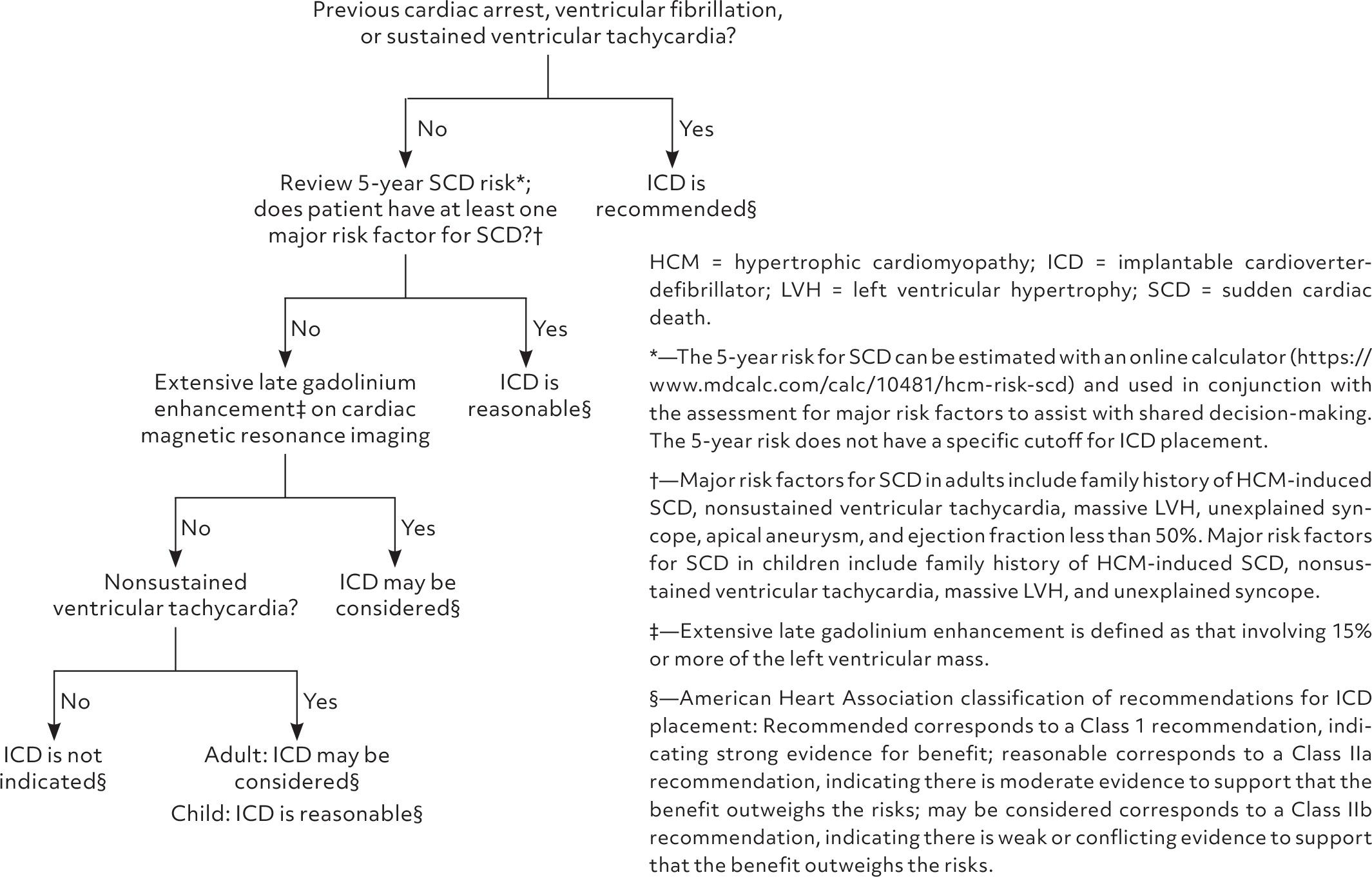
The following algorithm from the AHA/ACC guidelines outlines the ICD decision-making framework:
ECG
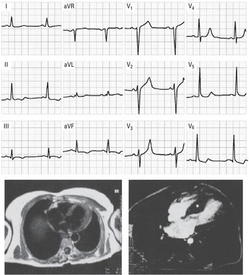
ECG is abnormal in ~90–95% of HCM patients: [1][4-5][14]
- LV hypertrophy voltage criteria
- Prominent Q waves — especially in inferior and lateral leads (septal depolarization)
- ST-segment depression and T-wave inversions — particularly in lateral/anterolateral leads
- Left axis deviation
- P-wave abnormalities (left atrial enlargement)
- Giant negative T waves in precordial leads — suggest apical variant HCM [5][15]
- A normal ECG does not exclude HCM but is present in only 5–10% of cases [5][13]
Dangerous patterns to recognize:
- Sustained or nonsustained ventricular tachycardia
- Atrial fibrillation with rapid ventricular response
- Pre-excitation (WPW) — associated with some HCM subtypes
Assessment
- HOCM is a chronic, progressive genetic cardiomyopathy with dynamic LVOTO that is highly variable throughout daily life [6]
- Severity stratification is based on: NYHA functional class, resting/provoked LVOT gradient, LV wall thickness, EF, presence of SCD risk markers [6][12]
- Most patients achieve normal longevity with appropriate management; annual SCD rate ~1% before ICD era [2]
- Atypical presentations include isolated AF, embolic stroke, or incidental murmur/ECG finding [4-5]
- Complications: SCD, progressive heart failure, atrial fibrillation with stroke, infective endocarditis (rare), end-stage (burned-out) phase with EF <50% [2][7]
Treatment Plan
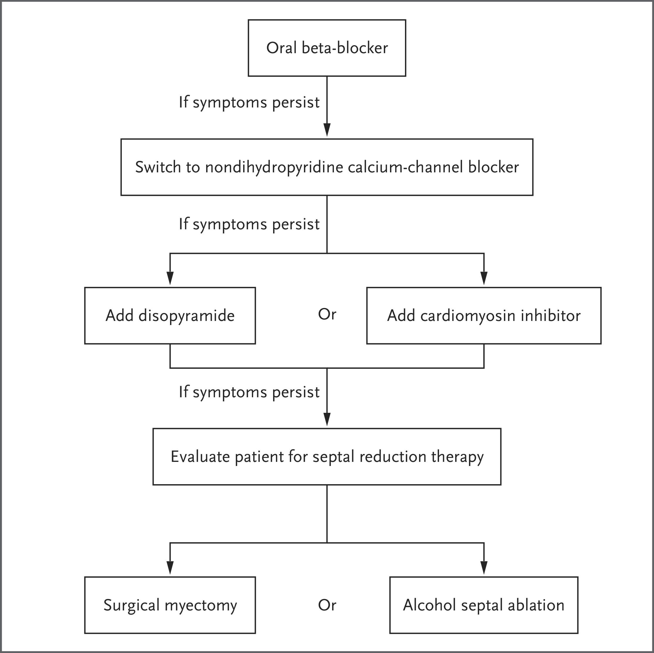
The following figure from Braunwald's 2025 NEJM review illustrates the stepwise treatment algorithm:
Initial stabilization (ED setting): [6]
- Acute hypotension is a medical urgency: IV fluids for volume resuscitation, IV phenylephrine (pure alpha-agonist to increase afterload), IV beta-blocker to reduce contractility and HR
- Avoid vasodilators, inotropes, and aggressive diuresis
- Trendelenburg positioning; passive leg raise
Chronic pharmacologic management (per 2024 AHA/ACC): [6]
- Nonvasodilating beta-blockers — first-line; titrate to resting HR 50–60 bpm
- Verapamil or diltiazem — if beta-blockers fail or are not tolerated
- Escalation if persistent symptoms: add disopyramide (with AV nodal blocker), or cardiac myosin inhibitor (mavacamten or aficamten), or refer for septal reduction therapy (SRT)
Septal reduction therapy (at experienced HCM centers): [6]
- Surgical myectomy — gold standard; preferred when concomitant cardiac surgery needed
- Alcohol septal ablation — alternative for non-surgical candidates
- ICD for secondary prevention (prior cardiac arrest/sustained VT)
- ICD for primary prevention based on individual risk marker assessment (≥1 major risk factor)
- Reassess SCD risk every 1–2 years
Atrial fibrillation management: [1][7]
- Anticoagulation is strongly recommended for all HCM patients with AF regardless of CHA₂DS₂-VASc score
- Rhythm control preferred for symptomatic AF
Disposition
Admit for
- Acute hemodynamic instability / hypotension
- Syncope with suspected arrhythmic etiology
- New sustained ventricular arrhythmia or cardiac arrest
- Acute decompensated heart failure (NYHA III–IV)
- New-onset rapid atrial fibrillation with hemodynamic compromise
- Post-septal reduction therapy
Observation for
- Presyncope with borderline hemodynamics
- New AF with controlled rate but uncertain stability
Discharge with close follow-up if
- Stable, known HOCM with mild symptom exacerbation attributable to identifiable trigger (dehydration, medication non-adherence)
- No syncope, no hemodynamic instability, no new arrhythmia
- Adequate oral intake and stable vital signs
Specialist consultation triggers: All patients with HCM should be managed in conjunction with a comprehensive HCM center — these centers have better outcomes. [4][12] Urgent cardiology consultation for new diagnosis, syncope, arrhythmia, or refractory symptoms.
Follow-Up / Return Precautions
- Cardiology re-evaluation every 12 months (or up to 24 months if stable)
- Annual: echocardiogram + 12-lead ECG
- Every 1–3 years: ambulatory ECG monitoring (Holter or wireless patch)
- Every 3–5 years: contrast CMR (or sooner if clinical concern)
- SCD risk reassessment every 1–2 years [6]
- First-degree family member screening: ECG + echocardiography every 1–3 years (pediatric relatives) or 3–5 years (adult relatives) [4]
Return precautions — instruct patients to seek immediate care for:
- Syncope or near-syncope
- Sustained palpitations or irregular heartbeat
- Worsening dyspnea at rest or with minimal exertion
- Chest pain not relieved by rest
- ICD shocks
Patient counseling
- Maintain adequate hydration at all times; avoid dehydration [8][11]
- Avoid sudden intense exertion; individualized exercise guidance per shared decision-making with cardiologist [7]
- Avoid vasodilators, stimulants, and excessive alcohol/caffeine [8]
- Educate on medication adherence and side effects
- Reassure that most patients with HCM achieve normal longevity with appropriate management [12]
References
1. Cardiomyopathy: A Guide for Primary Care. — Coppiano J, Carrasco M, Asif I. American Family Physician. 2026.
2. Hypertrophic Cardiomyopathy. — Braunwald E. The New England Journal of Medicine. 2025.
3. Randomized Trial of Metoprolol in Patients With Obstructive Hypertrophic Cardiomyopathy. — Dybro AM, Rasmussen TB, Nielsen RR, et al. Journal of the American College of Cardiology. 2021.
4. Diagnosis and Management of Hypertrophic Cardiomyopathy: Updated Guidelines From the ACC/AHA. — Leggit JC, Whitaker D. American Family Physician. 2022.
5. Hypertrophic Obstructive Cardiomyopathy. — Veselka J, Anavekar NS, Charron P. Lancet. 2017.
6. 2024 AHA/ACC/AMSSM/HRS/PACES/SCMR Guideline for the Management of Hypertrophic Cardiomyopathy: A Report of the American Heart Association/American College of Cardiology Joint Committee on Clinical Practice Guidelines. — Ommen SR, Ho CY, Asif IM, et al. Journal of the American College of Cardiology. 2024.
7. Current Management of Hypertrophic Cardiomyopathy. — Sikand N, Stendahl J, Sen S, Lampert R, Day S. BMJ. 2025.
8. Management of Hypertrophic Cardiomyopathy: JACC State-of-the-Art Review. — Maron BJ, Desai MY, Nishimura RA, et al. Journal of the American College of Cardiology. 2022.
9. FDA Orange Book. — FDA Orange Book. 2026.
10. Aficamten or Metoprolol Monotherapy for Obstructive Hypertrophic Cardiomyopathy. — Garcia-Pavia P, Maron MS, Masri A, et al. The New England Journal of Medicine. 2025.
11. Comprehensive Evaluation of Hypertrophic Cardiomyopathy: European Journal of Heart Failure Expert Consensus Document. — Martens P, Olivotto I, Garcia-Pavia P, et al. European Journal of Heart Failure. 2026.
12. Diagnosis and Evaluation of Hypertrophic Cardiomyopathy: JACC State-of-the-Art Review. — Maron BJ, Desai MY, Nishimura RA, et al. Journal of the American College of Cardiology. 2022.
13. Hypertrophic Cardiomyopathy: A Practical Approach to Guideline Directed Management. — Ommen SR, Semsarian C. Lancet. 2021.
14. The Adolescent Athlete and the Team Physician: A Consensus Statement. 2025 Update. — Putukian M, Leclere LE, Herring SA, et al. Medicine and Science in Sports and Exercise. 2026.
15. Advanced Imaging Insights in Apical Hypertrophic Cardiomyopathy. — Hughes RK, Knott KD, Malcolmson J, et al. JACC. Cardiovascular Imaging. 2020.
16. Inherited Heart Diseases. — Antoni Bayés De Luna, Miquel Fiol‐Sala, Antoni Bayés‐Genís, et al. Clinical Electrocardiography. 2021.
Figures
More from Deep Dive
Reviewed by Dr. Lucas Mastropaolo. SymptomDx is an educational tool for medical professionals — it does not replace clinical judgment. Verify all clinical data and drug dosages with authoritative sources.