Hypocalcemia
Hypocalcemia is defined as a serum calcium level <8.5 mg/dL (2.12 mmol/L) or ionized calcium <4.8 mg/dL (1.20 mmol/L). It ranges from asymptomatic to life-threatening and requires a systematic approach to identify the underlying etiology and guide management. [1-2]
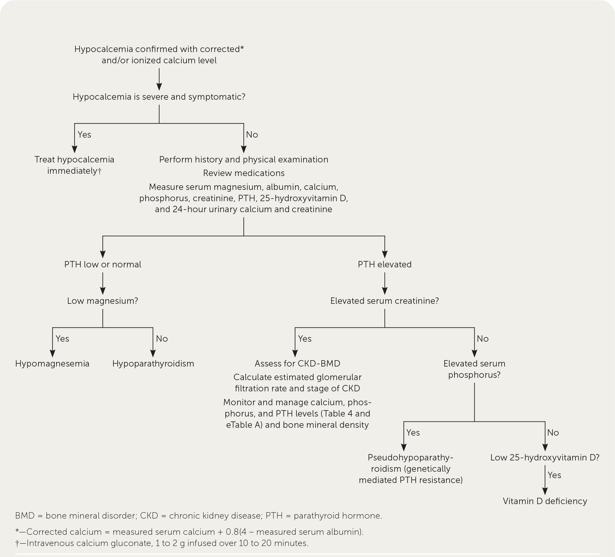
The following algorithm outlines a practical approach to evaluating hypocalcemia:
History
- Onset and acuity: Acute (post-surgical, massive transfusion, critical illness) vs. chronic (vitamin D deficiency, CKD, hypoparathyroidism)
- Symptom characterization: Perioral/fingertip numbness and tingling (earliest symptoms), muscle cramps, stiffness, spasms, carpopedal spasm [4]
- Surgical history: Recent anterior neck surgery (thyroidectomy, parathyroidectomy) — the most common cause of hypoparathyroidism (75% of cases) [3]
- Dietary intake: Calcium and vitamin D intake, dairy avoidance, malabsorptive conditions (celiac disease, gastric bypass, short bowel)
- Medication review: Bisphosphonates, denosumab, PPIs, loop diuretics, calcimimetics, cisplatin, aminoglycosides, foscarnet [5-6]
- Associated symptoms: Seizures, confusion, irritability, depression, dyspnea, palpitations, wheezing [3]
- Important negatives: No neck surgery, no renal disease, no malignancy, no alcohol use
Alarm Features
- Tetany (carpopedal spasm, laryngospasm, generalized muscle rigidity) [4]
- Seizures — may be the presenting symptom, especially in neonates and children [1][7]
- Laryngospasm/stridor — airway emergency [4]
- QTc prolongation → risk of torsades de pointes and ventricular fibrillation [4][8]
- Hypocalcemic cardiomyopathy — reversible heart failure with severely depressed LVEF [9-10]
- Serum calcium <7.0 mg/dL — requires immediate IV calcium and cardiac monitoring [4]
- Refractory hypocalcemia despite oral supplementation — consider hypomagnesemia or underlying malignancy [4][11]
Medications
Medications that cause hypocalcemia
- Denosumab — highest risk among antiresorptives; FDA black box warning for severe hypocalcemia in advanced CKD/dialysis patients (cumulative incidence ~41% in dialysis patients) [12-13]
- Bisphosphonates (IV zoledronate > oral) [14]
- Calcimimetics (cinacalcet) [5]
- Loop diuretics (furosemide) — increase renal calcium excretion [5]
- PPIs — via hypomagnesemia and impaired calcium absorption [6]
- Cisplatin, aminoglycosides, foscarnet [5]
- Massive blood transfusion — citrate chelates ionized calcium
Treatment medications
- IV calcium gluconate (acute), oral calcium carbonate/citrate, calcitriol, cholecalciferol, magnesium supplementation [4][15]
- Thiazide diuretics — adjunctive in refractory cases (enhance renal calcium reabsorption) [4]
- rhPTH(1-84) (Natpara) — for chronic hypoparathyroidism refractory to conventional therapy [2]
Cautions
- Calcium inhibits levothyroxine absorption — separate by ≥1 hour before or 3 hours after [4]
- Calcium gluconate is incompatible with phosphate- or bicarbonate-containing fluids [16]
Diet
- Calcium-rich diet: Dairy products, fortified foods, leafy greens; recommended daily intake 1000–1300 mg elemental calcium for general population [17]
- CKD patients: Limit to 800–1000 mg/day elemental calcium from all sources [17]
- Vitamin D sources: Fortified foods, fatty fish, sunlight exposure
- Avoid excess phosphorus: Processed foods, colas — phosphorus binds calcium and worsens hypocalcemia
- Hydration: Adequate fluid intake to prevent nephrolithiasis from calcium supplementation
- Calcium carbonate requires acidic environment — take with meals; calcium citrate preferred in patients on PPIs, post-gastric bypass, or with achlorhydria [4]
Review of Systems
- Neurologic: Paresthesias, numbness, seizures, confusion, headache, impaired vision [3]
- Musculoskeletal: Cramps, spasms, stiffness, tetany
- Cardiovascular: Palpitations, syncope, dyspnea, chest pain [3]
- Psychiatric: Anxiety, depression, emotional instability, irritability [3-4]
- Pulmonary: Wheezing, bronchospasm, stridor (laryngospasm) [3]
- GI: Abdominal pain, diarrhea (malabsorption clue)
- Chronic features: Dry skin, brittle nails, dental abnormalities, cataracts (chronic hypoparathyroidism) [15]
Collateral History and Family History
- Family history of endocrine disorders: Autoimmune polyendocrinopathy (APECED), MEN syndromes, 22q11.2 deletion syndrome (DiGeorge) [7][15]
- Family history of hypocalcemia or seizures in childhood — consider genetic causes (CaSR mutations, ADH1) [15]
- Autoimmune conditions in patient or family: Chronic candidiasis, adrenal insufficiency, thyroid disease → consider autoimmune hypoparathyroidism [15]
- Social history: Alcohol use (hypomagnesemia), dietary restrictions (veganism), sun avoidance (vitamin D deficiency)
Risk Factors
- Post-thyroidectomy/parathyroidectomy — most common cause of hypoparathyroidism [2-3]
- Chronic kidney disease (especially stages 4–5) — impaired 1,25(OH)₂D production, hyperphosphatemia [18-19]
- Vitamin D deficiency — most common cause of hypocalcemia overall [3]
- Malabsorption: Celiac disease, inflammatory bowel disease, gastric bypass, short bowel syndrome [20]
- Hypomagnesemia — impairs PTH secretion and end-organ response [4]
- Active malignancy — tumor lysis syndrome, osteoblastic metastases, treatment-related [11]
- Critical illness/sepsis, massive transfusion, pancreatitis
- Hungry bone syndrome — post-parathyroidectomy or post-thyroidectomy for Graves' disease [4]
- Advanced age, institutionalization, dark skin pigmentation (vitamin D deficiency risk)
Differential Diagnosis
The differential is organized by PTH level, which is the critical first branch point: [3][21]
Low/inappropriately normal PTH (hypoparathyroidism)
- Postsurgical hypoparathyroidism (most common) [2]
- Autoimmune hypoparathyroidism (isolated or APECED)
- Genetic: 22q11.2 deletion, CaSR activating mutations (ADH1), HDR syndrome
- Infiltrative: Hemochromatosis, Wilson disease, metastatic disease
Elevated PTH (secondary hyperparathyroidism)
- Vitamin D deficiency (most common cause of hypocalcemia overall) [3]
- CKD-mineral bone disorder [18]
- Pseudohypoparathyroidism (PTH resistance) — elevated PTH + elevated phosphorus + normal renal function [3]
Other mechanisms
- Hypomagnesemia — functional hypoparathyroidism [4]
- Acute pancreatitis (calcium saponification)
- Tumor lysis syndrome / rhabdomyolysis (calcium-phosphate deposition)
- Massive transfusion (citrate chelation)
- Hungry bone syndrome
- Medication-induced (denosumab, bisphosphonates, calcimimetics) [5][12]
Past Medical History
- Prior neck surgery (thyroidectomy, parathyroidectomy, radical neck dissection)
- CKD stage and dialysis status
- Malabsorptive conditions (celiac, IBD, bariatric surgery)
- Autoimmune diseases (thyroid, adrenal, vitiligo)
- Prior episodes of hypocalcemia or seizures
- Malignancy (especially with bone metastases or recent chemotherapy)
- History of radiation to the neck
- Pancreatitis
Physical Exam
- Vital signs: Hypotension, tachycardia (if severe)
- Chvostek sign: Twitching of facial muscles upon tapping the facial nerve anterior to the ear (present at baseline in up to 25% of normals — limited specificity) [4]
- Trousseau sign: Carpopedal spasm after inflating BP cuff above systolic for 3 minutes (more specific than Chvostek) [4]
- Neuromuscular: Hyperreflexia, muscle fasciculations, tetany
- Neck: Surgical scars (prior thyroidectomy)
- Skin/nails: Dry skin, brittle nails, alopecia (chronic hypocalcemia)
- Eyes: Cataracts (chronic hypoparathyroidism) [15]
- Dysmorphic features: Consider 22q11.2 deletion (micrognathia, low-set ears, cleft palate) [7]
Lab Studies
Initial workup
- Serum calcium (total) and albumin (for corrected calcium) or ionized calcium
- Intact PTH — the critical branch point for differential diagnosis [3][15]
- Serum magnesium — must correct hypomagnesemia before calcium will respond [4]
- Phosphorus — elevated in hypoparathyroidism and CKD; low in vitamin D deficiency [3]
- 25-hydroxyvitamin D [7]
- BMP/CMP (creatinine, BUN for renal function)
Additional labs as indicated
- 1,25-dihydroxyvitamin D (if CKD or suspected vitamin D metabolism disorder)
- 24-hour urine calcium and creatinine (monitoring chronic supplementation) [7]
- Alkaline phosphatase (bone turnover)
- Lipase (if pancreatitis suspected)
- Uric acid, LDH, potassium (if tumor lysis suspected)
Corrected calcium formula: [22-23]
$$\text{Corrected Ca (mg/dL)} = \text{Measured Ca} + 0.8 \times (4.0 - \text{Albumin in g/dL})$$
Important caveat: Recent evidence suggests albumin-adjusted calcium correlates poorly with ionized calcium in critically ill patients, those on dialysis, and the elderly. When in doubt, measure ionized calcium directly. [24-25]
Imaging
- Not routinely required for the initial evaluation of hypocalcemia
- Renal ultrasound: If chronic hypoparathyroidism on supplementation — monitor for nephrocalcinosis/nephrolithiasis [7]
- CT head: If basal ganglia calcifications suspected (chronic hypoparathyroidism) [15]
- Neck ultrasound: If post-surgical etiology unclear or parathyroid pathology suspected
- DEXA scan: In chronic hypoparathyroidism for bone density assessment
Special Tests
- Corrected calcium calculator
- Chvostek and Trousseau signs — bedside provocative tests [4]
- Genetic testing: Targeted gene panels for nonsurgical hypoparathyroidism (22q11.2 FISH/microarray, CASR, GNA11, GCM2, PTH gene) [15][26]
- Anti-CaSR antibodies: If autoimmune hypoparathyroidism suspected
- Point-of-care ionized calcium: Available on most ABG analyzers — useful in the ED for rapid assessment
ECG
Indications: All patients with symptomatic hypocalcemia or serum calcium <7.0 mg/dL [4]
Key findings
- Prolonged QTc interval — the hallmark ECG finding; due to prolonged ST segment (not T-wave changes) [4][8]
- T-wave changes: May see peaked or inverted T-waves
- Risk of torsades de pointes → ventricular fibrillation [4]
- Reversible cardiomyopathy with severely depressed LVEF has been reported [9-10]
- QTc prolongation correlates inversely with serum calcium levels [10]
Pearl: Hypocalcemia prolongs the ST segment specifically, whereas hypokalemia prolongs the QT by flattening the T-wave — this distinction can help identify the electrolyte abnormality on ECG.
Assessment
Severity stratification
- Mild: Ca 8.0–8.4 mg/dL, asymptomatic or mild paresthesias
- Moderate: Ca 7.0–7.9 mg/dL, symptomatic (cramps, paresthesias, Chvostek/Trousseau positive)
- Severe: Ca <7.0 mg/dL, or any level with tetany, seizures, laryngospasm, QTc prolongation, or hemodynamic instability [4]
Key clinical pearls
- Always correct for albumin or check ionized calcium — pseudohypocalcemia from hypoalbuminemia is common, especially in hospitalized patients [24]
- Hypocalcemia is frequently underrecognized in the ED — identified and documented in only 35% of cases in one study, and calcium replacement was administered in less than half of severe cases [11]
- 41.9% of patients with severe hypocalcemia re-presented to the ED within 1 year [11]
Treatment Plan
Acute/Severe symptomatic hypocalcemia (tetany, seizures, QTc prolongation, Ca <7.0 mg/dL):
- IV calcium gluconate 1–2 g (10–20 mL of 10% solution = 93–186 mg elemental calcium) in 50 mL D5W infused over 10–20 minutes via peripheral IV [4][15]
- Follow with continuous calcium gluconate infusion to prevent recurrence while initiating oral therapy [15]
- Cardiac monitoring is mandatory during IV calcium administration [4][15]
- Calcium chloride (270 mg elemental Ca per 10 mL) — use only via central line due to risk of tissue necrosis [4]
- Check and correct magnesium — hypocalcemia will be refractory until Mg is repleted (Mg oxide 400 mg PO BID or IV MgSO₄) [4]
- Start calcitriol 0.25–0.5 μg PO BID concurrently [4]
Mild-moderate/Chronic hypocalcemia
- Oral calcium: Calcium carbonate 1–3 g/day (400–1200 mg elemental calcium) in divided doses with meals, or calcium citrate equivalent [4]
- Calcitriol 0.25–0.5 μg BID (preferred active vitamin D in hypoparathyroidism) [15]
- Cholecalciferol/ergocalciferol supplementation to maintain 25-OH-D >30 ng/mL [15]
- Thiazide diuretics (HCTZ 12.5–50 mg daily) — adjunctive for refractory cases [4]
- Goal: Maintain serum calcium in the low-normal range to minimize hypercalciuria and renal complications [15]
Disposition
Admit (ICU or telemetry)
- Symptomatic severe hypocalcemia (tetany, seizures, laryngospasm)
- Serum calcium <7.0 mg/dL
- QTc prolongation or arrhythmia
- Requiring IV calcium infusion
- Hemodynamic instability
Observation
- Moderate hypocalcemia with symptoms responding to oral therapy
- Post-thyroidectomy patients with declining calcium trends
Discharge criteria
- Asymptomatic or mild symptoms controlled on oral calcium ± calcitriol
- Stable or improving calcium levels on serial checks
- Identified and addressed underlying cause
- Reliable follow-up arranged [4]
Specialist consultation triggers
- Endocrinology: New-onset hypoparathyroidism, genetic/autoimmune causes, refractory hypocalcemia
- Nephrology: CKD stages 4–5, dialysis patients, denosumab-related hypocalcemia in CKD [13]
- Surgery: Post-thyroidectomy complications
Follow Up / Return Precautions
Follow-up timing
- Post-discharge: Recheck calcium within 24–48 hours, then every 1–2 weeks until stable [4]
- Chronic hypoparathyroidism: Calcium and creatinine every 3–4 months; 24-hour urine calcium periodically; renal ultrasound intermittently [7]
- Calcitriol dose should be gradually reduced once steady state is achieved to avoid rebound hypercalcemia [4]
Return precautions — instruct patients to seek immediate care for:
- Numbness/tingling around the mouth or fingertips
- Muscle spasms, cramping, or stiffness
- Difficulty breathing or throat tightness (laryngospasm)
- Seizures, confusion, or fainting
- Palpitations or chest pain
Patient counseling
- Take calcium supplements with meals, in divided doses (not all at once)
- Separate calcium from levothyroxine by ≥1 hour [4]
- Maintain adequate vitamin D intake
- Avoid abrupt discontinuation of calcitriol
- Expected recovery: Post-surgical hypoparathyroidism is transient in ~75% of cases; permanent hypoparathyroidism requires lifelong supplementation [2-3]
References
1. Hypocalcemic Disorders. — Bove-Fenderson E, Mannstadt M. Best Practice & Research. Clinical Endocrinology & Metabolism. 2018.
2. Diagnosis and Management of Hypocalcemia. — Pepe J, Colangelo L, Biamonte F, et al. Endocrine. 2020.
3. Parathyroid Disorders. — Sell J, Ramirez S, Partin M. American Family Physician. 2022.
4. American Thyroid Association Statement on Postoperative Hypoparathyroidism: Diagnosis, Prevention, and Management in Adults. — Orloff LA, Wiseman SM, Bernet VJ, et al. Thyroid : Official Journal of the American Thyroid Association. 2018.
5. Denosumab-Induced Hypocalcemia: Detection of Drug-Drug Interactions Using Disproportionality Analysis in VigiBase®. — Batteux B, Gras-Champel V, Cohen-Solal M, Al Balkhi MH, Bennis Y. Bone. 2026.
6. Proton Pump Inhibitors, Bone and Phosphocalcic Metabolism. — Philippoteaux C, Paccou J, Chazard E, Cortet B. Joint Bone Spine. 2024.
7. Health Supervision for Children With 22q11.2 Deletion Syndrome: Clinical Report. — Scheuerle AE, Geleske TA, Merchant N, Goldenberg PC. Pediatrics. 2025.
8. Hypocalcemia-Induced QT Interval Prolongation. — Tang JKK, Rabkin SW. Cardiology. 2022.
9. Opposite Calcium Disorders, Similar Electrical Risk: Reversible Cardiomyopathy Due to Hypocalcemia and Brugada Syndrome Unmasked by Hypercalcemia. — Megid TBC, Ubida C, de Melo Sousa Ferro AC, et al. Journal of Electrocardiology. 2026.
10. Reversible Cardiac Dysfunction Associated With Hypocalcemia: A Systematic Review and Meta-Analysis of Individual Patient Data. — Newman DB, Fidahussein SS, Kashiwagi DT, et al. Heart Failure Reviews. 2014.
11. Severe Hypocalcemia in the Emergency Department: A Retrospective Cohort Study of Prevalence, Etiology, Treatment and Outcome. — Haidinger M, Putallaz E, Ravioli S, Exadaktylos A, Lindner G. Internal and Emergency Medicine. 2025.
12. Severe Hypocalcemia With Denosumab Among Older Female Dialysis-Dependent Patients. — Bird ST, Smith ER, Gelperin K, et al. The Journal of the American Medical Association. 2024.
13. Denosumab and Fracture Prevention in Primary Care Practice. — Bauer DC, Ensrud KE. JAMA Internal Medicine. 2025.
14. Risks of Hypocalcemia and Other Bone Mineral Disorders for Denosumab Versus Zoledronate Across the Spectrum of Kidney Function: A Target Trial Emulation. — Xiao R, Faucon AL, He N, et al. American Journal of Kidney Diseases : The Official Journal of the National Kidney Foundation. 2026.
15. Hypoparathyroidism. — Gafni RI, Collins MT. The New England Journal of Medicine. 2019.
16. FDA Drug Label. — Updated date: 2024-05-06. Food and Drug Administration.
17. Nutritional Management of Chronic Kidney Disease. — Kalantar-Zadeh K, Fouque D. The New England Journal of Medicine. 2017.
18. Chronic Kidney Disease. — Webster AC, Nagler EV, Morton RL, Masson P. Lancet. 2017.
19. Stage IV Chronic Kidney Disease. — Abboud H, Henrich WL. The New England Journal of Medicine. 2010.
20. Osteoporosis Management in the Era of COVID-19. — Yu EW, Tsourdi E, Clarke BL, Bauer DC, Drake MT. Journal of Bone and Mineral Research : The Official Journal of the American Society for Bone and Mineral Research. 2020.
21. Causes and Differential Diagnosis of Hypocalcemia--Recommendation Proposed by Expert Panel Supported by Ministry of Health, Labour and Welfare, Japan. — Fukumoto S, Namba N, Ozono K, et al. Endocrine Journal. 2008.
22. Cancer-Associated Hypercalcemia. — Guise TA, Wysolmerski JJ. The New England Journal of Medicine. 2022.
23. Treatment of Hypercalcemia of Malignancy in Adults: An Endocrine Society Clinical Practice Guideline. — El-Hajj Fuleihan G, Clines GA, Hu MI, et al. The Journal of Clinical Endocrinology and Metabolism. 2023.
24. Use of Albumin-Adjusted Calcium Measurements in Clinical Practice. — Desgagnés N, King JA, Kline GA, Seiden-Long I, Leung AA. JAMA Network Open. 2025.
25. A Contemporary Review of the Limitations of Adjusted Calcium in Clinical Practice. — Choy KW, Hickey S, Loh TP. Pathology. 2025.
26. Hypoparathyroidism: Genetics and Diagnosis. — Mannstadt M, Cianferotti L, Gafni RI, et al. Journal of Bone and Mineral Research : The Official Journal of the American Society for Bone and Mineral Research. 2022.
Figure
More from Deep Dive
Reviewed by Dr. Lucas Mastropaolo. SymptomDx is an educational tool for medical professionals — it does not replace clinical judgment. Verify all clinical data and drug dosages with authoritative sources.