Impetigo
Impetigo is the most common bacterial skin infection in children, caused by Group A Streptococcus (GAS) and/or Staphylococcus aureus. It presents in two forms: nonbullous (70%) and bullous (30%). Diagnosis is clinical; treatment is primarily topical antibiotics for mild disease and oral antibiotics for severe or multilesional cases. [1-3]
History
- Onset and progression: Erythematous papules progressing to vesicles/pustules or bullae that rupture over 4–6 days, forming characteristic honey-colored crusts [1]
- Location: Face and extremities (nonbullous); trunk and intertriginous areas such as axillae/groin (bullous) [1][3]
- Pruritus/pain: Lesions can be pruritic and painful; scratching promotes autoinoculation and satellite lesions [1]
- Preceding skin disruption: Insect bites, eczema, abrasions, herpetic lesions, scabies — impetigo commonly secondarily infects these [3-4]
- Contacts: Ask about similar lesions in household members, daycare, or school contacts (highly contagious) [1][5]
- Important negatives: Absence of fever and lymphadenopathy is typical; their presence should raise concern for deeper infection [1]
Alarm Features
- Rapidly spreading erythema, warmth, or induration beyond the crusted lesion → concern for cellulitis or erysipelas [1]
- Deep ulceration with adherent crust and scarring → ecthyma (deeper infection requiring oral antibiotics) [2]
- Systemic toxicity (high fever, tachycardia, hypotension) → concern for necrotizing soft tissue infection or sepsis (rare) [1]
- Periorbital involvement → risk of orbital cellulitis
- Cola-colored urine, edema, or hypertension 1–3 weeks after streptococcal impetigo → poststreptococcal glomerulonephritis [1][6]
- Widespread bullous impetigo in neonates → consider staphylococcal scalded skin syndrome (SSSS)
Medications
Topical (mild, localized disease — first line)
- Mupirocin 2% ointment — apply BID × 5 days (first-line topical) [1-2]
- Retapamulin 1% ointment — apply BID × 5 days (≥9 months; note: rising resistance) [1-2]
Oral (severe, multilesional, or topical failure after 3–5 days):
- Cephalexin — Adults: 250 mg q6h; Children: 25–50 mg/kg/day divided TID-QID × 7–10 days (first-line oral in children) [1]
- Dicloxacillin — Adults: 250 mg q6h × 7–10 days (first-line oral in adults) [1]
- If MRSA suspected: Clindamycin 300 mg q6h (adults) or 20 mg/kg/day divided TID (children); TMP-SMX; or doxycycline (≥8 years) [1-2]
Contraindicated/Ineffective
- Oral penicillin alone is NOT effective for impetigo [3][6]
- Topical disinfectants are inferior to antibiotics and should not be used [3][6]
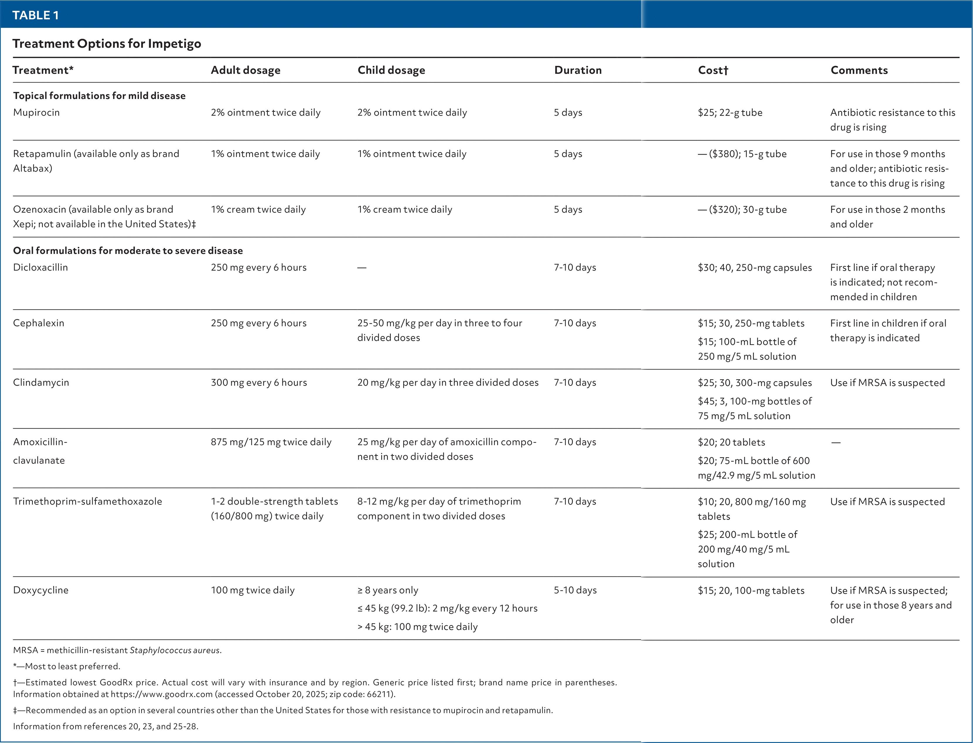
The following table from the AAFP summarizes treatment options with dosing:
Diet
- No specific dietary triggers or restrictions
- Adequate hydration and nutrition support immune function; malnutrition is a recognized risk factor for impetigo [1]
Review of Systems
- Skin: New lesions elsewhere (autoinoculation), underlying eczema or dermatitis, recent insect bites or trauma
- Renal: Dark/cola-colored urine, decreased urine output, facial/periorbital edema (poststreptococcal glomerulonephritis)
- Constitutional: Fever, malaise (uncommon in uncomplicated impetigo — if present, consider deeper infection) [1]
- MSK: Joint pain or swelling (rare; rheumatic fever complication) [4]
Collateral History and Family History
- Contacts with similar skin lesions — daycare, school, household members, sports teams [1][5]
- History of recurrent skin infections in the patient or family (consider MRSA colonization, immunodeficiency, or underlying dermatosis)
- Social context: Crowded living conditions, homelessness, incarceration — all increase transmission risk [1]
- Atopic dermatitis in the family — predisposes to secondary impetiginization
Risk Factors
- Age 2–5 years (peak incidence) [1][3]
- Disrupted skin barrier: Eczema, insect bites, abrasions, scabies, varicella [3-4]
- Poor hygiene and crowded living environments [1]
- Hot, humid climates (streptococcal forms predominate in tropical settings) [6]
- Malnutrition and diabetes [1]
- Contact sports (e.g., wrestling — "mat herpes" and impetigo outbreaks)
Differential Diagnosis
- Herpes simplex virus (HSV): Grouped vesicles on an erythematous base; Tzanck smear or PCR distinguishes
- Varicella: Diffuse vesicles in various stages ("crops"), systemic symptoms
- Contact dermatitis: Pruritic, well-demarcated; no honey-colored crust; history of exposure
- Tinea corporis: Annular, scaly plaque with central clearing; KOH positive
- Scabies with secondary impetiginization: Burrows, interdigital involvement, intense nocturnal pruritus [4]
- Staphylococcal scalded skin syndrome (SSSS): Widespread erythema and desquamation in neonates/young children; Nikolsky sign positive
- Ecthyma: Deeper ulcerative lesions with thick crust and scarring (a complication/extension of impetigo) [2]
- Pemphigus/bullous pemphigoid: Consider in adults with bullous lesions unresponsive to antibiotics
Past Medical History
- Atopic dermatitis/eczema — most common predisposing condition for secondary impetigo [3]
- Previous episodes of impetigo — recurrence suggests persistent colonization (consider nasal mupirocin decolonization)
- Immunocompromised states — HIV, chemotherapy, chronic corticosteroid use
- Diabetes mellitus [1]
- History of MRSA infection or colonization — guides empiric antibiotic choice [2]
Physical Exam
- Nonbullous: Erythematous papules → vesicles/pustules → honey-colored crusts on an erythematous base, typically on face and extremities [1-2]
- Bullous: Large, flaccid bullae with clear-to-yellow fluid; collarettes of scale at periphery of ruptured bullae; favors trunk and intertriginous areas [1][3]
- Satellite lesions from autoinoculation [1]
- Regional lymphadenopathy: Uncommon; if present, consider deeper or more extensive infection [1]
- Assess for: Surrounding cellulitis (spreading erythema, warmth, induration), signs of ecthyma (deep ulceration with scarring), and underlying dermatosis (eczema, scabies) [1][4]
- In skin of color: Erythema may be subtle; look for crusting, erosions, and post-inflammatory dyspigmentation [4]
Lab Studies
- Routine cases: No labs needed; diagnosis is clinical [1]
- Culture and Gram stain of vesicle fluid/exudate: Recommended by IDSA but not required for typical presentations; obtain if treatment failure, recurrence, or diagnostic uncertainty [1-2]
- If poststreptococcal glomerulonephritis suspected: Urinalysis (hematuria, proteinuria), BMP (creatinine, BUN), complement levels (low C3), anti-DNase B (more sensitive than ASO for skin streptococcal infections) [1]
- ASO titer: Not helpful for diagnosing impetigo itself, but useful if poststreptococcal complications are suspected [1]
Imaging
- Not indicated for uncomplicated impetigo
- Ultrasound: Consider if concern for underlying abscess (fluctuant area beneath crusted lesion)
- CT/MRI: Only if concern for necrotizing soft tissue infection or deep space infection (rare)
Special Tests
- Wound culture with sensitivities: Most useful special test when empiric therapy fails or MRSA is suspected [2]
- KOH preparation: To rule out tinea if annular or scaly morphology
- Tzanck smear or HSV PCR: If vesicular lesions raise concern for herpes simplex
- Dermoscopy: Can help differentiate from other vesiculobullous conditions
ECG
- Not routinely indicated
- Consider if concern for acute rheumatic fever (prolonged PR interval) as a rare complication of streptococcal impetigo [4]
Assessment
Severity stratification
- Mild: ≤5 localized lesions, no systemic symptoms → topical therapy [1]
- Moderate-severe: >5–6 lesions, multilesional, or outbreak setting → oral antibiotics [1-2]
- Complicated: Progression to ecthyma, cellulitis, abscess, or systemic toxicity → escalate care [1]
Typical presentation: Afebrile child with honey-colored crusted lesions on the face, often with a history of preceding skin break. Resolves within 2–3 weeks without scarring even untreated, though ~25% show no improvement without antibiotics. [1][3]
Complications (rare but important)
- Ecthyma, cellulitis, erysipelas, abscess, necrotizing fasciitis [1]
- Poststreptococcal glomerulonephritis (antibiotics may not prevent this) [1][6]
- Acute rheumatic fever (antibiotics do prevent this) [1][4]
- Guttate psoriasis, scarlet fever [6]
Treatment Plan
Initial management
- Gentle wound care: Soak and remove crusts with warm water and mild soap before applying topical antibiotics
- Mild disease: Mupirocin 2% ointment BID × 5 days [1-2]
- Moderate-severe/multilesional: Cephalexin (children) or dicloxacillin (adults) × 7–10 days [1-2]
- MRSA suspected: Clindamycin, TMP-SMX, or doxycycline (≥8 years) [1-2]
- Ecthyma: Always requires oral antibiotics [2]
Infection control
- Good hand hygiene; wash fomites (towels, linens) [1]
- Keep lesions covered
- Exclude from school/daycare until 12–24 hours after initiating antibiotics or until clinical improvement [1][5]
Outbreak setting: Use systemic (oral) antibiotics to decrease transmission; if poststreptococcal glomerulonephritis outbreak, systemic antibiotics are recommended to eliminate nephritogenic strains [2]
Disposition
- Discharge (vast majority): Uncomplicated impetigo is managed entirely as an outpatient
- Observation/admission criteria:
- Extensive bullous impetigo in neonates (concern for SSSS)
- Signs of systemic toxicity or sepsis
- Rapidly progressive cellulitis or suspected necrotizing fasciitis
- Immunocompromised patients with extensive disease
- Specialist consultation:
- Dermatology: Atypical presentation, treatment failure, recurrent disease
- Infectious disease: Suspected MRSA, recurrent infections, outbreak management
- Nephrology: If poststreptococcal glomerulonephritis develops
Follow Up / Return Precautions
- Follow-up: Reassess in 3–5 days if no improvement on topical therapy; switch to oral antibiotics if worsening or no response [1]
- Expected course: Lesions typically resolve within 7–10 days with treatment; 2–3 weeks untreated [3]
- Return precautions — advise return if:
- Lesions spreading despite treatment
- New fever, increasing redness/swelling, or pain
- Dark or bloody urine, facial swelling (glomerulonephritis — typically 1–3 weeks post-infection)
- Joint pain or new rash (rheumatic fever)
- Recurrence prevention: Treat underlying dermatoses (eczema, scabies); consider nasal mupirocin decolonization for recurrent staphylococcal impetigo; maintain good hygiene practices [1]
References
1. Impetigo: Rapid Evidence Review. — Trang DT, Buck EC, Schoenherr DT. American Family Physician. 2026.
2. Practice Guidelines for the Diagnosis and Management of Skin and Soft Tissue Infections: 2014 Update by the Infectious Diseases Society of America. — Stevens DL, Bisno AL, Chambers HF, et al. Clinical Infectious Diseases : An Official Publication of the Infectious Diseases Society of America. 2014.
3. Impetigo: Diagnosis and Treatment. — Hartman-Adams H, Banvard C, Juckett G. American Family Physician. 2014.
4. Healthy Skin for Children and Young People With Skin of Colour Starts With Clinician Knowledge and Recognition: A Narrative Review. — Ricciardo BM, Kessaris HL, Cherian S, et al. The Lancet. Child & Adolescent Health. 2025.
5. Efficacy and Safety of Ozenoxacin Cream for Treatment of Adult and Pediatric Patients With Impetigo: A Randomized Clinical Trial. — Rosen T, Albareda N, Rosenberg N, et al. JAMA Dermatology. 2018.
6. Interventions for Impetigo. — Koning S, van der Sande R, Verhagen AP, et al. The Cochrane Database of Systematic Reviews. 2012.
Figure
More from Deep Dive
Reviewed by Dr. Lucas Mastropaolo. SymptomDx is an educational tool for medical professionals — it does not replace clinical judgment. Verify all clinical data and drug dosages with authoritative sources.