Liver Laceration
The liver is the most commonly injured abdominal solid organ in both blunt and penetrating trauma. [1] The majority of injuries (80–90%) are low-grade (AAST I–III), and 85–90% of blunt liver injuries can be managed nonoperatively in hemodynamically stable patients, regardless of grade. [1-2] Management is driven primarily by hemodynamic status, not injury grade alone. [2]
History
- Mechanism of injury: motor vehicle collision (most common), falls, bicycle handlebar injuries, assault, penetrating trauma (stab wound, gunshot wound)
- Speed, seatbelt use, airbag deployment, steering wheel deformity, ejection from vehicle
- Direct blow or compression to the right upper quadrant/lower right chest
- Timing of injury and symptom onset
- Abdominal pain — location, severity, radiation (right shoulder pain = Kehr sign suggesting diaphragmatic irritation/hemoperitoneum)
- Lightheadedness, syncope, or near-syncope suggesting hemorrhage
- Associated injuries: rib fractures, pelvic fractures, other solid organ injuries
- Anticoagulant/antiplatelet use (increases bleeding risk)
- Last oral intake (relevant for potential operative management)
Alarm Features
- Hemodynamic instability: SBP <90 mmHg, HR >120 bpm, poor capillary refill, altered mental status [2]
- Signs of peritonitis (diffuse abdominal rigidity, rebound, guarding) — mandate emergent laparotomy [3]
- Rapidly expanding abdominal distension
- Persistent tachycardia despite fluid resuscitation (transient responder or non-responder)
- Ongoing transfusion requirement (>4 units pRBC in 24 hours is a predictor of NOM failure) [4]
- Kehr sign (left shoulder pain from diaphragmatic irritation by hemoperitoneum)
- Signs of hemorrhagic shock (Class III/IV)
Medications
- Relevant contributors: Anticoagulants (warfarin, DOACs) and antiplatelets (aspirin, clopidogrel) increase hemorrhage risk and may lower threshold for intervention
- Acute treatment:
- Tranexamic acid (TXA) — administer within 3 hours of injury per CRASH-2 protocol (1 g IV bolus then 1 g over 8 hours)
- Massive transfusion protocol activation when indicated (1:1:1 ratio pRBC:FFP:platelets)
- Reversal agents for anticoagulants as appropriate (4-factor PCC for warfarin, idarucizumab for dabigatran, andexanet alfa for factor Xa inhibitors)
- Avoid: NSAIDs in the acute setting (platelet dysfunction, renal injury risk in hypovolemia)
- Pain management: Acetaminophen should be used cautiously given hepatic injury; opioids are the mainstay for acute pain control
Diet
- NPO on presentation — anticipate potential operative intervention or angiography
- Resume diet as tolerated once hemodynamically stable and operative management is not anticipated
- No specific long-term dietary restrictions related to liver laceration itself
- Avoid alcohol during recovery to minimize hepatic stress
Review of Systems
- Cardiovascular: Lightheadedness, syncope, palpitations (hemorrhage)
- Respiratory: Dyspnea, pleuritic chest pain (associated hemothorax, diaphragmatic injury, right lower lobe atelectasis)
- GI: Nausea, vomiting, hematemesis or melena (hemobilia from pseudoaneurysm rupture is a delayed complication) [5]
- GU: Hematuria (concomitant renal injury)
- Neurologic: Altered mental status (hemorrhagic shock or concomitant head injury)
- MSK: Rib pain, pelvic pain (associated fractures)
Collateral History and Family History
- Witnesses or EMS report of mechanism, estimated blood loss at scene, extrication time
- Pre-hospital vitals and interventions (fluids, blood products)
- Prior abdominal surgeries (adhesions may complicate operative management)
- Bleeding disorders or family history of coagulopathy
- Hepatic comorbidities: cirrhosis, hepatic steatosis (increased friability and bleeding risk)
- Social context: alcohol use, substance use (may affect exam reliability and hepatic reserve)
Risk Factors
- Blunt trauma: MVC, falls, sports injuries, bicycle handlebar injuries (especially pediatric)
- Penetrating trauma: Stab wounds, gunshot wounds — penetrating injuries are more likely to require operative/endoscopic/percutaneous procedures [6]
- Pre-existing hepatomegaly (hepatitis, malignancy, congestion) increases susceptibility
- Cirrhosis/steatosis: Increased parenchymal friability and coagulopathy
- Anticoagulant/antiplatelet therapy
- Age >55 years (historically considered a risk factor, though no longer an absolute contraindication to NOM) [3]
- ISS >34 and transfusion >4 units pRBC are predictors of NOM failure [4]
Differential Diagnosis
- Splenic laceration/rupture — left-sided mechanism, Kehr sign, similar hemodynamic presentation
- Renal injury — flank pain, hematuria
- Hollow viscus injury (bowel perforation) — peritonitis, free air on imaging; incidence increases with multiple solid organ injuries [3]
- Mesenteric/omental injury — hemoperitoneum without solid organ injury on CT
- Diaphragmatic rupture — especially right-sided, may coexist with liver injury
- Retroperitoneal hemorrhage (pelvic fracture, aortic injury)
- Rib fractures with intercostal artery bleeding — right lower rib fractures commonly associated with liver injury
- Adrenal hemorrhage — right-sided, may mimic perihepatic hematoma on CT
Past Medical History
- Prior abdominal trauma or surgery
- Known liver disease (cirrhosis, hepatitis, steatosis) — affects parenchymal integrity and coagulation
- Coagulopathy or bleeding disorders
- Splenectomy (alters differential for hemoperitoneum)
- Chronic anticoagulation
- Prior angioembolization (altered hepatic arterial anatomy)
Physical Exam
- Vitals: Tachycardia, hypotension, tachypnea — signs of hemorrhagic shock; ATLS defines unstable as SBP <90, HR >120 with skin vasoconstriction and altered consciousness [2]
- Inspection: Seatbelt sign across RUQ/chest, abrasions, ecchymosis (Grey Turner, Cullen signs are late findings)
- Palpation: RUQ tenderness, guarding, rigidity; diffuse peritonitis mandates laparotomy [3]
- Percussion: Dullness in flanks (hemoperitoneum)
- Auscultation: Decreased breath sounds right base (associated hemothorax/diaphragmatic injury)
- Lower right rib tenderness: Fractures of ribs 9–12 on the right are strongly associated with liver injury
- Rectal exam: Assess for melena (delayed hemobilia)
- Serial exams are critical during NOM — any change in abdominal pain pattern or new peritonitis warrants reassessment [3]
Lab Studies
- CBC with serial hemoglobin — cornerstone of NOM monitoring; unexplained Hgb drop prompts repeat imaging [3]
- Type and screen/crossmatch — essential; activate MTP if indicated
- Comprehensive metabolic panel including hepatic function
- AST/ALT: Grossly elevated transaminases (AST >200, ALT >80–100) are strongly associated with liver laceration and should prompt CT imaging. Combined AST >100 + ALT >80 + WBC >10,000 yields ~90% sensitivity and 92% specificity for liver laceration [7-8]
- Coagulation studies (PT/INR, PTT, fibrinogen) — assess for coagulopathy and guide resuscitation
- Lactate — marker of tissue hypoperfusion/shock
- ABG/VBG — assess base deficit (marker of shock severity)
- Lipase — evaluate for concurrent pancreatic injury
Imaging
- E-FAST (Extended Focused Assessment with Sonography for Trauma): Rapid bedside detection of intra-abdominal free fluid; first-line in hemodynamically unstable patients (GoR 1A). Sensitivity is limited (42–52% in pediatrics); a negative FAST does not exclude liver injury [5]
- CT abdomen/pelvis with IV contrast: Gold standard in hemodynamically stable patients (sensitivity/specificity 96–100%). Identifies laceration grade, hemoperitoneum volume, active contrast extravasation ("blush"), pseudoaneurysm, and associated injuries [5][9]
- Routine follow-up CT is NOT recommended — repeat imaging only for clinical deterioration (fever, increasing pain, Hgb drop, jaundice, SIRS). Surveillance imaging in asymptomatic patients has a 0% complication detection rate vs. 39% when prompted by clinical deterioration [5][10]
- Ultrasound: Useful for follow-up assessment of bilomas in grade IV–V injuries [2]
Special Tests
- AAST Organ Injury Scale (OIS)[4][11]
- WSES Classification: Integrates AAST grade with hemodynamic status — Minor (WSES I = AAST I–II, stable), Moderate (WSES II = AAST III, stable), Severe (WSES III = AAST IV–V, stable; WSES IV = any grade, unstable) [5]
- Diagnostic peritoneal lavage (DPL): Largely replaced by FAST/CT but may be used in resource-limited settings
- Angiography: Both diagnostic and therapeutic; indicated for contrast blush on CT or ongoing hemorrhage [3][12]
- ERCP: For biliary complications (bile leak, hemobilia, biliary fistula) [2][5]
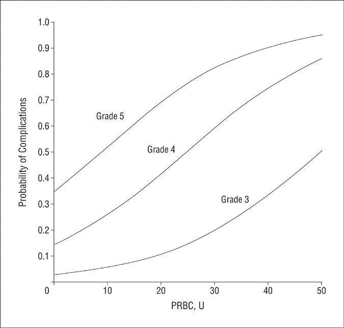
The following figure illustrates the probability of hepatic complications based on injury grade and 24-hour pRBC transfusion volume, a useful risk stratification tool:
ECG
- ECG is part of standard trauma workup
- Evaluate for sinus tachycardia (hemorrhage), arrhythmias (electrolyte derangements from massive resuscitation)
- Rule out myocardial contusion in high-energy blunt chest/abdominal trauma (ST changes, new bundle branch block)
- Pulseless electrical activity (PEA) in the setting of massive hemoperitoneum or tension pneumothorax
Assessment
- Severity stratification is based on AAST grade AND hemodynamic status, not grade alone [2]
- 80–90% of liver injuries are grade I–III (minor); 12–20% are grade IV–V [1]
- Overall mortality is ~5%; mortality after operative management for severe injuries reaches 30–68% [14-15]
- Complications occur in 12–14% of high-grade injuries managed nonoperatively and include: [2][5]
- Delayed hemorrhage/pseudoaneurysm rupture (1.7–5.9%)
- Biliary complications (bile leak, biloma, hemobilia, biliary fistula) — up to 30%
- Hepatic abscess (0–7%)
- Abdominal compartment syndrome
- Hepatic necrosis (especially post-angioembolization)
Treatment Plan
Initial Stabilization
- ATLS primary and secondary survey
- Large-bore IV access, massive transfusion protocol if indicated (1:1:1 pRBC:FFP:platelets)
- TXA within 3 hours of injury
- Correct hypothermia, acidosis, coagulopathy (lethal triad)
Nonoperative Management (NOM) — standard of care for hemodynamically stable patients [3][5]
- Serial clinical exams, hemoglobin monitoring q4–6h initially
- ICU admission for AAST grade III–V injuries [5]
- Floor admission may suffice for grade I–II injuries
- Angiography with embolization for arterial blush on CT in stable patients [3][12]
- An observation-first strategy (even with CT blush) is safe and effective in stable patients, with only 14% requiring delayed intervention; empiric angioembolization is associated with higher complication rates (abscess, biloma) [16]
Operative Management — indicated for: [3][5]
- Hemodynamic instability/non-responders (WSES grade IV)
- Diffuse peritonitis
- Failed NOM
- Operative strategy: "6 Ps" — Push, Pack, Pringle, Put Back, Phone, Pivot [1]
Complications Management [5]
- Delayed hemorrhage → angioembolization first-line
- Pseudoaneurysm → angioembolization to prevent rupture
- Abscess → percutaneous drainage
- Biloma → percutaneous drainage ± ERCP with stenting
- Biliary fistula → laparoscopic lavage/drainage + endoscopic stenting
Disposition
- Emergent OR: Hemodynamic instability, non-responder to resuscitation, peritonitis [3][5]
- ICU admission: AAST grade III–V injuries undergoing NOM; transient responders; patients requiring close hemodynamic monitoring, serial exams, and immediate OR availability [5]
- Floor/step-down: Hemodynamically stable grade I–II injuries without other significant injuries
- Transfer to higher level of care: If facility lacks ICU monitoring, angiography capability, immediate OR access, or blood bank resources. Notably, 92% of transferred patients undergo NOM and only 3.4% fail NOM [5][15]
- Discharge: Once hemodynamically stable, tolerating diet, pain controlled, hemoglobin stable, and no evidence of complications
Specialist consultation triggers
- Trauma surgery — all liver lacerations
- Interventional radiology — arterial blush on CT, ongoing hemorrhage amenable to embolization
- Hepatobiliary surgery — complex biliary injuries, hepatic necrosis
- Gastroenterology/ERCP — biliary complications (bile leak, hemobilia)
Follow Up / Return Precautions
- Follow-up timing: Outpatient trauma surgery follow-up within 1–2 weeks of discharge; repeat labs (CBC, LFTs) as clinically indicated
- Routine follow-up imaging is NOT necessary — imaging should be driven by clinical symptoms [5][10]
- Most liver lesions heal within approximately 4 months [5]
- Patients may resume normal physical activities after 3–4 months for moderate-to-severe injuries [5]
- Activity restrictions: Avoid contact sports and heavy lifting during recovery; duration depends on injury grade
Return precautions — instruct patients to return immediately for: [5]
- Increasing abdominal pain
- Lightheadedness, dizziness, or fainting
- Nausea or vomiting
- Fever or chills
- Dark/tarry stools or vomiting blood (hemobilia)
- Jaundice
- Patients should not remain alone for extended periods during recovery [5]
References
1. Liver Injury: What You Need to Know. — Reed CR, Brown JB, Peitzman AB. The Journal of Trauma and Acute Care Surgery. 2025.
2. WSES Classification and Guidelines for Liver Trauma. — Coccolini F, Catena F, Moore EE, et al. World Journal of Emergency Surgery : WJES. 2016.
3. Nonoperative Management of Blunt Hepatic Injury: An Eastern Association for the Surgery of Trauma Practice Management Guideline. — Stassen NA, Bhullar I, Cheng JD, et al. The Journal of Trauma and Acute Care Surgery. 2012.
4. Non-Operative Management Versus Operative Management in High-Grade Blunt Hepatic Injury. — Cirocchi R, Trastulli S, Pressi E, et al. The Cochrane Database of Systematic Reviews. 2015.
5. Liver Trauma: WSES 2020 Guidelines. — Coccolini F, Coimbra R, Ordonez C, et al. World Journal of Emergency Surgery : WJES. 2020.
6. The American Association for the Surgery of Trauma (AAST) Liver Injury Grade Does Not Equally Predict Interventions in Blunt and Penetrating Trauma. — Brigode W, Adra A, Capron G, et al. World Journal of Surgery. 2022.
7. Combination of White Blood Cell Count With Liver Enzymes in the Diagnosis of Blunt Liver Laceration. — Lee WC, Kuo LC, Cheng YC, et al. The American Journal of Emergency Medicine. 2010.
8. Hepatic Enzymes Have a Role in the Diagnosis of Hepatic Injury After Blunt Abdominal Trauma. — Tan KK, Bang SL, Vijayan A, Chiu MT. Injury. 2009.
9. Hepatobiliary Trauma Imaging Update. — Stephens J, Yu HS, Uyeda JW. Radiologic Clinics of North America. 2022.
10. Surveillance Imaging Following Liver Trauma Has a Low Detection Rate of Liver Complications. — Fischer N, Bartlett A. Injury. 2022.
11. Grading Abdominal Trauma: Changes in and Implications of the Revised 2018 AAST-OIS for the Spleen, Liver, and Kidney. — Dixe de Oliveira Santo I, Sailer A, Solomon N, et al. Radiographics : A Review Publication of the Radiological Society of North America, Inc. 2023.
12. Society of Interventional Radiology Position Statement on Endovascular Intervention for Trauma. — Padia SA, Ingraham CR, Moriarty JM, et al. Journal of Vascular and Interventional Radiology : JVIR. 2020.
13. Risk Factors for Hepatic Morbidity Following Nonoperative Management: Multicenter Study. — Kozar RA, Moore FA, Cothren CC, et al. Archives of Surgery. 2006.
14. Management of Severe Blunt Hepatic Injury in the Era of Computed Tomography and Transarterial Embolization: A Systematic Review and Critical Appraisal of the Literature. — Melloul E, Denys A, Demartines N. The Journal of Trauma and Acute Care Surgery. 2015.
15. Contemporary Epidemiologic Overview of Adult Liver Trauma Management Across the United States: Analysis of the American College of Surgeons Trauma Quality Improvement Program Database. — Obaid O, Torres-Ruiz T, Rady E, et al. Surgery. 2025.
16. An Observation-First Strategy for Liver Injuries With "Blush" on Computed Tomography Is Safe and Effective. — Samuels JM, Carmichael H, McIntyre R, et al. The Journal of Trauma and Acute Care Surgery. 2023.
Figure
More from Deep Dive
Reviewed by Dr. Lucas Mastropaolo. SymptomDx is an educational tool for medical professionals — it does not replace clinical judgment. Verify all clinical data and drug dosages with authoritative sources.