Malignant Otitis Externa
Malignant otitis externa (MOE) — also termed necrotizing otitis externa (NOE) or osteomyelitis of the temporal bone — is a rare, life-threatening invasive infection of the external auditory canal (EAC) that extends to the temporal bone and skull base, predominantly caused by Pseudomonas aeruginosa (>90% of cases). [1-3] It carries historically reported mortality as high as one-third overall and up to 80% when cranial nerves are involved. [4]
History
- Severe, unrelenting otalgia — classically described as "excruciating" or "pain out of proportion to exam," often worse at night [1][5-6]
- Persistent purulent otorrhea unresponsive to standard topical therapy for ≥2 weeks [1-2]
- Duration and progression: diagnosis is often delayed 6–8 weeks when symptoms are mistakenly attributed to simple otitis externa [5]
- Hearing loss (conductive or sensorineural) [5]
- Headache, trismus, or preauricular swelling suggesting extension [7]
- Ask about prior ear canal manipulation (cotton swabs, cerumen irrigation — a reported iatrogenic trigger) [2][6]
- Ask about water exposure (swimming, ear irrigation) [2]
- Important negatives: Fever is often absent in early disease [5]
Alarm Features
- Pain out of proportion to exam findings in a diabetic or immunocompromised patient [1]
- Otitis externa failing topical therapy after 2+ weeks [1]
- Cranial nerve deficits — facial nerve (CN VII) most common; glossopharyngeal (CN IX) and spinal accessory (CN XI) less frequently involved [8-9]
- Periauricular edema, bone exposure, or TMJ pain [7]
- Signs of intracranial extension: altered mental status, seizures, meningismus, new neurological deficits [10]
- Trismus (suggests TMJ or infratemporal fossa involvement) [11]
Medications
Causative/contributing medications
- Prior topical fluoroquinolone use may select for ciprofloxacin-resistant Pseudomonas [2]
- Immunosuppressants (chemotherapy, biologics, chronic corticosteroids) increase susceptibility [9-10]
Treatment antibiotics
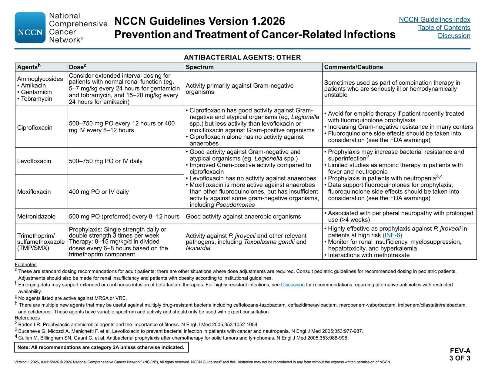
- First-line empiric: IV antipseudomonal therapy — commonly ciprofloxacin 750 mg PO BID (if susceptible) or IV ceftazidime + ciprofloxacin combination [2][12-13]
- One protocol suggests 3 weeks initial combination therapy (ceftazidime + ciprofloxacin, high doses) followed by 3 weeks ciprofloxacin monotherapy [12]
- Alternative IV agents: piperacillin-tazobactam, meropenem (for resistant organisms or ciprofloxacin failure) [11][14]
- Duration: 6–8 weeks of systemic antipseudomonal therapy, guided by culture/sensitivity [2][5]
- Topical adjuncts: antipseudomonal or acetic acid otic drops [5]
Cautions
- Ciprofloxacin resistance is emerging — always obtain cultures with antibiogram [2][9]
- Aminoglycosides carry ototoxicity risk in this population
- Avoid ear canal irrigation in diabetic/immunocompromised patients — may predispose to NOE [8]
Diet
- Strict glycemic control is essential and directly impacts outcomes [9][15]
- Diabetic diet optimization; endocrinology consultation for poorly controlled diabetes
- Adequate hydration and nutrition to support immune function
- No specific dietary triggers, but uncontrolled hyperglycemia is a major modifiable risk factor
Review of Systems
- ENT: Otalgia severity, otorrhea character/duration, hearing changes, tinnitus, vertigo
- Neurological: Facial asymmetry/weakness, dysphagia, hoarseness, shoulder weakness (cranial nerves VII, IX, X, XI) [8]
- Constitutional: Fever (often absent early), night sweats, weight loss (consider malignancy mimic)
- MSK: Jaw pain, trismus, TMJ tenderness [7][11]
- Vascular: Headache pattern changes (consider sigmoid sinus thrombosis)
Collateral History and Family History
- Diabetes control: HbA1c, medication adherence, recent glucose trends
- Immunosuppression history: HIV status, chemotherapy, organ transplant, chronic steroid use [2][10]
- Prior ear interventions: Recent cerumen removal, ear irrigation, hearing aid use [2][6]
- Prior episodes of otitis externa or MOE (recurrence rate is notable) [11]
- Family history is generally not contributory, though familial diabetes risk is relevant
Risk Factors
- Diabetes mellitus (poorly controlled) — present in 65–90% of cases [7][16]
- Advanced age — mean age ~62–77 years [16-17]
- Male sex — ~62–74% male predominance [7][16]
- Immunosuppression: HIV/AIDS, chemotherapy, organ transplant, chronic corticosteroids [2][10]
- History of radiation therapy to the head/neck [8]
- Ear canal trauma: cotton swab use, hearing aids, ear irrigation [2][6]
- Water exposure (swimming, ear irrigation converting acidic EAC pH to alkaline) [10]
- Chronic kidney disease, hypertension, and ischemic heart disease are common comorbidities [16]
Differential Diagnosis
- Squamous cell carcinoma of the EAC — the most critical mimic; tissue biopsy is recommended to rule out malignancy [5-6]
- Uncomplicated acute otitis externa — responds to topical therapy, no granulation tissue or bone involvement [18]
- Chronic otitis externa — itching predominant, lasts >3 months, no osteomyelitis [18]
- Ramsay Hunt syndrome — herpetic vesicles in canal, facial paralysis, loss of taste; treat with antivirals [18]
- Cholesteatoma of the EAC — can mimic on imaging [17]
- Chronic suppurative otitis media — chronic otorrhea with nonintact tympanic membrane [18]
- Otomycosis (Aspergillus, Candida) — itching predominant, fungal elements on otoscopy; can coexist with MOE [10]
- Referred pain (dental, TMJ, pharyngeal) — normal ear exam [18]
- Wegener granulomatosis (GPA) — granulomatous inflammation, may involve temporal bone
Past Medical History
- Diabetes mellitus — type and control (HbA1c), complications
- Prior episodes of otitis externa or MOE
- HIV/AIDS status and CD4 count
- Malignancy and chemotherapy history
- Prior head/neck radiation
- Chronic kidney disease (affects antibiotic dosing)
- Prior ear surgeries, tympanostomy tubes [8]
- Hearing aid use
Physical Exam
Vital signs
- Fever may be absent early; when present, suggests advanced disease [5]
- Tachycardia may indicate sepsis
Otoscopic exam
- Granulation tissue at the bony-cartilaginous junction — hallmark finding, though not always present [3][8]
- Edematous, erythematous EAC with purulent otorrhea [1]
- Necrotic tissue in the canal [18]
- Assess tympanic membrane integrity
Focused exam
- Cranial nerve exam — especially CN VII (facial nerve): ask patient to smile, raise eyebrows, close eyes tightly [1][8]
- CN IX, X, XI, XII assessment (gag reflex, voice quality, shoulder shrug, tongue protrusion) [8]
- Tragal and pinnal tenderness
- Periauricular and preauricular lymphadenopathy/swelling
- TMJ tenderness, trismus [7]
- Parotid gland assessment
- Mastoid tenderness
Lab Studies
- ESR — highly useful; ESR >26 mm/h has a positive likelihood ratio of 10.15 for NOE (AUC 0.92) [19]
- CRP — CRP >10 mg/L has a positive likelihood ratio of 8.25 [19]
- If both ESR and CRP are normal, an alternative diagnosis should be sought [19]
- EAC culture with Gram stain — essential; send for aerobic bacterial culture; consider fungal culture [20]
- Tissue biopsy of granulation tissue — for culture and to rule out squamous cell carcinoma [5-6]
- CBC — leukocytosis (may be absent in immunocompromised)
- BMP — renal function (affects antibiotic dosing), glucose
- HbA1c — assess diabetic control
- Blood cultures — if febrile or septic
Imaging
First-line
- CT temporal bone with IV contrast — most commonly used initial imaging; excellent for demonstrating bone erosion, soft tissue changes at skull base [1][10]
- CT was used in 73% of cases as the primary diagnostic modality [10]
- However, CT can be negative in early disease — 50% of patients with negative CT were ultimately diagnosed with NOE on further imaging [17]
Gold standard for diagnosis
- Tc-99m bone scintigraphy — highly sensitive (positive in 100% of NOE cases); useful when CT is negative or equivocal [10][21]
- Limitation: remains positive after clinical resolution (cannot be used to confirm cure) [10]
For treatment monitoring
- Gallium-67 scintigraphy — normalizes with infection resolution; used to guide antibiotic cessation [10]
- FDG-PET/CT — emerging as an alternative for both diagnosis and treatment monitoring [22]
For intracranial complications
When imaging is unnecessary
Special Tests
Diagnostic criteria (combination of)
- Clinical findings (severe otalgia, otorrhea, granulation tissue in immunocompromised host)
- Elevated ESR [2]
- Positive EAC culture (Pseudomonas in >90%) [2]
- Radiographic evidence of soft tissue/bone involvement [2]
Point-of-care
- Bedside otoscopy — look for granulation tissue at bony-cartilaginous junction
- Bedside glucose/HbA1c
Procedures
ECG
- ECG is not routinely indicated for MOE itself
- Consider ECG if:
- Fluoroquinolone therapy planned (QTc prolongation risk, especially with concurrent QT-prolonging medications)
- Elderly patient with cardiac comorbidities (common in this population) [16]
- Sepsis or hemodynamic instability
Assessment
MOE is a medical emergency with high morbidity and potential mortality. [1][18] The typical patient is an elderly diabetic male presenting with severe, unrelenting otalgia and purulent otorrhea unresponsive to topical therapy. The diagnosis requires a high index of suspicion, as early disease may appear similar to uncomplicated otitis externa.
Severity stratification
- Early/localized: EAC inflammation with granulation tissue, no cranial nerve involvement, positive Tc-99m scan
- Advanced/extensive: Skull base osteomyelitis, cranial nerve palsies (facial nerve most common), periauricular extension [7]
- Intracranial: Meningitis, cerebral abscess, sigmoid sinus thrombosis — highest mortality [10]
Complications
- Facial nerve paralysis (most common CN deficit) [15]
- Skull base osteomyelitis [10]
- Meningitis, cerebritis, intracranial abscess [10]
- Jugular vein/sigmoid sinus thrombosis [4]
- Contralateral spread
- Recurrence (notable risk, especially with inadequate treatment duration) [11]
Treatment Plan
Initial stabilization (ED)
- IV access, pain management (often requires opioids given severity)
- Obtain EAC cultures before starting antibiotics
- Check glucose, initiate glycemic control
Antibiotic therapy
- Empiric IV antipseudomonal coverage — start immediately upon clinical suspicion [1]
- Preferred regimen: Combination of IV ceftazidime + oral/IV ciprofloxacin initially, transitioning to ciprofloxacin monotherapy after 3 weeks if susceptible (total 6 weeks) [12]
- Alternative: Ciprofloxacin 750 mg PO BID monotherapy for 6–8 weeks (for less severe, susceptible cases) [2][13]
- Resistant organisms: Meropenem, piperacillin-tazobactam, or antibiogram-guided therapy [9][11]
- Fungal MOE: Systemic antifungals (voriconazole, amphotericin B) if Aspergillus or Candida identified [10]
- Topical antipseudomonal or acetic acid drops as adjunct [5]
Surgical management
- Debridement of necrotic/granulation tissue by ENT [5][9]
- Extensive surgery reserved for refractory cases or facial nerve involvement [15]
Adjunctive
- Glycemic optimization — endocrinology consultation [9]
- Hyperbaric oxygen therapy — considered in refractory cases, though a Cochrane review found no RCTs to support its use [4][11]
- Correction of immunosuppression when possible [6]
Disposition
Admission criteria (most patients require admission): [1][18]
- All patients with suspected or confirmed MOE
- Cranial nerve deficits
- Need for IV antibiotics
- Poorly controlled diabetes requiring inpatient optimization
- Intracranial complications
- Hemodynamic instability or sepsis
Observation
Discharge considerations
Specialist consultation triggers
- ENT (otolaryngology) — consult early in all suspected cases for debridement, biopsy, and ongoing management [1][5]
- Infectious disease — for antibiotic guidance, especially resistant organisms or prolonged therapy [12]
- Endocrinology — for diabetic optimization
- Neurosurgery — if intracranial extension suspected
Follow Up / Return Precautions
Follow-up timing
- Close ENT follow-up within 1 week of discharge
- Serial ESR/CRP monitoring to assess treatment response [19]
- Gallium-67 scan or FDG-PET/CT after 6 weeks of therapy to confirm resolution before stopping antibiotics [10][22]
- Follow-up for at least 1 year post-treatment given recurrence risk [15]
Return precautions — instruct patients to return immediately for:
- Worsening ear pain despite antibiotics
- New facial weakness or drooping
- Difficulty swallowing, hoarseness, or new neurological symptoms
- Fever, rigors, or altered mental status
- Inability to tolerate oral antibiotics (nausea/vomiting)
Patient counseling
- Avoid ear canal manipulation (no cotton swabs, no ear irrigation) [6]
- Minimize water exposure to the ear canal [6]
- Strict medication adherence — full antibiotic course is critical
- Diabetic control is essential to prevent recurrence [9][15]
Expected recovery
- Clinical improvement typically within 1–2 weeks of appropriate therapy, but full treatment course (6–8 weeks) is mandatory [2][12]
- Cranial nerve palsies may or may not recover fully
- Recurrence is possible, particularly with inadequate treatment duration or persistent immunosuppression [11]
Images
References
1. An Emergency Medicine-Focused Review of Malignant Otitis Externa. — Long DA, Koyfman A, Long B. The American Journal of Emergency Medicine. 2020.
2. The Changing Face of Malignant (Necrotising) External Otitis: Clinical, Radiological, and Anatomic Correlations. — Rubin Grandis J, Branstetter BF, Yu VL. The Lancet. Infectious Diseases. 2004.
3. Otitis Externa and Malignant Otitis Externa-for the Hospitalist/Internist. — Patel S, Owen GS, Vivas EX. The Medical Clinics of North America. 2026.
4. Hyperbaric Oxygen as an Adjuvant Treatment for Malignant Otitis Externa. — Phillips JS, Jones SE. The Cochrane Database of Systematic Reviews. 2013.
5. Infections in Patients with Diabetes Mellitus. — Joshi N, Caputo GM, Weitekamp MR, Karchmer AW. The New England Journal of Medicine. 1999.
6. Necrotizing (Malignant) External Otitis. — Handzel O, Halperin D. American Family Physician. 2003.
7. Epidemiology and Risk Factors for Extension of Necrotizing Otitis Externa. — Krawiec E, Brenet E, Truong F, et al. European Archives of Oto-Rhino-Laryngology : Official Journal of the European Federation of Oto-Rhino-Laryngological Societies : Affiliated With the German Society for Oto-Rhino-Laryngology - Head and Neck Surgery. 2024.
8. Clinical Practice Guideline: Acute Otitis Externa. — Rosenfeld RM, Schwartz SR, Cannon CR, et al. Otolaryngology--Head and Neck Surgery : Official Journal of American Academy of Otolaryngology-Head and Neck Surgery. 2014.
9. Patient Cases With Malignant Otitis Externa at the University Clinic of Ludwig Maximilians University Munich From 2009 Until 2020. — Stocker M, Hempel JM. Scientific Reports. 2025.
10. ACR Appropriateness Criteria® Inflammatory Ear Disease. — Agarwal M, Juliano AF, Hagiwara M, et al. Journal of the American College of Radiology : JACR. 2025.
11. Isolated Temporomandibular Joint Involvement in a Rare Case of Malignant Otitis Externa. — Celleoğlu S, Yazici D. The Journal of Craniofacial Surgery. 2025.
12. Antibiotic Therapy in Necrotising External Otitis: Case Series of 32 Patients and Review of the Literature. — Pulcini C, Mahdyoun P, Cua E, et al. European Journal of Clinical Microbiology & Infectious Diseases : Official Publication of the European Society of Clinical Microbiology. 2012.
13. Ciprofloxacin Treatment of Malignant External Otitis. — Sadé J, Lang R, Goshen S, Kitzes-Cohen R. The American Journal of Medicine. 1989.
14. Necrotising Otitis Externa: Clinical Profile and Management Protocol. — Lambor DV, Das CP, Goel HC, et al. The Journal of Laryngology and Otology. 2013.
15. Malignant Otitis Externa: An Updated Review. — Treviño González JL, Reyes Suárez LL, Hernández de León JE. American Journal of Otolaryngology. 2021.
16. Evaluation of the Clinical and Radiological Features of Patients With Malignant Otitis Externa (MOE). — Tabashiri A, Allahverdi Nazhand H, Fathy M, et al. BMC Infectious Diseases. 2025.
17. Refining Necrotising Otitis Externa Management: A Follow-Up Study on a Departmental Algorithm and the Role of Nuclear Medicine Imaging. — Haleem AH, Shahidi S, Hilton JM, et al. American Journal of Otolaryngology. 2025.
18. Acute Otitis Externa: Rapid Evidence Review. — Jackson EA, Geer K. American Family Physician. 2023.
19. Necrotizing External Otitis: Diagnostic Clues in the Emergency Department. — Vaca M, Medina MM, Cordero AI, et al. European Archives of Oto-Rhino-Laryngology : Official Journal of the European Federation of Oto-Rhino-Laryngological Societies : Affiliated With the German Society for Oto-Rhino-Laryngology - Head and Neck Surgery. 2024.
20. Guide to Utilization of the Microbiology Laboratory for Diagnosis of Infectious Diseases: 2024 Update by the Infectious Diseases Society of America (IDSA) and the American Society for Microbiology (ASM). — Miller JM, Binnicker MJ, Campbell S, et al. Clinical Infectious Diseases : An Official Publication of the Infectious Diseases Society of America. 2024.
21. Malignant External Otitis: Early Scintigraphic Detection. — Strashun AM, Nejatheim M, Goldsmith SJ. Radiology. 1984.
22. FDG-PET/CT for Diagnosis and Follow-Up of Necrotizing (Malignant) External Otitis. — Stern Shavit S, Bernstine H, Sopov V, Nageris B, Hilly O. The Laryngoscope. 2019.
Figures
More from Deep Dive
Reviewed by Dr. Lucas Mastropaolo. SymptomDx is an educational tool for medical professionals — it does not replace clinical judgment. Verify all clinical data and drug dosages with authoritative sources.