Mallet Finger
Mallet finger (also called "drop finger" or "baseball finger") is a disruption of the terminal extensor tendon at the distal interphalangeal (DIP) joint, resulting in an inability to actively extend the distal phalanx. It may involve a pure tendon rupture or a bony avulsion fracture at the dorsal base of the distal phalanx. [1-3]
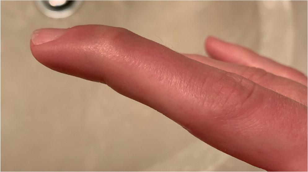
The following figure demonstrates the characteristic clinical presentation:
History
- Mechanism: Forced flexion of an extended DIP joint — classically a ball striking the fingertip, but the majority occur during non-sports activities (bed-making, pulling on socks, tucking in sheets) [5]
- Ask about the specific activity, hand dominance, and which finger is involved (middle, ring, and little fingers most common; index and thumb rare) [5]
- Timing of injury — acute vs. delayed presentation; notably, delayed presentation still responds well to splinting [2]
- Severity of initial trauma — significant force in younger males vs. trivial force in older females [5]
- Ability to straighten the fingertip since injury
- Occupation and hand-use requirements (impacts splint compliance and treatment planning) [6]
- Prior finger injuries or surgeries
Alarm Features
- Open wound over the DIP joint (open mallet — requires surgical referral) [2-3]
- Inability to passively extend the DIP joint (suggests interposed tissue or significant injury) [1]
- Volar subluxation of the distal phalanx on lateral radiograph [2][7]
- Fracture fragment involving >30% of the articular surface [1]
- Neurovascular compromise — loss of distal sensation or perfusion [1]
- Signs of infection (open injury with erythema, warmth, purulence)
Medications
- Acute pain management: NSAIDs (ibuprofen 400–600 mg PO q6–8h or naproxen 250–500 mg PO q12h) are first-line; acetaminophen as adjunct
- Ice and elevation for initial swelling
- No specific medications are contraindicated
- Caution with anticoagulants if surgical intervention is being considered
- No role for antibiotics in closed injuries; consider prophylactic antibiotics for open mallet injuries
Diet
- No specific dietary modifications required
- Adequate protein and calcium intake supports tendon and bone healing
- Smoking cessation counseling — smoking impairs tendon healing
Review of Systems
- MSK: Other finger/hand injuries, joint stiffness, prior tendon injuries
- Neuro: Numbness or tingling distal to injury (assess digital nerve integrity)
- Vascular: Color changes, capillary refill of the affected fingertip
- Rheumatologic: History of inflammatory arthritis (may predispose to tendon weakening)
- Dermatologic: Skin integrity over the DIP joint (open vs. closed injury)
Collateral History and Family History
- Witness account of mechanism if unclear
- Occupational demands — manual labor, musicians, athletes (impacts compliance and urgency)
- Family history is generally not contributory
- Social context: ability to comply with 6–8 weeks of continuous splinting, hand dominance, ADL impact
Risk Factors
- Sports participation — ball-handling sports (basketball, baseball, volleyball, cricket) [5]
- Occupational: Manual labor, housework
- Demographics: More common in males overall; older females sustain injuries from more trivial mechanisms [5]
- Dominant hand affected in approximately two-thirds of cases [5]
- Middle, ring, and little fingers most commonly affected [5][8]
- Rheumatoid arthritis or other connective tissue disorders (tendon weakening)
Differential Diagnosis
- Jersey finger (FDP avulsion) — inability to flex the DIP joint (opposite mechanism: forced extension of a flexed finger); most common in ring finger; requires urgent surgical referral [1]
- DIP joint dislocation — obvious deformity, typically reducible
- Distal phalanx fracture (non-avulsion) — crush mechanism, may have subungual hematoma; DIP extension preserved [1]
- Boutonnière deformity — central slip injury at PIP joint (flexion at PIP, hyperextension at DIP — opposite pattern)
- Trigger finger — intermittent locking/catching, not trauma-related
- DIP joint osteoarthritis — chronic stiffness, Heberden's nodes, no acute traumatic onset
- Pseudo-mallet — extensor tendon laceration proximal to DIP (open wound present)
Past Medical History
- Prior mallet finger or other tendon injuries
- Rheumatoid arthritis, psoriatic arthritis, or connective tissue disorders
- Diabetes (impaired healing)
- Previous hand/finger surgeries
- Chronic steroid use (tendon weakening)
Physical Exam
- Pathognomonic finding: DIP joint resting in flexion (droop) with inability to actively extend [1][3]
- Assess active and passive ROM at the DIP joint — passive extension should be possible in uncomplicated cases
- Swelling, ecchymosis, and tenderness over the dorsal DIP joint
- Evaluate for swan-neck deformity (PIP hyperextension with DIP flexion) — indicates chronic or severe injury [2]
- Test FDP function (ability to flex DIP with PIP held in extension) to rule out jersey finger
- Assess skin integrity — open vs. closed injury
- Neurovascular exam: capillary refill, two-point discrimination of the fingertip
- Evaluate for malrotation by having the patient make a fist (all fingers should point toward the scaphoid)
Lab Studies
- No routine labs required for uncomplicated mallet finger
- If open injury with concern for infection: CBC, CRP/ESR
- Pre-operative labs only if surgical intervention is planned
Imaging
- First-line: AP, lateral, and oblique radiographs of the affected finger [1][9]
- Key findings to assess on X-ray:
- Ultrasound: High-frequency ultrasound can identify tendon rupture vs. contusion vs. bony avulsion and may be useful as a point-of-care adjunct [10]
- MRI: Rarely needed; may be considered if diagnosis is uncertain or to evaluate soft-tissue mallet without bony involvement [10]
- Imaging is unnecessary to repeat unless there is concern for displacement during treatment or failure to improve
Special Tests
- Doyle Classification: [3][7]
- Type I: Closed injury, with or without small avulsion fracture
- Type II: Open injury (laceration)
- Type III: Open injury with loss of skin and tendon substance
- Type IV: (A) Epiphyseal plate fracture in children; (B) Fracture involving 20–50% of articular surface; (C) Fracture >50% with volar subluxation
- Crawford Criteria — used to assess treatment outcomes: [6][8]
- Excellent: Full extension, full flexion, no pain
- Good: 0–10° extension lag, full flexion, no pain
- Fair: 10–25° extension lag, any loss of flexion, no pain
- Poor: >25° extension lag or persistent pain
- Elson test — to rule out central slip injury (boutonnière) if PIP involvement is suspected
ECG
Assessment
Mallet finger is a clinical diagnosis confirmed by the inability to actively extend the DIP joint following trauma, with radiographs used to classify the injury and guide management. [2] Two subtypes exist:
- Tendinous mallet (soft-tissue only): Pure extensor tendon rupture; tends to have worse initial extensor lag (median ~28°) and slightly inferior outcomes with conservative treatment [8]
- Bony mallet (avulsion fracture): Tends to present in younger patients with less initial lag (median ~15°) and better outcomes with splinting [8]
Complications of untreated or poorly managed mallet finger include chronic extension lag and swan-neck deformity (compensatory PIP hyperextension). [2-3]
Treatment Plan
Conservative (standard of care for most cases)
- Continuous DIP splinting in full extension to slight hyperextension for 6–8 weeks, followed by 2–4 weeks of nighttime splinting [1-3]
- Splint options: dorsally padded aluminum splint, volar splint, thermoplastic stack splint, or prefabricated 3-point orthosis — all show comparable outcomes [5][11]
- Critical patient education: The DIP joint must never flex during the splinting period; if the splint is removed (e.g., for skin care), the finger must be held in extension on a flat surface. Any episode of flexion resets the clock [1]
- Follow-up every 2 weeks to assess skin integrity and splint fit [1]
- Splinting is effective even with delayed presentation (weeks to months after injury) [2][12]
Surgical indications: [2-3][7]
- Avulsion fracture involving >30% of the articular surface
- Volar subluxation of the distal phalanx
- Open mallet injury
- Failed conservative management (persistent significant extension lag after adequate splinting trial)
- Patient unable to comply with splinting (K-wire fixation is an alternative) [6]
Surgical options: K-wire fixation of DIP in extension, open reduction and internal fixation (ORIF) for large bony fragments, or extension block pinning. [3][6-7]
Disposition
- Discharge with splint application and follow-up for the vast majority of cases
- Orthopedic/hand surgery referral indications:
- No indication for admission for isolated mallet finger
Follow Up / Return Precautions
- Follow-up: Every 2 weeks during splinting to check skin (maceration, pressure sores) and splint integrity [1]
- Repeat radiograph at 2–4 weeks if bony mallet to assess alignment
- After splint removal at 6–8 weeks, begin gentle active ROM; continue nighttime splinting for an additional 2–4 weeks [3]
- Total recovery: 6–10 weeks; some residual extension lag (<10°) is common and generally well-tolerated [1][8]
- Return precautions — advise patients to return immediately for:
- Skin breakdown, blistering, or maceration under the splint
- Increasing pain, swelling, or redness (concern for infection)
- Worsening droop or loss of extension after splint removal
- Numbness or color change in the fingertip
- Counsel that premature removal or flexion of the DIP joint during treatment can prolong recovery or lead to treatment failure [1]
- If untreated, chronic mallet finger can lead to swan-neck deformity, which may require surgical correction [2]
References
1. Common Finger Fractures and Dislocations. — Childress MA, Olivas J, Crutchfield A. American Family Physician. 2022.
2. Approach to Mallet Finger Injury: Practical Guide for Canadian Primary Care Physicians. — Dinh V, Market M, Cheung K. Canadian Family Physician Medecin De Famille Canadien. 2026.
3. Mallet Finger. — Bendre AA, Hartigan BJ, Kalainov DM. The Journal of the American Academy of Orthopaedic Surgeons. 2005.
4. Fingertip Injuries. — Hilgefort J, Becker J, Chu J. American Family Physician. 2025.
5. Interventions for Treating Mallet Finger Injuries. — Handoll HH, Vaghela MV. The Cochrane Database of Systematic Reviews. 2004.
6. A Single K-Wire to Prevent Poor Outcomes in Closed Soft-Tissue Mallet Finger Management Due to Patient Non-Compliance. — Aksan T, Öztürk MB, Özçelik B. Archives of Orthopaedic and Trauma Surgery. 2021.
7. Current Concepts in the Evaluation and Treatment of Mallet Finger Injury. — Bloom JMP, Khouri JS, Hammert WC. Plastic and Reconstructive Surgery. 2013.
8. Outcome Differences Between Conservatively Treated Acute Bony and Tendinous Mallet Fingers. — Rubin G, Ammuri A, Mano UD, et al. Journal of Clinical Medicine. 2023.
9. ACR Appropriateness Criteria Acute Hand and Wrist Trauma. — Expert Panel on Musculoskeletal Imaging:, Torabi M, Lenchik L, et al.' Journal of the American College of Radiology : JACR. 2019.
10. The Role of High Frequency Ultrasonography in Diagnosis of Acute Closed Mallet Finger Injury. — Wang T, Qi H, Teng J, Wang Z, Zhao B. Scientific Reports. 2017.
11. Prospective Randomized Clinical Trial Comparing 3-Point Prefabricated Orthosis and Elastic Tape Versus Cast Immobilization for the Nonsurgical Management of Mallet Finger. — Algar L, Backe H, Richer R, et al. The Journal of Hand Surgery. 2023.
12. Treatment Options for Mallet Finger: A Review. — Smit JM, Beets MR, Zeebregts CJ, Rood A, Welters CFM. Plastic and Reconstructive Surgery. 2010.
Figure
More from Deep Dive
Reviewed by Dr. Lucas Mastropaolo. SymptomDx is an educational tool for medical professionals — it does not replace clinical judgment. Verify all clinical data and drug dosages with authoritative sources.