Median Arcuate Ligament Syndrome
Median arcuate ligament syndrome (MALS), also known as celiac artery compression syndrome, is a rare condition caused by extrinsic compression of the celiac artery and/or celiac ganglion by the median arcuate ligament and diaphragmatic crura, resulting in foregut ischemia and neuropathic pain. [1-2] It is a diagnosis of exclusion with a female predominance (4:1), typically affecting patients aged 30–50 with a thin body habitus. [2] The median time from symptom onset to surgical referral is approximately 10.5–24 months. [3-4]
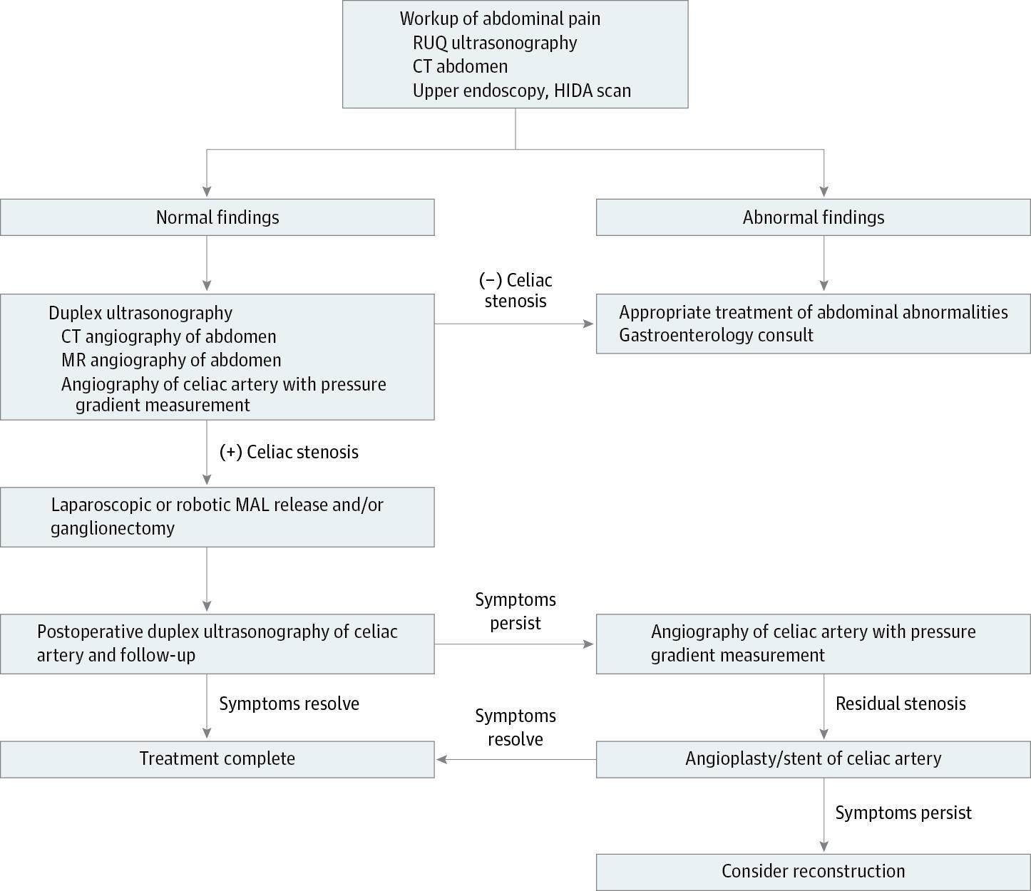
The following algorithm from JAMA Surgery outlines the recommended diagnostic and management approach:
History
- Key HPI questions: Characterize abdominal pain — location (epigastric), timing (postprandial vs. constant), relation to meals, exercise, and body position [1-2]
- Symptom characterization: Chronic epigastric pain (94%), postprandial pain (80%), nausea/vomiting (56%), unintentional weight loss (50%), bloating (39%), diarrhea (30%), exercise-induced pain (8%) [2][5]
- Timing/triggers: Pain worsens after eating (increased mesenteric blood demand) and with exercise; may be mitigated by leaning forward or drawing knees to chest [2]
- Progression: Typically chronic and progressive over months to years; median symptom duration before diagnosis is ~24 months [3]
- Important negatives: Ask about bloody stools, fever, jaundice, prior abdominal surgeries, and history of atherosclerotic disease to exclude other etiologies
Alarm Features
- Acute severe abdominal pain with hemodynamic instability (consider acute mesenteric ischemia or celiac artery thrombosis) [6]
- Rapid, severe weight loss (>25 lbs reported in 35.6% of one cohort) suggesting significant foregut ischemia or malignancy [7]
- Signs of peritonitis — suggests bowel infarction, not typical MALS
- GI bleeding — not characteristic of MALS; should prompt evaluation for alternative diagnoses
- Celiac trunk thrombosis has been reported as a rare complication, particularly in patients with prothrombotic conditions such as antiphospholipid syndrome [6]
Medications
- No specific pharmacologic treatment for MALS; management is primarily surgical [1-2]
- Symptomatic relief: Analgesics (NSAIDs, acetaminophen), antiemetics for nausea, proton pump inhibitors if concurrent GERD/gastritis suspected
- Celiac plexus block (percutaneous) is used both diagnostically and therapeutically — a positive response may predict surgical success [2][8]
- Avoid: Vasoconstrictors (ergotamines, triptans, cocaine) that could worsen mesenteric ischemia
- Caution: Opioid dependence is a risk given chronic pain; psychiatric comorbidities are common and may predict poorer surgical outcomes [9]
Diet
- Small, frequent, low-fat meals to reduce postprandial mesenteric blood flow demand and minimize pain [10]
- Adequate hydration and nutritional supplementation, especially in patients with significant weight loss
- Avoid large meals and high-fat foods that maximally stimulate splanchnic blood flow
- Nutritional support (enteral or parenteral) may be necessary in severely malnourished patients preoperatively
- Nonoperative management includes dietary modifications, though outcomes are worse compared with surgical treatment [1][10]
Review of Systems
- GI: Postprandial pain, nausea, vomiting, diarrhea, bloating, sitophobia (fear of eating), weight loss
- Vascular: Symptoms of other vascular compression syndromes (nutcracker syndrome, May-Thurner syndrome — especially in patients with connective tissue disorders) [7]
- Psychiatric: Depression, anxiety — psychiatric comorbidities are prevalent and may impact surgical outcomes [9]
- Autonomic: Dysautonomia symptoms (POTS, orthostatic intolerance) — preliminary data suggest worse surgical outcomes [9]
- Musculoskeletal/Connective tissue: Joint hypermobility, skin hyperextensibility — association with hypermobile Ehlers-Danlos syndrome (hEDS) [7]
Collateral History and Family History
- Collateral: Duration and extent of weight loss, dietary intake, prior GI workup results, prior imaging, and number of specialists seen (diagnosis is frequently delayed)
- Family history: Connective tissue disorders (Ehlers-Danlos syndrome), hypercoagulable states (antiphospholipid syndrome) [6-7]
- Social context: Impact on quality of life, functional status, employment; assess for eating disorders as a differential; screen for psychiatric comorbidities [9]
Risk Factors
- Female sex (4:1 ratio) [2]
- Age 20–50 years (median age ~38–40 in surgical series) [3][11]
- Thin body habitus / low BMI — less periaortic fat allows greater ligament compression; higher BMI inversely associated with symptom improvement after surgery [8][12]
- Connective tissue disorders — particularly hypermobile Ehlers-Danlos syndrome [7]
- Anatomic variant: Low insertion of the median arcuate ligament crossing the celiac axis origin [2][13]
- Mast cell activation syndrome (MCAS) — may overlap with and confound MALS symptoms; correlated with persistent symptoms after surgery [14]
Differential Diagnosis
- Peptic ulcer disease / gastritis — most common mimic; rule out with EGD
- Biliary disease (cholelithiasis, biliary dyskinesia) — RUQ ultrasound, HIDA scan [2]
- Chronic pancreatitis — lipase, CT abdomen
- Gastroparesis — gastric emptying study
- Functional dyspepsia / disorders of gut-brain interaction (DGBIs) — significant symptom overlap; must be excluded before MALS diagnosis [15]
- Chronic mesenteric ischemia (atherosclerotic) — typically older patients with cardiovascular risk factors; CTA distinguishes from MALS [13]
- Superior mesenteric artery syndrome — duodenal compression
- Malignancy (pancreatic, gastric) — CT, EGD
- Hyperkinetic gallbladder dyskinesia — GBEF >80%; overlapping presentation with MALS [16]
- Cannot-miss: Acute mesenteric ischemia, celiac trunk thrombosis, pancreatic malignancy [6]
Past Medical History
- Prior extensive GI workup (EGD, colonoscopy, HIDA scan, CT) — typically negative in MALS [2]
- Previous cholecystectomy or appendectomy without symptom resolution
- History of connective tissue disorders (hEDS), dysautonomia, MCAS [7][9][14]
- Psychiatric history (depression, anxiety) — impacts prognosis [9]
- Prior abdominal surgeries (adhesions may complicate surgical approach)
Physical Exam
- Vital signs: Usually normal; tachycardia or hypotension suggests acute complication
- Abdominal exam: Epigastric tenderness; epigastric bruit that is amplified with expiration (present in ~30% of younger patients, but not specific — found in 16% of asymptomatic individuals) [2]
- General: Thin habitus, signs of malnutrition/cachexia, temporal wasting
- Focused maneuvers: Auscultate epigastrium during deep expiration (bruit accentuation); assess for joint hypermobility if connective tissue disorder suspected
- Expected vs. concerning: Exam is often unremarkable; peritoneal signs or hemodynamic instability should prompt emergent evaluation
Lab Studies
- Recommended labs: CBC, CMP, lipase, lactate, hepatic function panel — primarily to exclude other diagnoses
- Expected abnormalities: Labs are typically normal in MALS [2]
- Rule-out labs: Lactate (if acute ischemia suspected), amylase/lipase (pancreatitis), celiac disease panel, H. pylori testing
- Nutritional markers: Albumin, prealbumin, vitamin levels in malnourished patients
- Hypercoagulability workup if thrombosis is identified [6]
Imaging
- First-line: Mesenteric duplex ultrasound with respiratory maneuvers (inspiration and expiration) — recommended initial diagnostic modality [9][15]
- Celiac artery peak systolic velocity (PSV) >200 cm/s during expiration; end-diastolic velocity ≥350 cm/s with deflection angle ≥50° has 83% sensitivity and 100% specificity [2]
- Velocities normalize with deep inspiration (pathognomonic dynamic finding)
- Confirmatory: CT angiography (CTA) with 3D reconstruction — demonstrates the characteristic "J-hook" or "hook sign" of the celiac artery, focal proximal narrowing, and poststenotic dilatation [9][12-13]
- Alternatives: MR angiography (useful in contrast allergy); lateral mesenteric angiography with breathing maneuvers is the criterion standard [2]
- When imaging is unnecessary: Incidental celiac compression on CT without symptoms does not warrant further workup — asymptomatic celiac compression is found in ~7–21% of the population [2]
Special Tests
- Celiac plexus block (percutaneous): Performed preoperatively in ~75–78% of surgical candidates; a positive response (pain relief) may predict favorable surgical outcome [3][8]
- Gastric exercise tonometry: Elevated gastric-arterial PCO₂ gradient after exercise suggests gastric ischemia; used in some centers for diagnosis and postoperative follow-up [2]
- Provocative mesenteric angiography: Performed in 12–36% of patients; demonstrates dynamic compression with respiratory maneuvers [8]
- Intraoperative duplex ultrasound: Used during surgery to confirm adequate celiac artery decompression [2]
- No validated clinical scoring system exists for MALS
ECG
- ECG is not directly relevant to MALS diagnosis
- Obtain ECG if acute abdominal pain presentation to rule out inferior MI (which can mimic epigastric pain)
- Consider ECG if dysautonomia or POTS is suspected as a comorbidity [9]
Assessment
MALS is a rare, controversial diagnosis of exclusion characterized by chronic postprandial epigastric pain, nausea, vomiting, and weight loss due to celiac artery compression by the median arcuate ligament. [1-2] The pathophysiology involves both vascular (foregut ischemia) and neuropathic (celiac ganglion compression) components. [2] Diagnosis is typically delayed by months to years. Asymptomatic celiac compression is common (7–21% of the population), making clinical correlation essential. [2]
- Severity stratification: Degree of celiac stenosis (grade I–IV), weight loss severity, nutritional status, and response to celiac plexus block
- Atypical presentations: Constant (non-postprandial) pain, exercise-induced pain only, concurrent vascular compression syndromes
- Complications: Celiac artery thrombosis, chronic malnutrition, functional GI disorders, opioid dependence [6]
Treatment Plan
Initial/Conservative Management
- Small, frequent, low-fat meals; nutritional optimization [10]
- Analgesics, antiemetics, PPI trial
- Celiac plexus block for diagnostic and therapeutic purposes [2][8]
- Multidisciplinary evaluation (gastroenterology, vascular surgery, pain management, psychology) [9]
Definitive Treatment — Surgical Decompression
- Median arcuate ligament release (MALR) ± celiac ganglionectomy ± revascularization [1-2]
- Approaches:
- Laparoscopic: Shorter hospital stay (1.7 ± 1.3 days), lower morbidity, 4.3–4.6% conversion rate; symptom improvement in 71.5–88.7% [3][8]
- Open: Greater pain improvement (84.2% vs. 71.5%) and nausea improvement (89.7% vs. 78.5%), but longer hospital stay (4.6 ± 1.7 days) and higher ileus rate (17.5%) [8]
- Robotic: Increasingly used; comparable outcomes to laparoscopic [5]
- Overall surgical success rate: 70–85% [9][11]
- Recurrence: Symptom recurrence in up to 45%, often within 3 months, frequently without radiographic correlate [3]
- Adjunctive procedures: Angioplasty/stenting for recalcitrant cases or fixed stenosis; vascular reconstruction (patch angioplasty, aortoceliac bypass) for chronic intimal damage [2][11]
Disposition
- Admission criteria: Acute mesenteric ischemia, celiac thrombosis, hemodynamic instability, severe malnutrition requiring nutritional support, postoperative monitoring
- Discharge criteria (postoperative): Tolerating oral intake, pain controlled, no surgical complications; median hospital stay is 2–3.5 days for laparoscopic, 4–5 days for open [3-4][8]
- Observation: Patients with equivocal diagnosis awaiting celiac plexus block results or further imaging
- Specialist consultation triggers: Vascular surgery (definitive management), gastroenterology (exclusion of other diagnoses), interventional radiology (celiac plexus block, angiography), pain management, psychology/psychiatry [9]
Follow Up / Return Precautions
- Postoperative follow-up: Duplex ultrasound at 1–3 months to assess celiac artery flow; repeat imaging if symptoms recur [2][9]
- Symptoms requiring immediate reassessment: Acute severe abdominal pain, hematemesis, melena, fever, signs of peritonitis
- Expected recovery: Most patients report initial symptom improvement within weeks; however, up to 45% may experience recurrence [3]
- Persistent symptoms postoperatively: Re-evaluate for residual celiac stenosis (may need revascularization), celiac plexus block if flow is normalized, or consider functional GI disorder requiring medical management [9]
- Patient counseling: MALS is a complex condition; surgical success is 70–85% but not guaranteed; psychiatric comorbidities and MCAS may predict poorer outcomes; long-term follow-up data beyond 5 years are limited [1][9][14]
References
1. Median Arcuate Ligament Syndrome. — Goodall R, Langridge B, Onida S, et al. Journal of Vascular Surgery. 2020.
2. Median Arcuate Ligament Syndrome—Review of This Rare Disease. — Kim EN, Lamb K, Relles D, et al. JAMA Surgery. 2016.
3. Minimally Invasive Surgery for Median Arcuate Ligament Syndrome: Clinical Outcomes and Recurrence Patterns. — Boaz E, Aeschbacher P, Okida F, et al. Surgery. 2026.
4. A Diagnostic Workup and Laparoscopic Approach for Median Arcuate Ligament Syndrome. — Diab J, Diab V, Berney CR. ANZ Journal of Surgery. 2022.
5. Short and Longterm Outcome of Minimally Invasive Therapy of Median Arcuate Ligament Syndrome. — Butz F, Haase O, Martin F, et al. Langenbeck's Archives of Surgery. 2024.
6. Celiac Trunk Thrombosis in a Patient With Antiphospholipid Syndrome Induced by Median Arcuate Ligament Compression: A Case Presentation and Literature Review. — Janiak P, Smoleńska Ż, Skotarczak M, Zdrojewski Z. Rheumatology International. 2024.
7. Abdominal Compression Syndromes in the Hypermobile Ehlers-Danlos Syndrome. — Milunsky A, Milunsky JM, Hsu R. American Journal of Medical Genetics. Part A. 2025.
8. Balancing Operative Risk and Symptom Relief: Outcomes of Open Versus Laparoscopic Release for Median Arcuate Ligament Syndrome: A Retrospective Study. — Alsabbagh Y, Erben Y, Lanka SP, et al. Journal of Vascular Surgery. 2026.
9. Median Arcuate Ligament Syndrome - Current State of Management. — Skelly CL, Mak GZ. Seminars in Pediatric Surgery. 2021.
10. ACR Appropriateness Criteria® Radiologic Management of Mesenteric Ischemia: 2022 Update. — Expert Panel on Interventional Radiology, Lam A, Kim YJ, et al. Journal of the American College of Radiology : JACR. 2022.
11. Open Surgical Decompression and Revascularization of the Celiac Axis for Median Arcuate Ligament Syndrome in the Modern Era of Minimally Invasive Surgery. — West CA, Vieira EJ, Crawford JL, et al. Annals of Vascular Surgery. 2025.
12. Utility of Hook Sign in the Diagnosis of Median Arcuate Ligament Syndrome. — Chan SM, Weininger G, Kozhimala M, et al. Annals of Vascular Surgery. 2023.
13. Median Arcuate Ligament Syndrome: Evaluation With CT Angiography. — Horton KM, Talamini MA, Fishman EK. Radiographics : A Review Publication of the Radiological Society of North America, Inc. 2005.
14. Prognostic Factors for the Long Term Outcome After Surgical Celiac Artery Decompression in MALS. — Woestemeier A, Semaan A, Block A, et al. Orphanet Journal of Rare Diseases. 2023.
15. Median Arcuate Ligament Syndrome: The Past and the Future. — Ratnasamy K, Sanghavi R. Gastroenterology Clinics of North America. 2025.
16. Median Arcuate Ligament Syndrome With Concomitant Hyperkinetic Gallbladder. — Hardy JT, Brandeis BO, Shiva A, Kavic SM, Nagarsheth K. The American Surgeon. 2025.
Figure
More from Deep Dive
Reviewed by Dr. Lucas Mastropaolo. SymptomDx is an educational tool for medical professionals — it does not replace clinical judgment. Verify all clinical data and drug dosages with authoritative sources.