Melioidosis
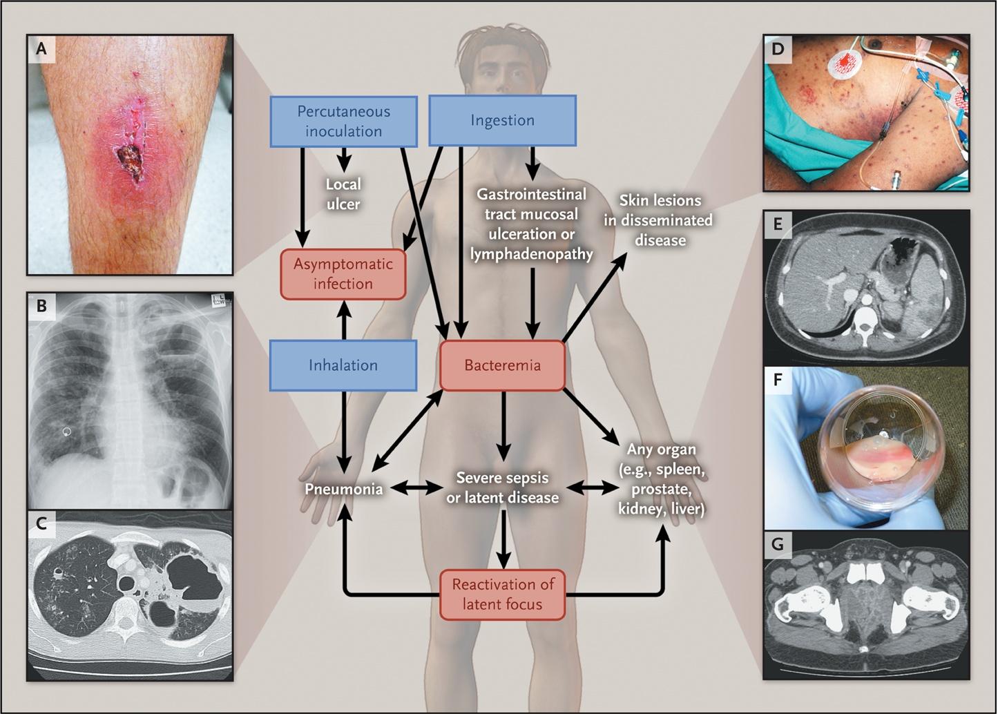
Melioidosis is a life-threatening infection caused by the Gram-negative environmental saprophyte Burkholderia pseudomallei, endemic to Southeast Asia, northern Australia, and increasingly recognized in the southern United States and other tropical regions. [1-2] Often called "the great mimicker," it has protean clinical manifestations ranging from localized skin ulcers to fulminant septic shock with multi-organ abscesses, and carries a mortality of 9–70% depending on resources and time to diagnosis. [1][3] The following figure from the NEJM illustrates the pathogenesis and clinical spectrum of disease:
History
- Exposure history: Contact with soil or surface water in endemic regions (Southeast Asia, northern Australia, southern US, South America, Africa); occupational exposure (farming, construction, military); recreational muddy/wet environments [1-2]
- Travel history: Any travel to or residence in tropical/subtropical regions, even decades prior — latency with reactivation up to 62 years has been documented [4]
- Onset and timing: Acute (<2 months, ~89% of cases) vs. chronic (≥2 months, ~11%); seasonal peak during monsoon/wet season (80% of cases) [5-6]
- Symptom characterization: Fever (present in ~76%), productive cough ± hemoptysis, dyspnea, pleuritic chest pain, localized swelling/abscess, dysuria, bone/joint pain, neurologic deficits [4][7]
- Important negatives: Ask about weight loss, night sweats (mimics TB); prior episodes of melioidosis; near-drowning events (aspiration route) [4][8]
Alarm Features
- Septic shock — occurs in ~21% of cases and is the strongest predictor of mortality [5][7]
- Rapidly progressive bilateral pneumonia with respiratory failure requiring mechanical ventilation (~16%) [5]
- Bacteremia without evident focus — suggests disseminated disease [4]
- Neurologic signs: Brainstem encephalitis with cranial nerve palsies (especially CN VII), myelitis, altered mental status [4][9]
- Multi-organ abscess formation (lungs, liver, spleen, prostate) [4][9]
- Hemoptysis with cavitary lung disease [10]
- Failure to respond to standard empiric antibiotics for community-acquired pneumonia/sepsis — a critical clue, as common empiric regimens do not cover B. pseudomallei [4]
Medications
Intensive Phase (IV, ≥10–14 days)
- First-line: IV ceftazidime (50 mg/kg up to 2 g q6–8h) [4][11]
- Severe/ICU: IV meropenem (25 mg/kg up to 1 g q8h) or imipenem [4-5]
- Adjunctive TMP-SMX may be added for deep-seated infections [8]
Eradication Phase (oral, ≥3 months, often 3–6 months)
- First-line: Oral TMP-SMX (TMP 6–8 mg/kg/day in two divided doses) [4][11-12]
- Alternative (children, pregnancy): Amoxicillin-clavulanate — associated with higher relapse rates [4][12]
Intrinsic Resistance (do NOT use)
- Penicillin, ampicillin, 1st/2nd-gen cephalosporins, aminoglycosides (gentamicin, tobramycin, streptomycin), polymyxins [4]
- Ertapenem, tigecycline, and moxifloxacin have limited activity [4]
Cautions
- ~25% of patients on TMP-SMX ± doxycycline develop adverse drug reactions; switching to amoxicillin-clavulanate increases relapse risk [12]
- Median time to fever defervescence is 9 days — slow response is expected and does not necessarily indicate treatment failure [9]
Diet
- No specific dietary triggers for melioidosis
- Hydration: Aggressive IV fluid resuscitation in septic patients per standard sepsis protocols
- Alcohol cessation: Hazardous alcohol use is a major independent risk factor (present in ~40% of cases) and should be addressed [5]
- Long-term: Optimize glycemic control in diabetic patients, as diabetes is the single most important comorbidity [1][5]
Review of Systems
- Respiratory: Cough (productive), hemoptysis, dyspnea, pleuritic chest pain
- GU: Dysuria, perineal pain, urinary retention (prostatic abscess in ~20% of males in Australia) [4]
- MSK: Joint pain/swelling (septic arthritis), bone pain (osteomyelitis) — ~4% of cases [4]
- Neuro: Facial weakness, diplopia, limb weakness, gait disturbance (brainstem encephalitis/myelitis) [4]
- Skin: Localized ulcers, subcutaneous nodules, pustules, cellulitis [13]
- GI: Abdominal pain (hepatic/splenic abscesses), diarrhea
- Constitutional: Fever, rigors, night sweats, weight loss, fatigue [7]
- Head/neck (pediatric): Parotid swelling — up to 40% of pediatric cases in Thailand/Cambodia [4][9]
Collateral History and Family History
- Collateral: Occupational exposure (farming, rice paddies, construction), recreational soil/water contact, military service in endemic areas, recent natural disaster or flooding exposure [2][14]
- Family history: No hereditary predisposition, but household members with similar environmental exposures may also be at risk
- Social context: Socioeconomic disadvantage, rural residence in endemic areas, homelessness, injection drug use [2]
- Immigration/travel: Detailed lifetime travel history is essential — reactivation can occur decades after leaving an endemic area [4][8]
Risk Factors
- Diabetes mellitus — present in ~45% of cases; the single most important risk factor [1][5][15]
- Hazardous alcohol use — ~40% of cases [5]
- Chronic kidney disease [8][16]
- Chronic lung disease (including COPD, cystic fibrosis) [8][16]
- Immunosuppression: Corticosteroid use, malignancy, thalassemia [1][15]
- Male sex (~63% of cases) [5]
- Age 40–60 years (median age ~50) [5][14]
- Occupational/environmental: Farming, soil/water contact, wet season exposure [2][14]
- 84% of patients have ≥1 underlying comorbidity; only 2% of fatalities had no identified risk factor [5]
Differential Diagnosis
- Tuberculosis — the most common mimic, especially chronic melioidosis with upper lobe cavitary disease [8][16]
- Community-acquired pneumonia (Klebsiella, Staphylococcus, Streptococcus) — acute pneumonic presentation [3]
- Lung abscess / empyema from other organisms
- Malignancy — chronic melioidosis with mass-like lesions can mimic lung cancer or lymphoma [16-17]
- Deep fungal infections: Histoplasmosis, coccidioidomycosis, penicilliosis
- Other tropical infections: Leptospirosis, typhoid fever, scrub typhus
- Visceral abscess from other causes: Amebic liver abscess, pyogenic liver abscess
- Mycotic aneurysm — rare but described with B. pseudomallei [18]
Key distinguishing feature: Failure to respond to standard empiric antibiotics (which lack B. pseudomallei coverage) in a patient with risk factors and endemic exposure should raise immediate suspicion. [4][17]
Past Medical History
- Diabetes mellitus — screen HbA1c in all suspected cases; poorly controlled diabetes worsens outcomes [7][15]
- Prior melioidosis — recurrence in ~1 in 16 patients, often within the first year; ~75% are relapse, ~25% reinfection [4]
- Chronic renal disease, chronic liver disease, chronic lung disease [8][16]
- Immunosuppressive therapy or malignancy
- Splenectomy or functional asplenia
- Surgical history: Prior abscess drainage, prostatectomy
- Substance use: Alcohol use disorder, IV drug use
Physical Exam
Vital Signs
Focused Exam
- Lungs: Crackles, bronchial breath sounds, decreased breath sounds (consolidation, effusion, abscess)
- Abdomen: Hepatomegaly, splenomegaly, RUQ/LUQ tenderness (hepatic/splenic abscesses) [4][19]
- Skin: Localized ulcers with surrounding erythema, subcutaneous abscesses, pustules, cellulitis, disseminated papular/pustular lesions [4][13]
- Head/neck: Parotid gland swelling (children in SE Asia), cervical lymphadenopathy [9][20]
- GU: Perineal tenderness, prostatic tenderness/boggy prostate on DRE (prostatic abscess) [4]
- MSK: Joint effusion, warmth, limited ROM (septic arthritis); bony tenderness (osteomyelitis) [4]
- Neuro: Cranial nerve palsies (especially CN VII), limb weakness, flaccid paraparesis (myelitis), cerebellar signs [4][9]
Lab Studies
- Blood cultures (×2 minimum) — the gold standard; positive in ~56% of cases [4-5]
- Sputum culture — alert the microbiology lab to suspect B. pseudomallei to avoid misidentification [4][8]
- Throat swab culture — useful adjunct in endemic settings [8]
- Urine culture — especially in GU presentations
- Wound/abscess aspirate culture
- CBC: Leukocytosis or leukopenia; thrombocytopenia in severe sepsis
- CMP: Hyponatremia (common), elevated creatinine (renal impairment), hyperglycemia/new diabetes diagnosis [21]
- LFTs: Elevated if hepatic abscesses present
- Lactate, ABG: Metabolic acidosis in septic shock [21]
- HbA1c: Screen for undiagnosed or poorly controlled diabetes in all cases
- CRP/Procalcitonin: Elevated; nonspecific but useful for monitoring
- PCR for B. pseudomallei: More rapid than culture but less sensitive, especially on blood [4]
- Serology (IHA): Limited diagnostic value — background seropositivity >50% in endemic areas; not adequate for confirming diagnosis [4]
Imaging
First-line
- Chest X-ray: Acute — diffuse nodular infiltrates, alveolar consolidation (often multilobar/bilateral), rapid cavitation; chronic — upper lobe infiltrates with cavities mimicking TB, often sparing the apices [8][22]
- Pleural effusion/empyema in ~21% of acute cases [22]
Gold Standard / Advanced
- CT chest: Better delineates lung abscesses, cavitation, mediastinal lymphadenopathy [19][23]
- CT abdomen/pelvis (or ultrasound): Recommended in all cases to search for internal-organ abscesses — liver, spleen, prostate, kidney [4][19]
- MRI: For musculoskeletal involvement (osteomyelitis, septic arthritis) or neurologic melioidosis (brainstem encephalitis) [19]
When imaging is unnecessary
Special Tests
- Ashdown selective agar: Specialized culture medium that enhances B. pseudomallei recovery from non-sterile sites [1]
- MALDI-TOF mass spectrometry: Increasingly used for rapid identification, though databases must include B. pseudomallei [1]
- Latex agglutination test: Rapid bedside test for colony identification
- Indirect hemagglutination assay (IHA): Titer ≥1:160 suggestive but not confirmatory in endemic areas [25]
- Fine-needle aspiration (FNA) of abscesses with inoculation into blood culture bottles — improves diagnostic yield, especially for head/neck lesions [20]
- Prostate-specific antigen (PSA): May be elevated with prostatic abscess; DRE and imaging guide diagnosis
ECG
- No melioidosis-specific ECG findings; direct cardiovascular involvement by B. pseudomallei is rare [18]
- Indications for ECG: All patients with sepsis/septic shock — standard sepsis workup
- Findings in sepsis context: Sinus tachycardia (most common), QT prolongation, new atrial fibrillation/flutter — all associated with worse outcomes in sepsis generally [26]
- Rare cardiovascular complications: Pericardial involvement (~6% of acute pulmonary cases), mycotic aneurysm [18][22]
Assessment
Clinical Summary
Melioidosis is a potentially fatal tropical infection with an extraordinarily diverse clinical spectrum. It should be suspected in any patient with sepsis, pneumonia, or deep-seated abscesses who has risk factors (diabetes, alcohol use, CKD) and relevant environmental/travel exposure to endemic regions. [1][4][15]
Severity Stratification
- Mild/localized: Skin abscess, localized soft tissue infection without bacteremia — mortality <1% [5]
- Moderate: Pneumonia without shock, single organ abscess — mortality ~10–15%
- Severe: Bacteremic pneumonia, septic shock, multi-organ abscesses, neurologic involvement — mortality 20–50% depending on resources [5][7]
Typical vs. Atypical
- Typical: Febrile adult with diabetes presenting with community-acquired pneumonia during wet season in endemic area
- Atypical: Chronic cavitary lung disease mimicking TB; parotid abscess in a child; brainstem encephalitis; reactivation decades after exposure in a non-endemic country [4][8]
Complications
- Septic shock, ARDS, multi-organ failure
- Relapse (~6–10% even with full treatment; 16% within 10 years in Thailand) [9][12]
- Mycotic aneurysm (rare) [18]
- Chronic osteomyelitis
Treatment Plan
Initial Stabilization (ED)
- Standard sepsis resuscitation: IV fluids, vasopressors if needed, supplemental O₂/intubation
- Obtain cultures (blood ×2, sputum, urine, wound) before antibiotics — alert microbiology lab to suspect B. pseudomallei [4][8]
Intensive Phase (minimum 10–14 days IV)
- Ceftazidime 50 mg/kg (max 2 g) IV q6–8h — first-line [4][11]
- Meropenem 25 mg/kg (max 1 g) IV q8h — preferred for ICU patients, severe disease, or treatment failure [4-5]
- Consider adjunctive TMP-SMX for deep-seated infections (bone, CNS, prostate) [8]
Eradication Phase (minimum 3 months, up to 6 months oral)
- TMP-SMX (TMP 6–8 mg/kg/day divided BID) [4][11-12]
- Alternative: Amoxicillin-clavulanate (children, pregnancy) — higher relapse risk [12]
Adjunctive
- Abscess drainage: Surgical or percutaneous drainage of all drainable collections (liver, spleen, prostate, soft tissue) [1][4]
- Septic joint washout [4]
- Glycemic optimization in diabetic patients
- Alcohol cessation counseling
Disposition
Admission Criteria (all suspected/confirmed cases should be admitted):
- Any bacteremia, pneumonia, or deep-seated organ involvement
- Sepsis or septic shock — ICU admission (~24% require ICU) [5]
- Need for IV antibiotics (minimum 10–14 days)
- Mechanical ventilation required in ~16% [5]
Observation
Specialist Consultation Triggers
- Infectious Disease — mandatory for all cases; directs antibiotic therapy and duration [5]
- Surgery/IR — for abscess drainage (hepatic, splenic, prostatic, soft tissue)
- ICU/Critical Care — septic shock, respiratory failure, multi-organ dysfunction
- Neurology/Neurosurgery — brainstem encephalitis, cerebral abscess, myelitis
Discharge Criteria
- Clinical improvement, afebrile, tolerating oral medications
- Transition to oral eradication therapy (TMP-SMX)
- Infectious disease follow-up arranged
Follow Up / Return Precautions
Follow-up Timing
- Weekly clinic visits initially after discharge, then monthly during eradication therapy [5]
- Follow-up continued ideally ≥6 months after completion of therapy [5]
- Repeat imaging to document resolution of abscesses
Adherence
- half of patients do not complete eradication therapy[5]
Return Precautions — seek immediate care for
- Recurrence of fever, chills, or rigors
- New or worsening cough, dyspnea, hemoptysis
- New swelling, abscess, or skin lesion
- Neurologic symptoms (facial weakness, limb weakness, confusion)
- Abdominal pain (concern for new visceral abscess)
- Medication side effects (rash, GI intolerance from TMP-SMX)
Expected Recovery
- Slow clinical response is typical — median fever defervescence is 9 days on appropriate IV therapy [9]
- Recurrent melioidosis occurs in ~1 in 16 patients, most within the first year [4]
- Lifelong awareness needed in patients from endemic areas, as reactivation from latency is possible [4][8]
References
1. Human Melioidosis. — Gassiep I, Armstrong M, Norton R. Clinical Microbiology Reviews. 2020.
2. Burkholderia Pseudomallei and Melioidosis. — Meumann EM, Limmathurotsakul D, Dunachie SJ, Wiersinga WJ, Currie BJ. Nature Reviews. Microbiology. 2024.
3. Melioidosis: A Neglected Cause of Community-Acquired Pneumonia. — Virk HS, Mukhopadhyay C, Wiersinga WJ. Seminars in Respiratory and Critical Care Medicine. 2020.
4. Melioidosis. — Wiersinga WJ, Currie BJ, Peacock SJ. The New England Journal of Medicine. 2012.
5. The Darwin Prospective Melioidosis Study: A 30-Year Prospective, Observational Investigation. — Currie BJ, Mayo M, Ward LM, et al. The Lancet. Infectious Diseases. 2021.
6. Global Burden of Melioidosis in 2015: A Systematic Review and Data Synthesis. — Birnie E, Virk HS, Savelkoel J, et al. The Lancet. Infectious Diseases. 2019.
7. Improving the Clinical Recognition, Prognosis, and Treatment of Melioidosis Through Epidemiology and Clinical Findings: The Sabah Perspective. — Hussin A, Nor Rahim MY, Dalusim F, et al. PLoS Neglected Tropical Diseases. 2023.
8. Melioidosis: An Important Cause of Pneumonia in Residents of and Travellers Returned From Endemic Regions. — Currie BJ. The European Respiratory Journal. 2003.
9. Melioidosis. — White NJ. Lancet. 2003.
10. Locally Acquired Melioidosis Linked to Environment — Mississippi, 2020–2023. — Petras JK, Elrod MG, Ty MC, et al. The New England Journal of Medicine. 2023.
11. Treatment of Melioidosis. — Meumann EM, Rajahram G, Woolley SD, Smith S, Gassiep I. Infectious Disease Clinics of North America. 2026.
12. Trimethoprim-Sulfamethoxazole Versus Trimethoprim-Sulfamethoxazole Plus Doxycycline as Oral Eradicative Treatment for Melioidosis (MERTH): A Multicentre, Double-Blind, Non-Inferiority, Randomised Controlled Trial. — Chetchotisakd P, Chierakul W, Chaowagul W, et al. Lancet. 2014.
13. Cutaneous Melioidosis: An Updated Review and Primer for the Dermatologist. — Schwartzman G, Reddy SA, Berg SH, Currie BJ, Saavedra AP. Journal of the American Academy of Dermatology. 2023.
14. Missing Links of Melioidosis in India: A Cross-Sectional Analysis of Case Reports, Agrometeorological and Socioeconomic Factors. — Jha S, Mittal M, Prasad LR, et al. Scientific Reports. 2025.
15. Melioidosis. — Wiersinga WJ, Virk HS, Torres AG, et al. Nature Reviews. Disease Primers. 2018.
16. Management of Accidental Laboratory Exposure to Burkholderia Pseudomallei and B. Mallei. — Peacock SJ, Schweizer HP, Dance DA, et al. Emerging Infectious Diseases. 2008.
17. Pitfalls and Optimal Approaches to Diagnose Melioidosis. — Kingsley PV, Arunkumar G, Tipre M, Leader M, Sathiakumar N. Asian Pacific Journal of Tropical Medicine. 2016.
18. ACCF/AHA/CDC Conference Report on Emerging Infectious Diseases and Biological Terrorism Threats. Task Force I: Direct Cardiovascular Implications of Emerging Infectious Diseases and Biological Terrorism Threats. — Baddour LM, Zheng ZJ, Labarthe DR, O'Connor S. Journal of the American College of Cardiology. 2007.
19. Spectrum of Imaging Findings in Melioidosis. — Muttarak M, Peh WC, Euathrongchit J, et al. The British Journal of Radiology. 2009.
20. Fine-Needle Aspiration to Improve Diagnosis of Melioidosis of the Head and Neck in Children: A Study From Sarawak, Malaysia. — Mohan A, Podin Y, Liew DW, et al. BMC Infectious Diseases. 2021.
21. Bacteraemic Melioidosis Pneumonia: Impact on Outcome, Clinical and Radiological Features. — Mukhopadhyay A, Lee KH, Tambyah PA. The Journal of Infection. 2004.
22. Pulmonary Melioidosis: Clinical-Radiologic Correlation in 183 Cases in Northeastern Thailand. — Dhiensiri T, Puapairoj S, Susaengrat W. Radiology. 1988.
23. Clinical-Radiological Features and Diagnostic Modalities for Mediastinal Melioidosis. — Nyanti LE, Lee SSY, Shanmugam V, et al. The International Journal of Tuberculosis and Lung Disease : The Official Journal of the International Union Against Tuberculosis and Lung Disease. 2023.
24. Radiological Manifestations of Melioidosis. — Lim KS, Chong VH. Clinical Radiology. 2010.
25. Clinical Presentation and Treatment of Melioidosis in the Head and Neck Region. — Mahawerawat K, Kasemsiri P. The Journal of Laryngology and Otology. 2018.
26. Association of Electrocardiogram Abnormalities With Clinical Outcomes in Emergency Department Sepsis Patients. — Kotruchin P, Chuehongthong M, Tangpaisarn T, et al. The Western Journal of Emergency Medicine. 2026.
Figure
More from Deep Dive
Reviewed by Dr. Lucas Mastropaolo. SymptomDx is an educational tool for medical professionals — it does not replace clinical judgment. Verify all clinical data and drug dosages with authoritative sources.