Mesenteric Ischemia
Mesenteric ischemia is a vascular emergency caused by inadequate blood flow to the intestines, carrying a mortality rate of 50–80% when acute. [1-2] It encompasses four subtypes: arterial embolism (40–50%), arterial thrombosis (15–35%), nonocclusive mesenteric ischemia (NOMI) (5–15%), and mesenteric venous thrombosis (MVT) (5–15%). [1][3] The key to survival is a high index of clinical suspicion and early diagnosis. [2]
History
- Cardinal symptom: Severe, acute-onset abdominal pain — classically "pain out of proportion to physical examination" [2-3]
- Timing by subtype:
- Embolic: Sudden, severe onset; often periumbilical [4]
- Thrombotic: Acute worsening of chronic postprandial pain and weight loss ("acute-on-chronic") [2]
- NOMI: Diffuse, episodic pain in the setting of critical illness or low-flow state [2]
- MVT: More insidious onset with cramping, nausea, vomiting, diarrhea [2-3]
- Ask about atrial fibrillation, recent MI, heart failure, prior embolic events, recent cardiac or abdominal surgery, hypercoagulable history [3-4]
- Chronic mesenteric ischemia (CMI): Postprandial pain, food fear (sitophobia), and unintentional weight loss [5]
- 95% present with abdominal pain; 44% nausea; 35% vomiting; 35% diarrhea; 16% blood per rectum [1-2]
- Important negatives: Absence of prior cardiovascular disease or risk factors does not exclude the diagnosis, especially in younger patients with MVT [2][6]
Alarm Features
- Pain out of proportion to exam — assume AMI until disproven (WSES Grade 1C recommendation) [2]
- Peritoneal signs → likely irreversible bowel necrosis [2][7]
- Triad of abdominal pain + fever + heme-positive stool [7]
- Hemodynamic instability, septic shock, altered mental status
- Rapidly rising lactate, worsening metabolic acidosis
- Bloody diarrhea or frank GI hemorrhage
- Pneumatosis intestinalis or portal venous gas on imaging → late, ominous findings [2]
Medications
- Contributors/Causes:
- Acute treatment:
- Post-intervention:
- Contraindicated: Vasoconstrictors should be minimized/avoided when possible in suspected mesenteric ischemia
Diet
- Acute: NPO / bowel rest during active ischemia [2][10]
- Enteral nutrition should be withheld during ongoing ischemia-reperfusion injury; start at low rate only after revascularization is convincingly achieved [11]
- Parenteral nutrition may be required in patients with short bowel syndrome post-resection [2]
- Chronic mesenteric ischemia: Small, frequent, low-fat meals to reduce postprandial demand; nutritional supplementation for malnutrition from food avoidance [5][12]
Review of Systems
- GI: Postprandial pain, nausea, vomiting, diarrhea, bloody stool, weight loss, food aversion
- Cardiovascular: Palpitations (atrial fibrillation), chest pain, claudication, prior stroke/TIA, prior embolic events
- Vascular: History of DVT/PE, peripheral arterial disease
- Constitutional: Fever, malaise, recent weight loss
- Hematologic: Easy bruising, prior thrombotic events, family history of clotting disorders
Collateral History and Family History
- Confirm cardiac history (atrial fibrillation, recent MI, valvular disease, CHF) from records or family [3-4]
- Prior embolic events (stroke, limb ischemia) — ~1/3 of embolic AMI patients have prior arterial embolus [2]
- Family history of thrombophilia (Factor V Leiden, prothrombin gene mutation, protein C/S deficiency, antithrombin deficiency) [2]
- Social history: Smoking (atherosclerotic risk), cocaine use, oral contraceptive use
- Recent hospitalizations, surgeries (especially abdominal/bariatric/splenectomy → MVT risk) [2]
Risk Factors
- Age >70 with exponential increase >75 [1]
- COVID-19 (hypercoagulability and hypoperfusion) [2]
- End-stage renal disease / hemodialysis [9]
Differential Diagnosis
- Acute pancreatitis — lipase elevation, different CT findings
- Perforated viscus — free air on imaging
- Small bowel obstruction — transition point on CT, different vascular findings
- Acute cholecystitis / cholangitis — RUQ pain, Murphy's sign, biliary dilation
- Ruptured AAA — pulsatile mass, hypotension, retroperitoneal hemorrhage
- Aortic dissection — tearing pain, differential blood pressures
- Inflammatory bowel disease — chronic history, mucosal pattern on CT
- Infectious enterocolitis — fever, exposure history, different CT pattern [13]
- Ischemic colitis (distinct from AMI) — typically left-sided, self-limited, elderly with hypotension
- Angioedema of bowel wall — medication history (ACE inhibitors), wall thickening without vascular occlusion [13]
Past Medical History
- Prior episodes of postprandial pain / "intestinal angina" → suggests chronic mesenteric ischemia at risk for acute-on-chronic event [2]
- Previous mesenteric revascularization (stent or bypass) — restenosis accounts for 6–8% of late deaths [2]
- Cardiovascular disease: CAD, CHF, atrial fibrillation, valvular disease
- Hypercoagulable states, prior DVT/PE
- Malignancy (especially abdominal)
- Cirrhosis / portal hypertension
- Prior abdominal surgery (adhesions, altered anatomy)
Physical Exam
- Early: Abdomen may be soft and nontender despite severe pain — the hallmark "pain out of proportion to exam" [2-3]
- Late: Peritoneal signs (guarding, rigidity, rebound) → bowel necrosis [2]
- Vital signs: Tachycardia, hypotension, fever (late)
- Epigastric bruit (suggests chronic mesenteric arterial stenosis) [3]
- Assess for atrial fibrillation (irregular pulse)
- Signs of peripheral vascular disease (diminished pulses, bruits)
- Rectal exam: Heme-positive stool in ~25% [4]
- Abdominal distension (late finding)
- Mental status changes, especially in elderly [3]
Lab Studies
- No laboratory test can definitively confirm or exclude AMI [2]
- Lactate — elevated in advanced ischemia; lactate >2 mmol/L is an independent predictor of transmural necrosis; however, a normal lactate does not rule out early disease [9-10]
- WBC — leukocytosis in >90% of patients [2]
- D-dimer — elevated; may assist but nonspecific [2]
- Metabolic panel — metabolic acidosis (elevated anion gap), hyperkalemia
- Amylase — elevated in ~50% [4]
- LDH, CK — may be elevated with bowel necrosis
- Stool guaiac — occult blood in ~25% [4]
- Coagulation studies — PT/INR, aPTT (baseline before anticoagulation)
- Type and screen — anticipate potential surgical intervention
- Hypercoagulable workup (for MVT): Protein C/S, antithrombin III, Factor V Leiden, prothrombin gene mutation, antiphospholipid antibodies [8]
Imaging
- First-line / Gold standard: CT angiography (CTA) — triphasic (non-contrast + arterial + venous phase) [1][8-9]
- Key CTA findings:
- Vascular: SMA/SMV thrombus or embolus, arterial occlusion, dissection
- Bowel: Decreased/absent wall enhancement, wall thickening, bowel dilation >25 mm (suggests necrosis) [15]
- Ominous: Pneumatosis intestinalis, portal venous gas, free perforation [2]
- MVT: "Target sign" in SMV on venous phase [2]
- NOMI: Bowel ischemia with patent mesenteric vessels [2]
- Plain radiograph: Limited utility; only positive with late findings (pneumoperitoneum, pneumatosis) — a negative X-ray does not exclude AMI [2]
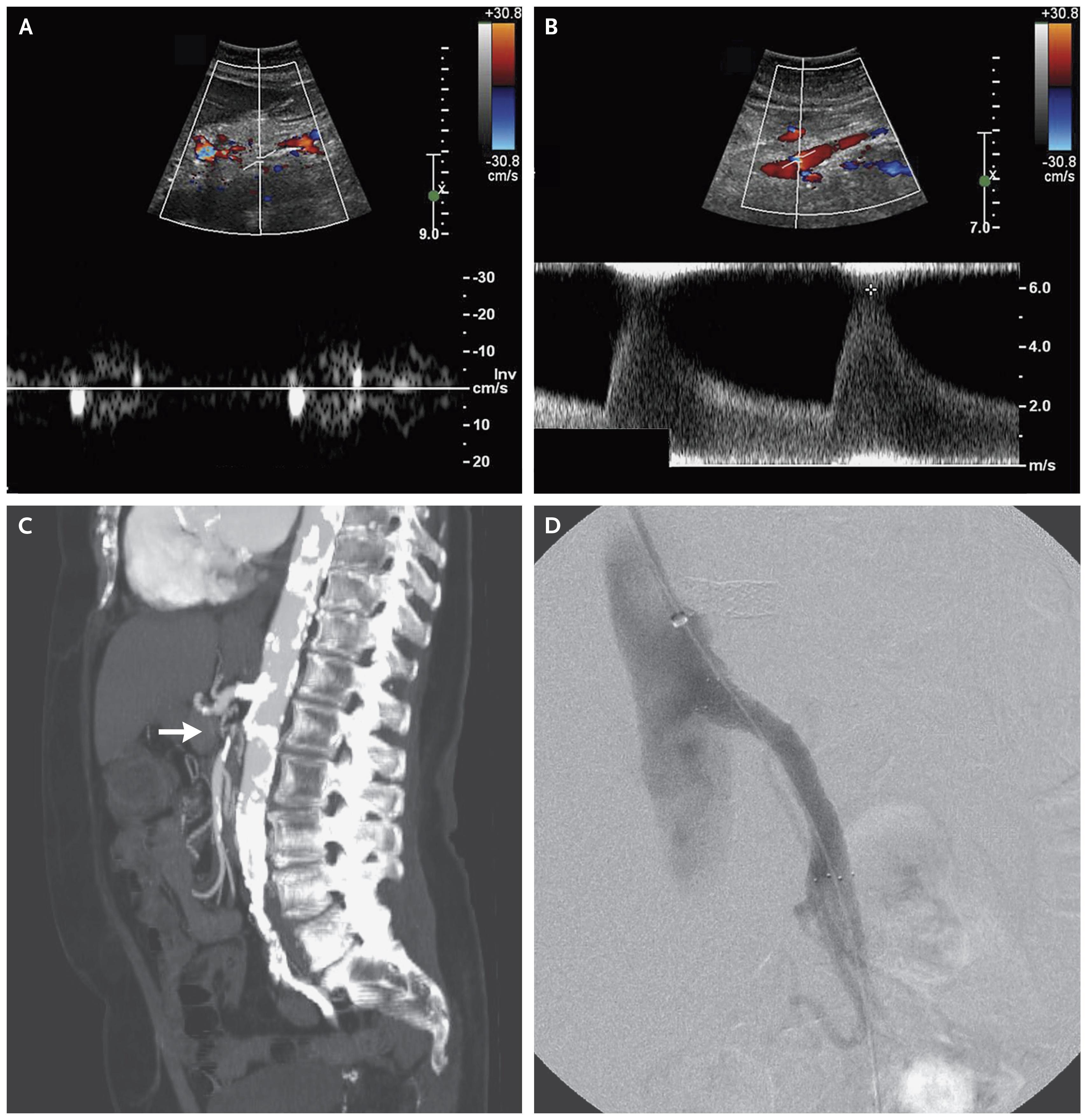
- Duplex ultrasound: Not appropriate for acute AMI (ACC/AHA Class III recommendation); useful for screening chronic disease [4-5]
- MRI: Overestimates stenosis, too slow for acute setting [1]
- Catheter angiography: Reserved for therapeutic intervention or when CTA is inconclusive [1]
Special Tests
- Point-of-care ultrasound (POCUS): May identify free fluid/ascites but cannot reliably diagnose AMI [6]
- Mesenteric duplex ultrasound: Preferred screening for chronic mesenteric ischemia; PSV >275 cm/s in SMA correlates with ≥70% stenosis (sensitivity 92%, specificity 96%) [8]
- Diagnostic laparoscopy: Limited role — early ischemia starts from mucosa and bowel may appear normal externally; can lead to false-negative results [16]
- Second-look laparotomy: Mandatory 24–48 hours after initial surgery in patients with extensive bowel involvement to reassess viability [2]
ECG
- Obtain ECG on all patients with suspected AMI to evaluate for:
- New atrial fibrillation + acute abdominal pain = high suspicion for embolic AMI
Assessment
- AMI is an abdominal vascular emergency with in-hospital mortality of ~49% and 90-day mortality of ~53% [17]
- Mortality by subtype: NOMI has the worst prognosis (72–75% mortality), followed by arterial occlusive AMI; venous AMI has the lowest mortality (~14–16%) [17-18]
- The "classic" presentation (pain out of proportion) is becoming less common; "acute-on-chronic" presentations are more typical and likely underdiagnosed [2]
- Median time from symptom onset to hospital admission is 24 hours; time from admission to diagnosis is 6 hours — both represent opportunities for improvement [17]
- Transmural necrosis predictors: Organ failure, lactate >2 mmol/L, bowel dilation >2.5 cm on CT [10]
- Complications: Bowel necrosis, perforation, peritonitis, sepsis, multiorgan failure, short bowel syndrome, death
Treatment Plan
Initial stabilization (ED)
- Aggressive IV fluid resuscitation
- IV unfractionated heparin — initiate immediately to prevent clot propagation [2][8]
- Broad-spectrum antibiotics (cover gram-negatives and anaerobes) for bacterial translocation [9]
- Nasogastric decompression, NPO [2]
- Correct metabolic acidosis, electrolyte derangements
- Adequate analgesia (do not withhold opioids)
Definitive management by subtype
- Arterial embolism: Open or endovascular embolectomy [8]
- Arterial thrombosis: Open bypass (antegrade or retrograde) or endovascular thrombectomy/stenting; single-vessel SMA revascularization usually sufficient acutely [2][19]
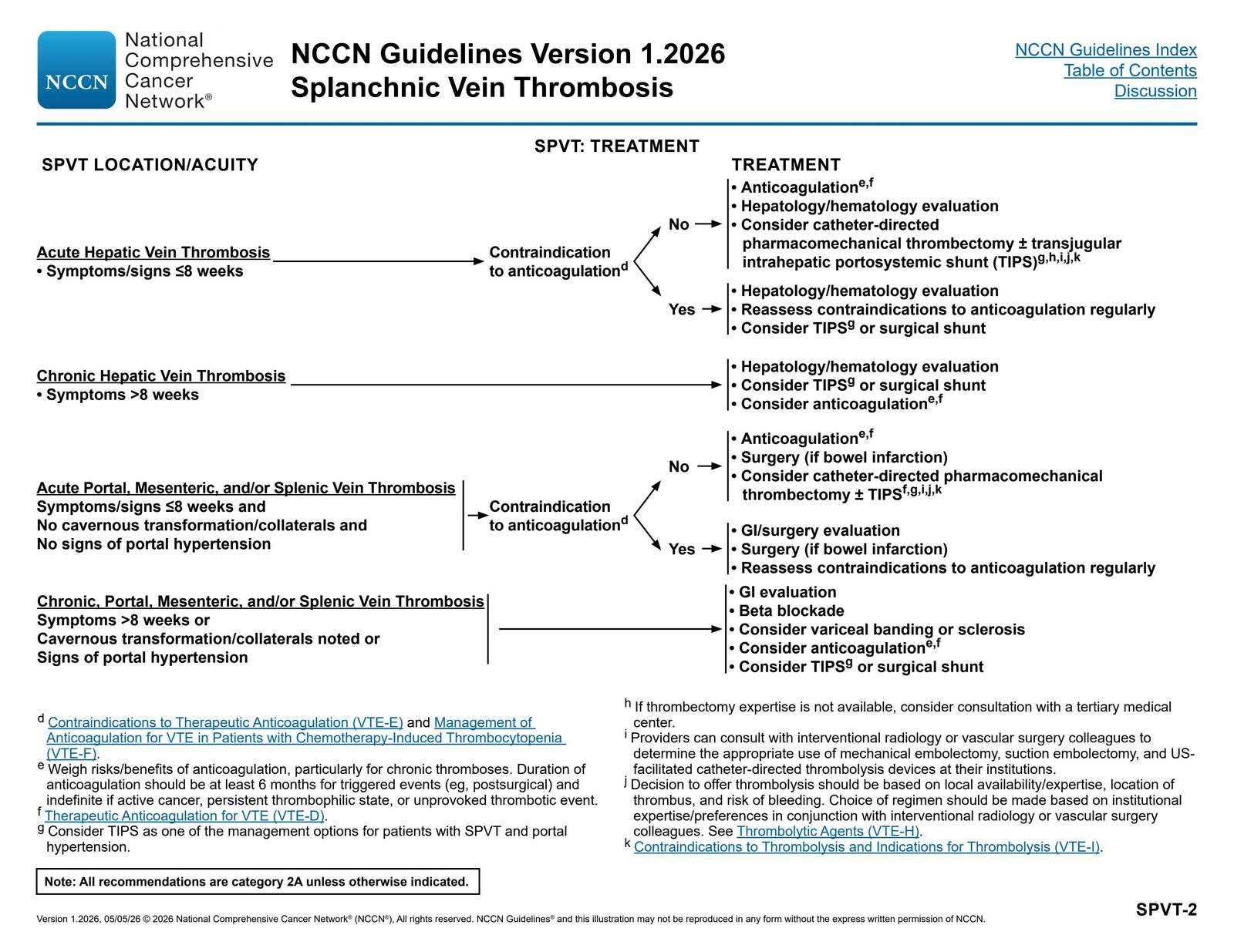
- MVT: First-line is anticoagulation with UFH; surgery reserved for peritonitis or clinical deterioration; resect only obviously necrotic bowel using damage control techniques [2]
- NOMI: Treat underlying cause (optimize cardiac output, reduce vasopressors); catheter-directed papaverine infusion; anticoagulation [3][8]
Surgical principles
- Resect non-viable bowel, preserve maximum viable bowel length
- Damage control laparotomy with planned second-look at 24–48 hours [2]
- Revascularization before resection when possible — revascularization is associated with significantly lower 30-day mortality (42% vs 62%) [2]
Post-intervention
- Lifelong anticoagulation/antiplatelet therapy for most patients [2]
- Surveillance imaging for stent/graft restenosis [2]
- Nutritional rehabilitation; parenteral nutrition if short bowel syndrome develops [2]
Disposition
- All confirmed AMI patients require admission — typically to the ICU [2]
- Emergent surgical or interventional radiology consultation for all confirmed cases [9]
- Peritonitis → emergent laparotomy [2]
- Hemodynamically stable, no peritoneal signs, arterial occlusion → endovascular revascularization if expertise available [16]
- MVT without peritonitis → may be managed with anticoagulation and close monitoring on a surgical service [2]
- Palliative care discussion should be considered in cases of extensive bowel necrosis in elderly/frail patients — shared decision-making is essential [2]
- Best outcomes are achieved at dedicated centers with 24-hour access to endovascular and surgical capabilities ("intestinal stroke centers") [2][20]
Follow Up / Return Precautions
- Post-discharge surveillance: Mesenteric duplex ultrasound or CTA at regular intervals to monitor for restenosis (6–8% of late deaths are from AMI after revascularization) [2]
- Long-term anticoagulation compliance is critical; most patients require lifelong therapy [2]
- Cardiovascular risk factor optimization: Statins, antihypertensives, smoking cessation, antiplatelet therapy [3]
- Return precautions: Recurrent abdominal pain (especially postprandial), nausea/vomiting, bloody stool, weight loss, food avoidance
- Nutritional follow-up for patients with bowel resection — monitor for short bowel syndrome, malabsorption, vitamin deficiencies
- Hypercoagulable workup completion and hematology follow-up for MVT patients [2][8]
- Expected recovery: Variable; depends on extent of bowel resection and comorbidities. Patients with chronic mesenteric ischemia treated with endovascular stenting have higher rates of symptom recurrence (29% vs 13% for open bypass) with faster onset [21]
Images
References
1. Gastrointestinal Surgical Emergencies Textbook. — Ashley E. Aaron, Andrea Amabile, Ciro Andolfi, et al American College of Surgeons (2021). 2021.
2. Acute Mesenteric Ischemia: Updated Guidelines of the World Society of Emergency Surgery. — Bala M, Catena F, Kashuk J, et al. World Journal of Emergency Surgery : WJES. 2022.
3. Mesenteric Ischemia. — Clair DG, Beach JM. The New England Journal of Medicine. 2016.
4. ACC/AHA 2005 Guidelines for the Management of Patients With Peripheral Arterial Disease (Lower Extremity, Renal, Mesenteric, and Abdominal Aortic): Executive Summary a Collaborative Report From the American Association for Vascular Surgery/Society for Vascular Surgery, Society for Cardiovascular Angiography and Interventions, Society for Vascular Medicine and Biology, Society of Interventional Radiology, and the ACC/AHA Task Force on Practice Guidelines (Writing Committee to Develop Guidelines for the Management of Patients With Peripheral Arterial Disease) Endorsed by the American Association of Cardiovascular and Pulmonary Rehabilitation; National Heart, Lung, and Blood Institute; Society for Vascular Nursing; TransAtlantic Inter-Society Consensus; And Vascular Disease Foundation. — Hirsch AT, Haskal ZJ, Hertzer NR, et al. Journal of the American College of Cardiology. 2006.
5. Chronic Mesenteric Ischemia: Clinical Practice Guidelines From the Society for Vascular Surgery. — Huber TS, Björck M, Chandra A, et al. Journal of Vascular Surgery. 2021.
6. Acute Mesenteric Ischemia in a Child: A Rare Case Report and Diagnostic Challenge in the Emergency Department. — Ashwin Kumar VA, Barik S, Hansda U, Prusty AV, Sahoo S. The American Journal of Emergency Medicine. 2026.
7. Acute Mesenteric Ischemia: Guidelines of the World Society of Emergency Surgery. — Bala M, Kashuk J, Moore EE, et al. World Journal of Emergency Surgery : WJES. 2017.
8. The Evaluation and Management of Intestinal Ischemia. — Theodore S, Xia T, Saillant N. NEJM Evidence. 2024.
9. High Risk and Low Prevalence Diseases: Mesenteric Ischemia. — Molyneux K, Beck-Esmay J, Koyfman A, Long B. The American Journal of Emergency Medicine. 2023.
10. Portal Vein Thrombosis: Diagnosis, Management, and Endpoints for Future Clinical Studies. — Elkrief L, Hernandez-Gea V, Senzolo M, et al. The Lancet. Gastroenterology & Hepatology. 2024.
11. A Clinical Approach to Acute Mesenteric Ischemia. — Reintam Blaser A, Acosta S, Arabi YM. Current Opinion in Critical Care. 2021.
12. Imaging and Endovascular Interventions in Mesenteric Ischaemia. — Agarwal D, Gupta S, Gadwal SK, et al. Abdominal Radiology. 2025.
13. Pearls, Pitfalls, and Conditions That Mimic Mesenteric Ischemia at CT. — Fitzpatrick LA, Rivers-Bowerman MD, Thipphavong S, et al. Radiographics : A Review Publication of the Radiological Society of North America, Inc. 2020.
14. Radiological Diagnosis of Acute Mesenteric Ischemia in Adult Patients: A Systematic Review and Meta-Analysis. — Reintam Blaser A, Koitmäe M, Laisaar KT, et al. Scientific Reports. 2025.
15. Insights Into Acute Mesenteric Ischaemia: An Up-to-Date, Evidence-Based Review From a Mesenteric Stroke Centre Unit. — Garzelli L, Ben Abdallah I, Nuzzo A, et al. The British Journal of Radiology. 2023.
16. Cesena Guidelines: WSES Consensus Statement on Laparoscopic-First Approach to General Surgery Emergencies and Abdominal Trauma. — Sermonesi G, Tian BWCA, Vallicelli C, et al. World Journal of Emergency Surgery : WJES. 2023.
17. Incidence, Diagnosis, Management and Outcome of Acute Mesenteric Ischaemia: A Prospective, Multicentre Observational Study (AMESI Study). — Reintam Blaser A, Mändul M, Björck M, et al. Critical Care. 2024.
18. Acute Mesenteric Ischemia: Pathophysiology-Based Approach to Imaging Findings and Diagnosis. — Lee MH, Pickhardt PJ, Sorensen AM, et al. Radiographics : A Review Publication of the Radiological Society of North America, Inc. 2025.
19. ACR Appropriateness Criteria® Radiologic Management of Mesenteric Ischemia: 2022 Update. — Expert Panel on Interventional Radiology, Lam A, Kim YJ, et al. Journal of the American College of Radiology : JACR. 2022.
20. Acute Mesenteric Ischaemia in the Elderly - Results of Combined Endovascular and Surgical Treatment. Primary Study. — Vrba R, Řezáč T, Špička P, et al. Scientific Reports. 2025.
21. Contemporary Management of Acute and Chronic Mesenteric Ischemia: 10-Year Experience From a Multihospital Healthcare System. — Andraska EA, Tran LM, Haga LM, et al. Journal of Vascular Surgery. 2022.
Figures
More from Deep Dive
Reviewed by Dr. Lucas Mastropaolo. SymptomDx is an educational tool for medical professionals — it does not replace clinical judgment. Verify all clinical data and drug dosages with authoritative sources.