Mucormycosis (Rhino-orbital-cerebral)
Rhino-orbital-cerebral mucormycosis is a rapidly progressive, angioinvasive fungal infection and a medical/surgical emergency with mortality rates of 50–80%, rising to >90% with disseminated or cerebral involvement. [1-2] Early recognition and immediate multidisciplinary intervention are the most critical determinants of survival. [3]
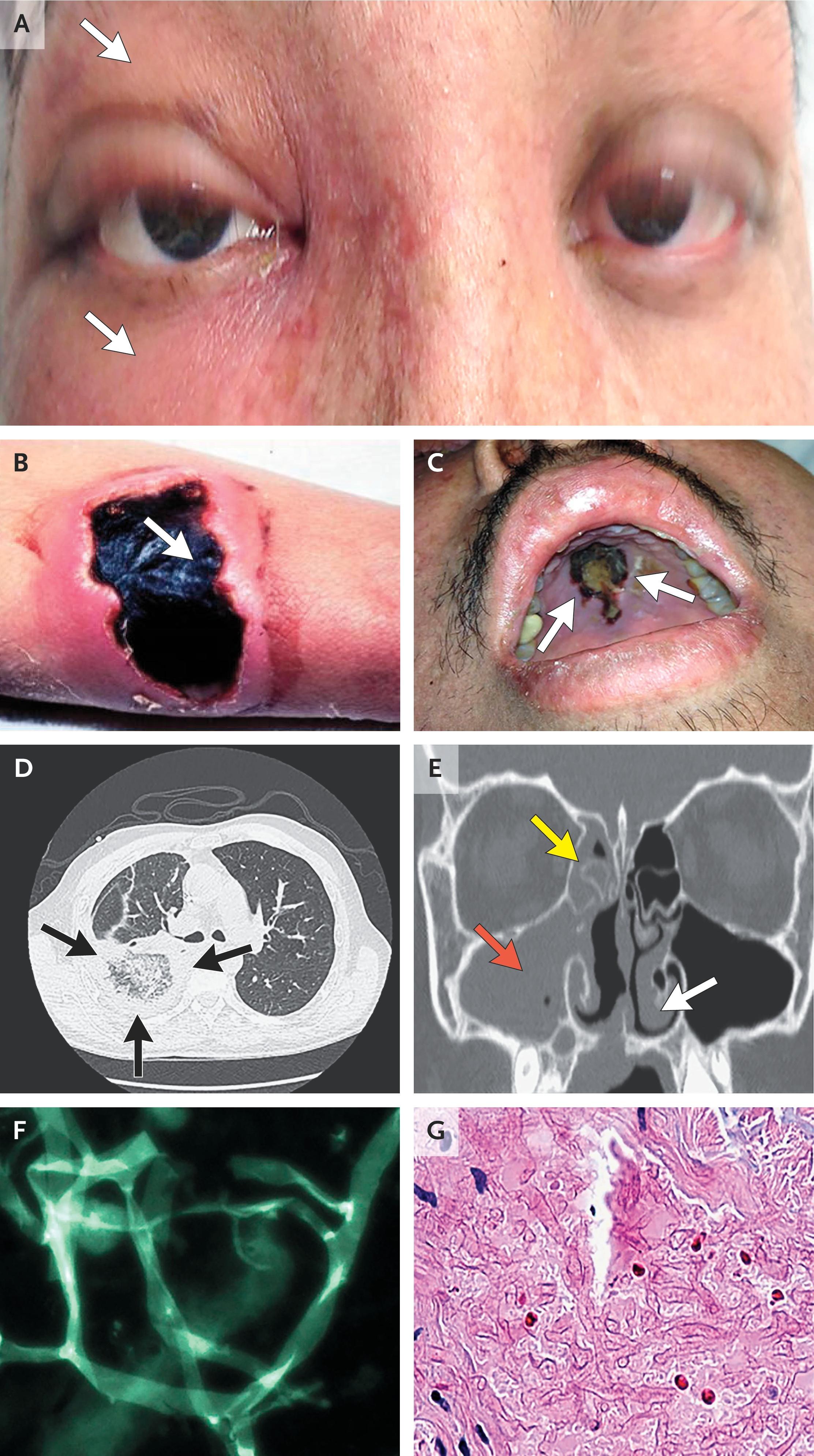
The following figure from a 2026 NEJM review illustrates the clinical and diagnostic spectrum of mucormycosis, including periorbital cellulitis, necrotic eschars, palatal necrosis, sinus CT findings, and the characteristic broad, ribbon-like, pauci-septate hyphae on histopathology:
History
- Key HPI questions: Onset and progression of facial pain, nasal congestion/discharge, headache, visual changes (diplopia, blurred vision, vision loss), facial numbness, tooth pain
- Symptom characterization: Unilateral facial pain/swelling → periorbital edema → visual changes; progression over hours to days [1][4]
- Timing: Rapid progression is the hallmark — symptoms can evolve from mild sinusitis to orbital/cerebral involvement within 24–72 hours [1]
- Triggers: Recent DKA episode, recent high-dose corticosteroid use, recent chemotherapy, recent COVID-19 illness [1]
- Associated symptoms: Fever, bloody/dark nasal discharge, anosmia, odontogenic pain, seizures, altered mental status [1][5]
- Important negatives: Absence of trauma (rules out cutaneous form), absence of cough/hemoptysis (rules out pulmonary form)
Alarm Features
- Black necrotic eschar on nasal mucosa, turbinates, or palate — pathognomonic for angioinvasive fungal infection [1][3]
- Periorbital cellulitis, proptosis, ophthalmoplegia — indicates orbital extension [1][6]
- Vision loss (especially no light perception) — associated with higher mortality [2]
- Cranial nerve palsies (CN III, IV, V1, V2, VI) — suggests cavernous sinus involvement [1]
- Impaired V2 nerve function — independently associated with higher mortality [2]
- Altered mental status, seizures, focal neurologic deficits — indicates cerebral extension, a surgical emergency [1][4]
- Rapid clinical deterioration despite antibiotics in an immunocompromised or diabetic patient [3]
Medications
- Relevant contributors:
- Prolonged high-dose glucocorticoids — major risk factor [1]
- Voriconazole prophylaxis — NOT active against Mucorales and may select for breakthrough mucormycosis [1]
- Ibrutinib, CAR-T therapy — recently identified risk factors [1]
- Deferoxamine (iron chelator) — increases iron availability to Mucorales; contraindicated [7]
- First-line treatment: Liposomal amphotericin B 5–10 mg/kg/day IV (10 mg/kg/day if CNS involvement) [3]
- Alternatives/step-down: Isavuconazole (FDA-approved for mucormycosis) or posaconazole delayed-release tablets — for renal failure, intolerance, or step-down maintenance [3][8-9]
- Contraindicated antifungals: Echinocandins, fluconazole, itraconazole, voriconazole — NO activity against Mucorales [1]
- Key caution: Monitor renal function closely on amphotericin B; hypokalemia is the most common adverse effect (50–70%) [5][10]
Diet
- Acute phase: NPO if surgical debridement planned; otherwise standard nutrition support
- DKA management: Aggressive IV fluid resuscitation, insulin infusion, electrolyte correction — metabolic correction is a pillar of treatment [1]
- Long-term: Strict glycemic control (HbA1c <8%) — elevated HbA1c is a significant mortality predictor [11]
- Iron supplementation: Avoid unnecessary iron supplementation, as iron overload states promote Mucorales growth [1][7]
Review of Systems
- HEENT: Nasal congestion, bloody/purulent discharge, facial pain/swelling, tooth pain, vision changes, eye pain, tearing
- Neurologic: Headache, facial numbness (V2 distribution), diplopia, seizures, altered mental status, focal weakness
- Pulmonary: Cough, dyspnea, hemoptysis, pleuritic chest pain (screen for sinopulmonary extension) [1]
- Constitutional: Fever, malaise, weight loss
- GI: Nausea, vomiting (may indicate DKA or GI mucormycosis)
- Skin: Any necrotic wounds or eschars at other sites (disseminated disease) [1]
Collateral History and Family History
- Collateral: Confirm diabetes control history, recent steroid use (dose and duration), recent hospitalization, COVID-19 history, chemotherapy regimen, transplant status and immunosuppression regimen
- Family history: Diabetes mellitus (type 1 or 2), hematologic malignancies
- Social context: IV drug use (risk for disseminated/endocarditis-associated mucormycosis), occupational exposure to soil/construction (environmental Mucorales exposure) [1]
Risk Factors
- Diabetes mellitus (especially with DKA) — the predominant risk factor for ROCM [1][12]
- Hematologic malignancies and prolonged neutropenia [1][3]
- Hematopoietic cell transplantation (median onset ~135 days post-HCT) [6]
- Solid organ transplantation [1]
- Prolonged high-dose glucocorticoid use [1]
- COVID-19 (particularly with concurrent DM and steroid use) [1][13]
- Iron overload states and deferoxamine therapy [1]
- Severe aplastic anemia [1]
- HbA1c >8%, age >60 years — independent mortality predictors [11]
Differential Diagnosis
- Invasive aspergillosis — most important mimic; septate, dichotomously branching hyphae (vs. broad, pauci-septate in Mucorales); galactomannan-positive [1][14]
- Bacterial orbital cellulitis — typically responds to antibiotics; lacks necrotic eschars
- Orbital apex syndrome from other causes (tumor, granulomatosis with polyangiitis, sarcoidosis)
- Cavernous sinus thrombosis — may coexist with ROCM; similar cranial nerve findings
- Invasive fusariosis or scedosporiosis — other angioinvasive molds; require culture/histopath for differentiation [1]
- Squamous cell carcinoma of the sinuses — can mimic destructive sinus disease
- Granulomatosis with polyangiitis (GPA) — midline destructive lesion, ANCA-positive
- Distinguishing pearl: Negative galactomannan and β-D-glucan in the setting of angioinvasive mold infection strongly favors mucormycosis over aspergillosis [6][15]
Past Medical History
- Critical to elicit: Diabetes (type, duration, control, DKA history), hematologic malignancy, transplant history, HIV/AIDS, chronic steroid use
- Previous episodes: Recurrence is possible, especially if underlying immunosuppression persists
- Surgical history: Prior sinus surgery, orbital surgery, dental procedures
- Chronic illnesses: Chronic kidney disease (affects amphotericin B dosing), liver disease, iron overload conditions
Physical Exam
- Vital signs: Fever (present in ~70% of cases), tachycardia; check for Kussmaul respirations (DKA) [5]
- Nasal exam: Black necrotic eschar on turbinates or septum, purulent/bloody discharge, septal ulceration [1][5]
- Oral exam: Palatal ischemia or necrosis (especially with maxillary sinus involvement), molar/premolar tenderness [1]
- Orbital exam: Periorbital edema/erythema, proptosis, chemosis, ophthalmoplegia, ptosis, decreased visual acuity, afferent pupillary defect [2][5]
- Cranial nerves: Test CN II–VI carefully — deficits suggest cavernous sinus invasion [1][6]
- Neurologic: Mental status, focal deficits, signs of meningismus
- Skin: Inspect for necrotic eschars elsewhere (disseminated disease)
- Key finding: Necrotic lesions in oral cavity/sinuses found in 87% of cases in one series [5]
Lab Studies
- Recommended labs:
- BMP (glucose, creatinine, potassium — baseline for amphotericin B monitoring)
- CBC with differential (assess for neutropenia)
- HbA1c
- ABG/VBG (assess for metabolic acidosis/DKA)
- Blood glucose, serum ketones
- LFTs, coagulation studies
- Serum iron studies (iron overload assessment)
- Expected abnormalities: Hyperglycemia, metabolic acidosis, elevated anion gap (DKA), hypokalemia (from amphotericin B) [5]
- Rule-out labs: Serum galactomannan and β-D-glucan are negative in mucormycosis (Mucorales lack these cell wall components) — a negative result in the setting of suspected invasive mold infection should raise suspicion for mucormycosis [6][15]
- Emerging diagnostics: Serum Mucorales qPCR (75–80% sensitivity; positive in 91% of hematogenous cases) — increasingly available commercially (MucorGenius, MycoGENIE) [6][16]
- Monitoring: Renal function and potassium at least every other day during amphotericin B therapy [10][15]
Imaging
- First-line: CT of paranasal sinuses (with contrast) — assess for mucosal thickening, bony erosion, peri-antral fat infiltration, orbital extension [3][17]
- Gold standard for orbital/cerebral involvement: MRI with gadolinium — substantially greater sensitivity than CT for detecting orbital soft tissue invasion, cavernous sinus involvement, meningeal enhancement, perineural spread, and cerebral extension [3][18]
- Important findings:
- Critical caveat: CT and MRI may be unremarkable in early disease — a normal scan does NOT rule out mucormycosis; repeat imaging and endoscopy are essential [1]
- Staging: Once ROCM is confirmed, obtain chest CT to evaluate for pulmonary involvement [1]
- Follow-up: Weekly CT/MRI recommended in unstable patients [3]
Special Tests
- Nasal endoscopy with biopsy — the most critical diagnostic procedure; brush biopsy of ulcerations and necrotic eschars [1][3]
- Histopathology: PAS and GMS stains showing broad (7–15 μm), ribbon-like, pauci-septate hyphae with non-dichotomous (right-angle) branching — pathognomonic [1]
- Direct microscopy: KOH wet mount or calcofluor/Blankophor fluorescence staining for rapid identification [1]
- Culture: Sabouraud's glucose agar at 25–37°C; grows rapidly but culture is positive in only ~50% of cases [1][16]
- MALDI-TOF MS: Rapid species identification from culture isolates [1]
- Molecular: Mucorales-specific PCR (serum, BAL, tissue) — emerging as a key early diagnostic tool [1][6][16]
- Metagenomic cell-free DNA sequencing: Promising but not yet validated for routine use [1]
ECG
- Indications: Baseline ECG before initiating amphotericin B (monitor for electrolyte-related arrhythmias, especially hypokalemia-induced QT prolongation)
- If isavuconazole is used: Note that isavuconazole can shorten the QTc interval — contraindicated in patients with familial short QT syndrome [3]
- Monitor for: Hypokalemia-related changes (U waves, ST depression, T wave flattening) during amphotericin B therapy
Assessment
- Clinical summary: ROCM is a medical and surgical emergency requiring immediate multidisciplinary intervention. The five pillars of management are: (1) early detection, (2) prompt antifungal therapy, (3) surgical debridement, (4) reversal of immunodeficiency, and (5) correction of metabolic abnormalities. [1]
- Severity stratification:
- Typical presentation: Diabetic patient in DKA with unilateral facial pain/swelling, bloody nasal discharge, periorbital edema progressing to proptosis and vision loss [4][12]
- Atypical presentations: May present as isolated headache, dental pain, or fever of unknown origin in neutropenic patients [1]
- Complications: Cavernous sinus thrombosis, internal carotid artery invasion with stroke, frontal lobe abscess, orbital exenteration, disseminated disease [1][18]
Treatment Plan
Initial stabilization
- ABCs, IV access, continuous monitoring
- Correct DKA aggressively: IV fluids, insulin drip, electrolyte repletion [1]
- Rapidly reduce glucocorticoid dose to the lowest effective dose [1]
Antifungal therapy (start empirically if high suspicion — do not wait for culture):
- Liposomal amphotericin B 5 mg/kg/day IV (standard dose) [3]
- Escalate to 10 mg/kg/day if CNS involvement (based on animal models and clinical data) [3][15]
- Alternative if renal failure/intolerance: Isavuconazole (loading: 200 mg IV/PO q8h × 6 doses, then 200 mg daily) or posaconazole delayed-release tablets (300 mg BID day 1, then 300 mg daily) [3][9]
- Duration: Continue until imaging resolution and immune reconstitution; no fixed duration — typically 4–6 weeks minimum of primary therapy, often longer [3][15]
Surgical intervention
- Urgent ENT/surgical consultation for debridement — perform as soon as feasible, in parallel with antifungal therapy [3][19]
- Endoscopic sinus debridement with removal of all necrotic tissue
- Second-look surgery at day 7 recommended to reassess margins [20]
- Orbital exenteration if globe is non-viable (NLP vision) with extensive orbital involvement — all exenteration patients survived in one 3-year cohort [2]
- Combined surgical + medical therapy reduces mortality from 88% to 32% compared to antifungals alone [19]
Adjunctive measures
- G-CSF or GM-CSF if neutropenic [1]
- Granulocyte transfusions in selected cases pending neutrophil recovery [1]
- Hyperbaric oxygen — may be beneficial in selected diabetic patients with sino-orbital/rhinocerebral disease [1]
- Deferasirox is NOT recommended (randomized pilot trial showed no benefit) [21]
Step-down/maintenance therapy
Disposition
- All suspected ROCM patients require immediate hospital admission, typically to an ICU or step-down unit [3]
- Admission criteria: Any patient with suspected invasive fungal sinusitis in the setting of immunosuppression or uncontrolled diabetes
- ICU indications: DKA, hemodynamic instability, altered mental status, cerebral involvement, post-operative monitoring after extensive debridement
- Specialist consultation triggers (all urgent):
- Infectious disease — antifungal management
- ENT/otolaryngology — endoscopic evaluation and surgical debridement
- Ophthalmology — orbital assessment, visual acuity monitoring
- Neurosurgery — if intracranial extension
- Endocrinology — DKA/glycemic management
- Discharge criteria: Clinical stability, resolving imaging, transition to oral step-down antifungal therapy, controlled underlying condition, reliable outpatient follow-up established
- ROCM is never an outpatient diagnosis — there is no role for observation or discharge from the ED [4]
Follow Up / Return Precautions
- Follow-up timing: Weekly imaging (CT or MRI) during active treatment; ENT endoscopic reassessment at 1–2 week intervals initially [3]
- Duration of antifungal therapy: Continue until complete resolution on imaging AND immune reconstitution — often months [3]
- Long-term monitoring: Renal function, electrolytes, hepatic function during prolonged antifungal therapy; ophthalmologic follow-up for visual outcomes
- Symptoms requiring immediate reassessment:
- New or worsening facial pain, swelling, or numbness
- Any visual changes (blurring, diplopia, vision loss)
- New headache, confusion, seizures, or focal neurologic deficits
- Fever recurrence
- Patient counseling:
- This is a life-threatening infection requiring prolonged treatment
- Strict glycemic control is essential to prevent recurrence
- Functional and cosmetic sequelae (vision loss, facial disfigurement) may be permanent
- Recovery is slow — imaging improvement in mucormycosis takes longer than in other fungal infections [15]
- Expected recovery: Partial or complete response typically achieved by 4–6 weeks of primary therapy, but total treatment duration is often months; recurrence is possible if immunosuppression persists [3][15]
References
1. Mucormycosis. — Kontoyiannis DP, Walsh TJ. The New England Journal of Medicine. 2026.
2. Survival and Prognostic Factors in Rhino-Orbito-Cerebral Mucormycosis: A 3-Year Cohort Study. — Zia Z, Sajadi MJ, Bazrafshan H, Khademi B, Janipour M. Scientific Reports. 2025.
3. Global Guideline for the Diagnosis and Management of Mucormycosis: An Initiative of the European Confederation of Medical Mycology in Cooperation With the Mycoses Study Group Education and Research Consortium. — Cornely OA, Alastruey-Izquierdo A, Arenz D, et al. The Lancet. Infectious Diseases. 2019.
4. Rhino-Orbital Cerebral Mucormycosis in a Diabetic Patient: An Emergency Medicine Case Report. — Sweet R, Hovenden M, Harvey CE, Peterson W, Lott I. The Journal of Emergency Medicine. 2023.
5. Retrospective Evaluation of Mucormycosis Cases at a Tertiary Care Center Between 2000 and 2020 in Türkiye. — Öztürk Belik H, Heper Y, Kazak E, et al. Mycopathologia. 2026.
6. American Society of Transplantation and Cellular Therapy Series: #8-Management and Prevention of Non-Aspergillus Molds in Hematopoietic Cell Transplantation Recipients. — Douglas AP, Lamoth F, John TM, et al. Transplantation and Cellular Therapy. 2025.
7. Mucormycosis in 2023: An Update on Pathogenesis and Management. — Alqarihi A, Kontoyiannis DP, Ibrahim AS. Frontiers in Cellular and Infection Microbiology. 2023.
8. FDA Orange Book. — FDA Orange Book. 2026.
9. Part 2: Mucormycosis: Focus on Therapy. — Lynch JP, Zhanel GG. Expert Review of Anti-Infective Therapy. 2023.
10. Prevention and Treatment of Cancer-Related Infections. — Updated 2026-03-11. National Comprehensive Cancer Network.
11. Analysis of COVID-19-associated Rhino-Orbital-Cerebral Mucormycosis Patients in a Tertiary Care Center in Northern India. — Yadav H, Sen S, Nath T, et al. Indian Journal of Ophthalmology. 2022.
12. Mucormycosis. — Czech MM, Cuellar-Rodriguez J. Infectious Disease Clinics of North America. 2025.
13. COVID-19 Associated Mucormycosis in Assiut University Hospitals: A Multidisciplinary Dilemma. — Farghly Youssif S, Abdelrady MM, Thabet AA, et al. Scientific Reports. 2022.
14. CT Findings for Differentiating Pulmonary Mucormycosis From Invasive Pulmonary Aspergillosis, Prior to Invasive Procedure Such as a Biopsy or Surgery: A 22-Year Single-Center Experience. — Jang HM, Kim MY, Lim SY, et al. Mycoses. 2025.
15. Definition, Diagnosis, and Management of COVID-19-associated Pulmonary Mucormycosis: Delphi Consensus Statement From the Fungal Infection Study Forum and Academy of Pulmonary Sciences, India. — Muthu V, Agarwal R, Patel A, et al. The Lancet. Infectious Diseases. 2022.
16. The Diagnosis of Mucormycosis by PCR in Patients at Risk: A Systematic Review and Meta-Analysis. — Brown L, Tschiderer L, Alanio A, et al. EClinicalMedicine. 2025.
17. Lung and Sinus Fungal Infection Imaging in Immunocompromised Patients. — Lamoth F, Prakash K, Beigelman-Aubry C, Baddley JW. Clinical Microbiology and Infection : The Official Publication of the European Society of Clinical Microbiology and Infectious Diseases. 2024.
18. Multicenter Analysis of General Presentations and Imaging Features in Cerebral MUCORmycosis (MAGICMUCOR): Towards Different Entities. — de La Porte C, Provost C, Coste A, et al. Medical Mycology. 2025.
19. Impact of Anatomical Extent and Combined Surgical-Medical Therapy on Survival in Sinonasal and Rhino-Orbito-Cerebral Mucormycosis: A 14-Year Retrospective ENT Cohort. — Kozan G, Dedeoğlu S, Ayral M, Akdağ M. Journal of Clinical Medicine. 2025.
20. Systematic Surgical Reassessment Combined With Liposomal Amphotericin B: The MICA Protocol. — Gervais M, Aubertin M, Verillaud B, et al. Acta Oto-Laryngologica. 2026.
21. Fourth European Conference on Infections in Leukaemia (ECIL-4): Guidelines for Diagnosis, Prevention, and Treatment of Invasive Fungal Diseases in Paediatric Patients With Cancer or Allogeneic Haemopoietic Stem-Cell Transplantation. — Groll AH, Castagnola E, Cesaro S, et al. The Lancet. Oncology. 2014.
22. Rhino-Orbito-Cerebral Mucormycosis in New-Onset Type 1 Diabetes Mellitus: A Case-Based Short Review. — Sharifi I, Razzeto A, Sacco SB, et al. The American Journal of the Medical Sciences. 2026.
Figure
More from Deep Dive
Reviewed by Dr. Lucas Mastropaolo. SymptomDx is an educational tool for medical professionals — it does not replace clinical judgment. Verify all clinical data and drug dosages with authoritative sources.