Nasal Septal Hematoma
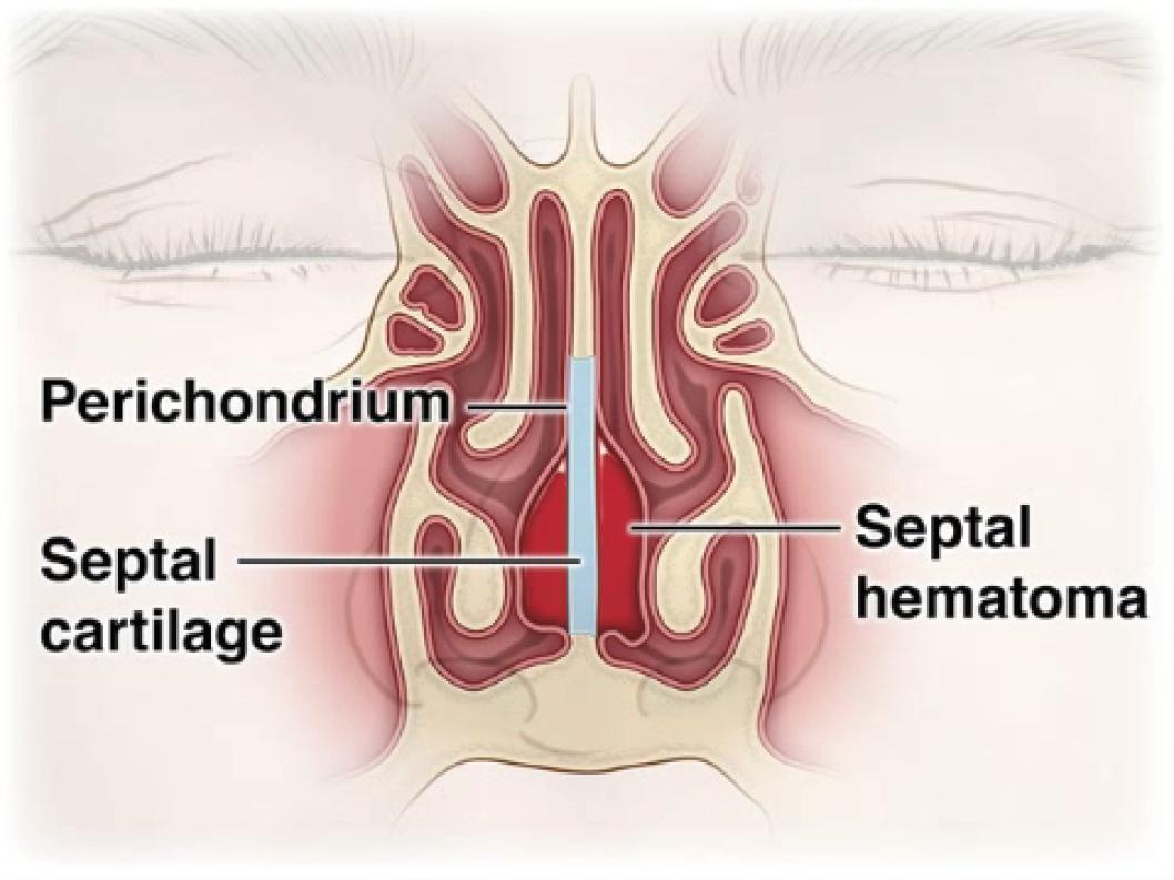
Nasal septal hematoma (NSH) is a subperichondrial collection of blood between the septal cartilage and its overlying mucoperichondrium. It is a rare but true ENT emergency — the avascular septal cartilage depends entirely on the mucoperichondrium for its blood supply, and separation by hematoma leads to cartilage ischemia and necrosis within days if untreated. [1-3] This condition is frequently missed: in one pediatric series, 82% of cases were initially missed in the ED, with a median time to diagnosis of 7 days and an average of 2.2 evaluations before diagnosis. [4]
The following figure illustrates the pathophysiology — blood accumulating in the subperichondrial space, stripping the cartilage from its sole blood supply.
History
- Mechanism of injury: Direct nasal trauma (assault, sports, fall, MVA) — present in ~85% of cases [4-5]
- Timing: Onset of progressive bilateral nasal obstruction and pain after trauma; may develop acutely or with delayed onset up to 48–72 hours post-injury [6]
- Key symptoms: Nasal congestion/obstruction (bilateral and progressive), nasal pain, pressure sensation, rhinorrhea, epistaxis [1][7-8]
- Symptom progression: Worsening obstruction despite decongestants; new-onset fever or purulent discharge suggests abscess formation [7]
- Important negatives: Absence of fever, purulent drainage, or facial cellulitis helps distinguish hematoma from abscess
Alarm Features
- Fever + purulent nasal discharge → suspect progression to septal abscess (mean symptom duration for abscess ~7.6 days vs ~4.1 days for hematoma) [7]
- Saddle nose deformity developing → cartilage necrosis has already occurred
- Headache, periorbital swelling, visual changes, altered mental status → intracranial complications: meningitis, cavernous sinus thrombosis, intracranial abscess, subdural empyema [2][9]
- Bilateral septal swelling in any post-trauma patient is a hematoma until proven otherwise
- Brain abscess has been reported as a fatal complication of infected septal hematoma [10]
Medications
- Prophylactic antibiotics: Generally recommended after drainage to prevent abscess formation; anti-staphylococcal coverage is standard (e.g., amoxicillin-clavulanate, first-generation cephalosporin) [2][11]
- If abscess suspected or confirmed: Parenteral broad-spectrum antibiotics covering Staphylococcus aureus (isolated in ~70% of cases), including MRSA coverage in at-risk patients [2][12]
- In children, also consider coverage for H. influenzae and S. pneumoniae [13]
- Medications to review: Anticoagulants, antiplatelets, NSAIDs (may contribute to hematoma formation, though one study found no significant correlation with coagulopathy) [1]
- Topical decongestants (oxymetazoline) may be used adjunctively for mucosal decongestion during examination
Diet
- No specific dietary triggers or recommendations
- Ensure adequate hydration, especially in pediatric patients
- Avoid hot liquids/foods that may promote vasodilation and re-bleeding in the immediate post-drainage period
Review of Systems
- ENT: Nasal obstruction (bilateral vs unilateral), epistaxis, rhinorrhea (clear vs purulent), anosmia, facial pain/pressure
- Neurologic: Headache, vision changes, altered mental status, facial numbness (may indicate intracranial extension) [14]
- Constitutional: Fever, chills, malaise (suggest infection/abscess)
- Dental: Recent dental procedures or infections (rare cause of septal abscess)
Collateral History and Family History
- Collateral: Witnesses to mechanism of injury; timing and force of trauma; prior nasal surgeries or procedures (septoplasty, rhinoplasty, cauterization for epistaxis) [14-15]
- Family history: Bleeding disorders (von Willebrand disease, hemophilia) — though coagulopathy was not significantly associated with NSH incidence in one large series [1]
- Social context: Sports participation (contact sports), interpersonal violence, child abuse (consider non-accidental trauma in pediatric cases with inconsistent history)
Risk Factors
- Nasal trauma (most common cause — ~85%) [4]
- Nasal/sinus surgery or procedures (septoplasty, rhinoplasty, cauterization) [14-15]
- Male sex (92% male in pediatric series) [4]
- Pediatric age group (children are more susceptible; mean age ~7 years in pediatric studies) [7]
- Anticoagulant/antiplatelet use (theoretical risk, though not statistically confirmed) [1]
- Diabetes mellitus (particularly for progression to abscess — present in 47% of adult abscess cases in one series) [16]
- Immunocompromised states [8]
- Nose-picking (44% of adult abscess cases in one series) [16]
Differential Diagnosis
- Nasal septal abscess — the most dangerous progression; distinguished by fever, purulent discharge, fluctuance, and longer symptom duration [2][7]
- Deviated nasal septum — chronic, non-tender, firm on palpation (not boggy/fluctuant)
- Nasal polyps — typically pale, glistening, non-tender, insensate
- Inferior turbinate hypertrophy — lateral nasal wall, not septal; a common misdiagnosis [17]
- Nasal foreign body (pediatric) — unilateral, foul-smelling discharge
- Nasal bone fracture without hematoma — external deformity, crepitus, but no septal swelling
- Sinonasal neoplasm — unilateral obstruction, epistaxis, consider in non-traumatic presentations
Past Medical History
- Prior nasal trauma or fractures
- Previous nasal/sinus surgery
- Bleeding disorders or coagulopathy
- Diabetes mellitus (risk factor for abscess formation) [12][16]
- Immunosuppression (HIV, chemotherapy, chronic steroids)
- History of recurrent epistaxis or cauterization procedures [14]
Physical Exam
- Anterior rhinoscopy (essential — use nasal speculum or otoscope with largest ear speculum):
- Pathognomonic finding: Bilateral, boggy, bluish-red, fluctuant swelling of the nasal septum that narrows or obliterates both nasal passages [1][18]
- Palpation with cotton-tipped applicator: soft, compressible, fluctuant mass (distinguishes from deviated septum, which is firm)
- Painful septal swelling combined with nasal obstruction was present in 100% of NSH patients [1]
- External nose: Swelling, ecchymosis, deformity, tenderness, crepitus (assess for concurrent nasal fracture)
- Assess for abscess: Erythema, warmth, purulent drainage, fever
- Vital signs: Fever suggests infection/abscess progression
- Neurologic exam: If concern for intracranial complications
Lab Studies
- Routine labs are generally not required for uncomplicated septal hematoma
- If abscess suspected or drained:
- Coagulation studies (PT/INR, PTT) if bleeding disorder suspected or patient on anticoagulants
Imaging
- Imaging is generally unnecessary for straightforward clinical diagnosis — NSH is a clinical diagnosis made by anterior rhinoscopy [1]
- CT face/sinuses (with contrast): Indicated if:
- Concern for concurrent facial fractures
- Suspected intracranial or orbital complications
- Posterior septal abscess suspected (not visible on anterior rhinoscopy) [9]
- MRI head with contrast: Superior for detecting intracranial complications (meningitis, cavernous sinus thrombosis, brain abscess) — 97% diagnostic accuracy vs 87% for CT [19]
- Key imaging findings: Subperichondrial fluid collection along the septum; rim enhancement suggests abscess
Special Tests
- Cotton-tipped applicator palpation test: Gently press on the septal swelling — fluctuance and compressibility confirm hematoma vs firm deviated septum (the single most important bedside maneuver)
- Needle aspiration: Can be both diagnostic and partially therapeutic — aspiration of non-clotted blood confirms hematoma; purulent material confirms abscess
- No validated scoring systems exist for NSH
ECG
- Not routinely indicated
- Consider if trauma mechanism raises concern for associated injuries (e.g., blunt chest trauma, syncope preceding fall)
Assessment
NSH is a rare but frequently missed ENT emergency. The critical clinical pearl is that every patient with nasal trauma must have an anterior rhinoscopy to evaluate the septum. [13][20] The condition was missed in 46% of cases across multiple clinical settings in one pediatric study. [4] Untreated hematoma progresses to abscess (typically within days), cartilage necrosis, and potentially life-threatening intracranial infection. Long-term sequelae are common: 61.7% of patients experienced minor or major sequelae in one long-term follow-up study (mean 8.7 years), including saddle nose deformity (the most common major complication). [7-8] In children, cartilage destruction also impairs midface growth. [2][21]
Treatment Plan
Initial stabilization
- Manage concurrent injuries (epistaxis control, facial fracture assessment)
- Analgesia as needed
Definitive treatment — Incision and Drainage (I&D)
- Urgent drainage is required — ideally within 24–48 hours of diagnosis [5][11]
- Technique: Hemitransfixion incision through the mucoperichondrium; evacuate clot; irrigate
- Prevent re-accumulation: Quilting sutures through the septum, placement of a small drain (e.g., Penrose), and/or bilateral anterior nasal packing [11]
- Re-collection rate after I&D is low (~3.6%) [11]
- In adults, can often be performed under local anesthesia in the ED; most pediatric cases require general anesthesia [11]
Antibiotics
- Prophylactic oral antibiotics post-drainage (anti-staphylococcal coverage: amoxicillin-clavulanate or first-generation cephalosporin) [11]
- If abscess: IV broad-spectrum antibiotics with MRSA coverage in at-risk patients [12]
If cartilage destruction has occurred (abscess)
Disposition
- Small, uncomplicated hematoma drained in the ED → may be discharged with close ENT follow-up within 24–48 hours for re-examination and packing removal
- Admit if:
- Septal abscess confirmed or suspected
- Need for IV antibiotics
- Pediatric patient requiring OR drainage under general anesthesia
- Signs of systemic infection or intracranial complications
- Significant comorbidities (uncontrolled diabetes, immunosuppression)
- ENT consultation: Should be obtained for all confirmed cases; urgent/emergent for abscess or complicated presentations [4]
Follow Up / Return Precautions
- Follow-up: ENT re-evaluation within 24–48 hours post-drainage for packing removal and assessment for re-collection [6]
- Consider re-evaluation at 48–72 hours post-trauma even if initial exam was negative, as delayed hematoma formation has been reported [6]
- Return precautions — instruct patients/parents to return immediately for:
- Worsening nasal obstruction
- New-onset fever
- Purulent nasal discharge
- Increasing facial pain or swelling
- Headache, vision changes, or confusion
- Long-term follow-up: Monitor for saddle nose deformity, septal perforation, and (in children) midface growth abnormalities [7][21]
- Expected course: With prompt drainage, most patients recover without sequelae; 50% of hematoma patients had no long-term sequelae vs only 7.7% of abscess patients [7]
References
1. Nasal Septal Hematoma Is a Rare and Self-Recognizable Complication of Nasal Bone Fracture: A Retrospective Study. — Guchlerner L, Ernst BP, Issing C, et al. American Journal of Otolaryngology. 2025.
2. Nasal Septal Abscess in Children: From Diagnosis to Management and Prevention. — Alshaikh N, Lo S. International Journal of Pediatric Otorhinolaryngology. 2011.
3. Treatment of Hematoma of the Nasal Septum. — Kass JI, Ferguson BJ. The New England Journal of Medicine. 2015.
4. Nasal Septal Hematoma in Children: Time to Diagnosis and Resulting Complications. — Ali HM, Zavala H, Chinnadurai S, Roby B. International Journal of Pediatric Otorhinolaryngology. 2021.
5. Nasal Injuries in Sports. — Marston AP, O'Brien EK, Hamilton GS. Clinics in Sports Medicine. 2017.
6. Late-Onset Posttraumatic Septal Hematoma and Abscess Formation in a Six-Year-Old Tamil Girl--Case Report and Literature Review. — Dubach P, Aebi C, Caversaccio M. Rhinology. 2008.
7. Nasal Septal Hematoma and Abscess in Children: An Uncommon Otorhinolaryngology Emergency Revisited. — Sayin I, Yazici ZM, Abakay MA, Saygan GB, Gunes S. The Journal of Craniofacial Surgery. 2021.
8. Nasal Septal Abscesses: A Systematic Review. — Shaari AL, Patil D, Youssef V, et al. The Journal of Craniofacial Surgery. 2025.
9. Spontaneous Abscess of the Posterior Nasal Septum: An Unusual Cause of Nasal Obstruction in Children. — Berlucchi M, Tomasoni M, Bosio R, Rampinelli V. The Annals of Otology, Rhinology, and Laryngology. 2021.
10. Nasal Septal Haematoma in Nigeria. — Chukuezi AB. The Journal of Laryngology and Otology. 1992.
11. Evaluation of the Management of Nasal Septal Haematoma and Abscess: A Systematic Review. — Jackson R, Jia W, Edafe O. The Journal of Laryngology and Otology. 2025.
12. Nasal Septal Abscess: A 10-Year Retrospective Study. — Cheng LH, Wu PC, Shih CP, et al. European Archives of Oto-Rhino-Laryngology : Official Journal of the European Federation of Oto-Rhino-Laryngological Societies : Affiliated With the German Society for Oto-Rhino-Laryngology - Head and Neck Surgery. 2019.
13. Abscess of the Nasal Septum After Trauma. — Close DM, Guinness MD. The Medical Journal of Australia. 1985.
14. Lipid Keratopathy and Septal Abscess: Case Report. — Heo SJ, Kim JS, Kwon SH, Kim JS. Medicine. 2019.
15. Unusual Causes of Nasal Septal Abscess Including a COVID-19 Swab Test After Nasal Surgery. — Im YH, Kim DH, Lee IH. The Journal of Craniofacial Surgery. 2023.
16. Nasal Septal Abscess in Adult Patients - A Single Center Study. — Ngo NH, Luong NVC, Le MTQ, Nguyen HMH, Tran LV. American Journal of Otolaryngology. 2023.
17. Nasal Septal Abscess--Retrospective Analysis of 14 Cases From University Hospital, Kuala Lumpur. — Jalaludin MA. Singapore Medical Journal. 1993.
18. Clinical Practice Guideline: Nosebleed (Epistaxis). — Tunkel DE, Anne S, Payne SC, et al. Otolaryngology--Head and Neck Surgery : Official Journal of American Academy of Otolaryngology-Head and Neck Surgery. 2020.
19. ACR Appropriateness Criteria® Sinonasal Disease: 2021 Update. — Expert Panel on Neurological Imaging, Hagiwara M, Policeni B, et al. Journal of the American College of Radiology : JACR. 2022.
20. Nasal Septal Trauma in Children. — Olsen KD, Carpenter RJ, Kern EB. Pediatrics. 1979.
21. Management of Nasal Septal Abscess in Childhood: Our Experience. — Dispenza C, Saraniti C, Dispenza F, Caramanna C, Salzano FA. International Journal of Pediatric Otorhinolaryngology. 2004.
Figure
More from Deep Dive
Reviewed by Dr. Lucas Mastropaolo. SymptomDx is an educational tool for medical professionals — it does not replace clinical judgment. Verify all clinical data and drug dosages with authoritative sources.