Nipah Virus Infection
Nipah virus (NiV) is a WHO Blueprint priority zoonotic paramyxovirus (genus Henipavirus) causing severe encephalitis and/or respiratory disease with a case fatality rate of 40–75%. [1-2] First identified in Malaysia in 1998, outbreaks continue in Bangladesh and India, with no approved vaccines or therapeutics. [3-4] The virus is classified as a BSL-4 pathogen. [5]
History
- Exposure history is paramount: Travel to or residence in endemic regions (Malaysia, Bangladesh, India — particularly Kerala, West Bengal) [6-7]
- Contact with Pteropus fruit bats, pigs, horses, or their excreta [8-9]
- Consumption of raw date palm sap (major transmission route in Bangladesh) [10-11]
- Close contact with a confirmed or suspected NiV patient — touching, feeding, nursing, or exposure to body fluids [12-13]
- Incubation period: Median 9.5–10 days (range 4–21 days) [5][12]
- Prodrome: 3–4 days of fever, headache, myalgia, dizziness, nausea/vomiting [14-15]
- Progression to altered sensorium, respiratory distress, seizures
- Ask about occupation (pig farming, abattoir work), recent funeral attendance (corpse-to-person transmission documented), and known outbreak clusters [3][11]
Alarm Features
- Rapidly declining consciousness or GCS deterioration
- Segmental myoclonus, areflexia, hypotonia — brainstem involvement signs [14]
- Abnormal doll's-eye reflex and tachycardia — independently associated with mortality [14]
- Intractable hypotension/bradycardia suggesting myocarditis [16]
- Acute respiratory distress (bilateral infiltrates, ARDS) [12][16]
- Seizures (39.2% of cases) [1]
- Viremia detectable on diagnosis — associated with 100% fatality in one series [17]
- Clustering of encephalitis cases in a community or healthcare setting should trigger immediate public health alert [7]
Medications
- No approved antiviral therapy exists [3-4]
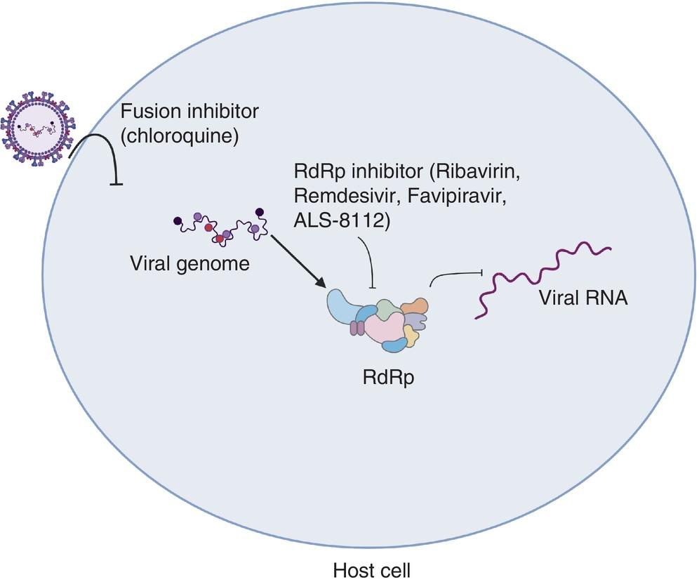
- Ribavirin: Only therapeutic with human outbreak data; associated with 36% mortality reduction in the Malaysian outbreak (open-label, historical controls) — used on compassionate basis. No statistically significant benefit confirmed in the 2018 Kerala outbreak [3][16][18]
- Remdesivir: Protective in African green monkey challenge model when initiated within 24 hours of exposure (IV, 12-day course); no human efficacy data [19-20]
- Favipiravir: Fully protective in hamster model; no human data [21]
- m102.4 monoclonal antibody: Phase 1 safety data in humans; targets henipavirus G glycoprotein; compassionate use protocol developed for future outbreaks; dosing: single 20 mg/kg IV dose (or two doses 48 hours apart for established disease) [18][20]
- Supportive care remains the standard: mechanical ventilation, vasopressors, seizure management, ICP management [3][22]
- Empiric acyclovir should be started pending HSV encephalitis exclusion [23]
The following figure illustrates the antiviral drug targets in the NiV life cycle, including fusion inhibitors and RdRp inhibitors:
Diet
- Avoid raw date palm sap — primary zoonotic transmission route in Bangladesh; bats contaminate sap collection pots [10-11][25]
- Avoid consumption of fruits partially eaten by bats or fallen fruit in endemic areas
- No specific dietary management once infected; standard ICU nutrition protocols apply
- Hydration management per encephalitis/ARDS protocols
Review of Systems
- Neurologic: Headache, dizziness, altered sensorium, confusion, hallucinations, focal deficits, seizures, drowsiness [14][23]
- Respiratory: Cough, dyspnea, respiratory distress (prominent in Bangladesh strain NiV-B) [12][26]
- Cardiovascular: Palpitations, chest pain (myocarditis) [16]
- GI: Nausea, vomiting (27–42.6%), abdominal pain [1][14]
- Constitutional: Fever (80–100%), myalgia (47%), fatigue [1][10]
- Other: Increased salivation (noted in Bangladesh surveillance) [10]
Collateral History and Family History
- Critical to obtain: Contact history with any febrile encephalitis patient in the preceding 3 weeks
- Household members, caregivers, and healthcare workers are at highest risk for person-to-person transmission [12-13]
- Spouses of case patients had 14% infection rate vs. 1.3% for other close family members [13]
- Duration of exposure >48 hours and exposure to body fluids significantly increase transmission risk [13]
- Funeral attendance and body preparation of deceased NiV patients — documented transmission route [3]
- No hereditary predisposition identified; family clustering reflects shared exposure
Risk Factors
- Geographic: Residence in or travel to Bangladesh, India (Kerala, West Bengal), Malaysia, Singapore, Philippines [6-7]
- Occupational: Pig farming, abattoir work, veterinary work [14][23]
- Behavioral: Consumption of raw date palm sap, contact with bat-contaminated fruit [10-11]
- Healthcare exposure: Nosocomial transmission is well-documented; healthcare workers caring for NiV patients without adequate PPE [12][27]
- Close contact: Touching, feeding, or nursing infected patients; exposure to respiratory secretions or body fluids [13]
- Seasonal: Outbreaks in Bangladesh peak December–April (date palm sap harvesting season) [10][25]
- Age ≥45 years with respiratory symptoms — highest predicted reproduction number (R₀ = 1.1) [13]
Differential Diagnosis
- Japanese encephalitis — historically confused with NiV in Malaysia; distinguished by MRI pattern (deep gray matter involvement vs. NiV's white matter lesions) and epidemiology (mosquito-borne, rice paddy exposure) [28-29]
- Herpes simplex encephalitis — temporal lobe predominance on MRI; CSF HSV PCR positive [30]
- Cerebral malaria — travel history, peripheral smear, rapid diagnostic test
- Bacterial meningitis/encephalitis — CSF Gram stain, culture, higher WBC counts
- Other arboviral encephalitides — West Nile, Eastern equine, dengue; serology-based diagnosis [30]
- Autoimmune encephalitis — subacute onset, psychiatric features, anti-NMDAR antibodies [30]
- Hendra virus — closely related henipavirus; Australia; horse exposure [8]
- Influenza-associated encephalopathy
- Rabies — animal bite history, hydrophobia
- Atypical pneumonia (if respiratory-predominant presentation) — Legionella, Mycoplasma, COVID-19
Past Medical History
- No specific comorbidities identified as major risk modifiers, though healthcare availability and quality affect outcomes [2]
- Prior NiV infection: Relapsed encephalitis occurs in 7.5% of acute encephalitis survivors; late-onset encephalitis in 3.4% of initially non-encephalitic cases, at a mean interval of 8.4 months [31]
- Immunosuppression status should be assessed (may affect viral clearance)
- Document vaccination history (to exclude vaccine-preventable encephalitides)
Physical Exam
- Vitals: Fever (80–100%), hypertension and tachycardia (brainstem dysfunction), or bradycardia and hypotension (myocarditis) [14][16]
- Neurologic:
- Respiratory: Tachypnea, crackles (bilateral infiltrates in ~82% on CXR in Kerala outbreak) [16]
- Cardiovascular: Signs of myocarditis — muffled heart sounds, gallop rhythm
- No exanthem typically seen
Lab Studies
- Confirmatory: Real-time RT-PCR (gold standard) — specimens: throat/nasal swab, blood, urine, CSF. Requires BSL-3 or BSL-4 laboratory [5][12][26]
- Serology: IgM capture ELISA (useful as primary screening tool; 99.3% specificity, 100% sensitivity); IgG ELISA for convalescent/seroprevalence studies [26][32]
- CSF: Pleocytosis in ~75% (mean ~40 cells/mm³, lymphocytic), elevated protein (mean 69 mg/dL), normal glucose. CSF virus isolation correlates with mortality [14-15][33]
- CBC: Lymphopenia (common), thrombocytopenia (30%) [14][23]
- Chemistry: Hyponatremia (45%), elevated AST (42%), elevated ALT (33%) [14][23]
- Prognostic: Detectable viremia at diagnosis → 100% fatality; IgG positivity at diagnosis → better survival [17]
- Standard encephalitis workup to exclude mimics: HSV PCR, arboviral serologies, blood cultures, malaria smear
Imaging
- MRI Brain (preferred modality):
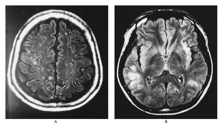
- Acute encephalitis: Multiple discrete 2–7 mm high-signal lesions on T2/FLAIR in subcortical and deep white matter of cerebral hemispheres; cortical and brainstem lesions also seen; no mass effect or edema [28][34-35]
- Lesions represent microinfarctions from small vessel vasculitis [28][35]
- DWI: Restricted diffusion (hyperintense) in acute phase, decreasing over time [29][34]
- Leptomeningeal enhancement in some cases [29]
- Relapsed encephalitis: Confluent cortical gray matter involvement — distinct from acute pattern [31][35]
- MRI is more sensitive than CSF examination for detecting disease [14]
The following MRI demonstrates the characteristic findings in acute versus relapsed Nipah encephalitis:
- CT Brain: Usually normal in acute phase — insufficient sensitivity [14]
- Chest X-ray: Bilateral interstitial infiltrates (common, especially NiV-B strain); may present as atypical pneumonia pattern [16][23]
Special Tests
- RT-LAMP assay (reverse transcription loop-mediated isothermal amplification): More sensitive than RT-PCR; potential for rapid point-of-care use in outbreak settings [3]
- Virus isolation: Vero cell culture with syncytial cell formation — requires BSL-4; not routinely attempted due to biosafety risk [14]
- Whole genome sequencing: For phylogenetic analysis and strain identification (Malaysia vs. Bangladesh lineage) [12][36]
- EEG: Nonspecific diffuse slowing; periodic complexes correlate with severity and poor outcome [14]
- SPECT: Shows evolution from focal hyperperfusion to hypoperfusion in relapsed cases [31]
- No validated clinical scoring system specific to NiV exists; GCS for encephalitis severity monitoring
ECG
- ECG indicated to evaluate for myocarditis — a recognized clinical prototype [16]
- Look for: Sinus bradycardia, ST-segment changes, conduction abnormalities, low voltage
- Tachycardia (sinus) is common and associated with brainstem dysfunction and poor prognosis [14]
- Continuous cardiac monitoring recommended in all hospitalized cases
Assessment
Three overlapping clinical phenotypes: [16]
- Encephalitis (most common, ~83%) — progressive brainstem dysfunction with high mortality
- Acute respiratory distress syndrome — prominent in Bangladesh strain (NiV-B)
- Myocarditis — hemodynamic instability requiring vasopressors
- Spectrum ranges from asymptomatic infection to rapidly fatal encephalitis [1][8]
- Case fatality rate: 40–75% overall; up to 91% in some outbreaks (Kerala 2018); 86% in recent Bangladesh outbreaks [12][17]
- Mortality predictors: Coma, abnormal doll's-eye reflex, tachycardia, hypertension, vomiting, segmental myoclonus, CSF virus isolation, viremia at diagnosis [14][17][33]
- Survivors: 53% full recovery, 15% persistent neurologic deficits [14]
- Relapsing/late-onset encephalitis: Unique complication occurring months after initial infection in 3.4–7.5% [31]
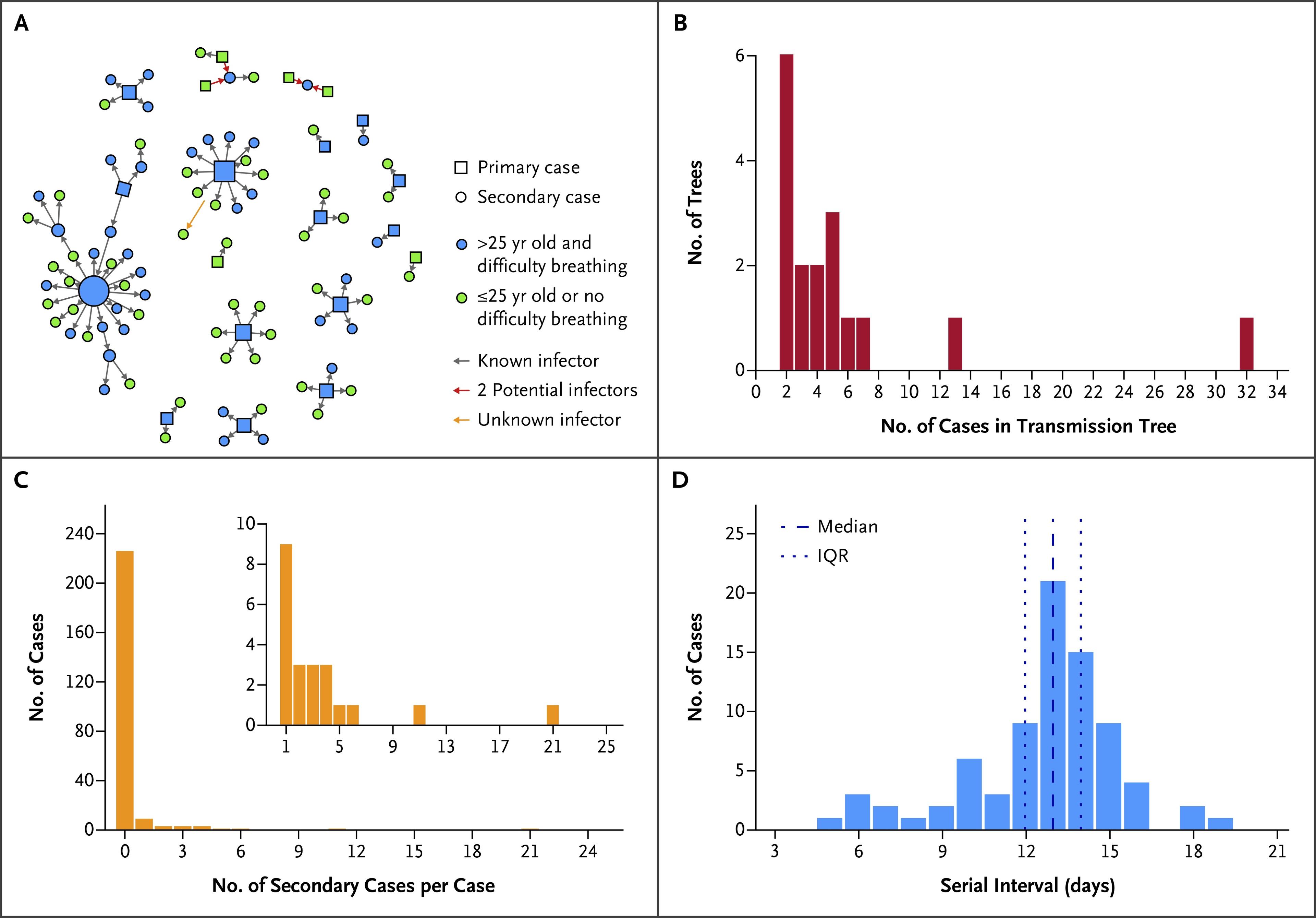
The following figure from an NEJM study illustrates person-to-person transmission dynamics in Bangladesh, showing that a minority of cases drive most transmission events:
Treatment Plan
Initial Stabilization
- Airborne isolation room immediately [37]
- ABCs with full PPE; intubation and mechanical ventilation for ARDS or GCS ≤8
- Vasopressor support for myocarditis-related hemodynamic instability [16]
- Seizure management with standard anticonvulsants
- Empiric IV acyclovir until HSV encephalitis excluded [23]
- ICP monitoring and management as indicated
Antiviral Therapy (compassionate/investigational)
- Ribavirin — may be considered on compassionate basis; limited evidence [3]
- Remdesivir — strongest preclinical evidence (NHP model); may be considered if available within 24 hours of exposure [19-20]
- m102.4 monoclonal antibody — available through compassionate use protocols in some settings; 20 mg/kg IV [18]
- Contact WHO, CDC, or national health authority for access to investigational therapeutics [22]
Supportive Care
- Intensive supportive therapy is the current standard of care [3]
- Mechanical ventilation (lung-protective strategy for ARDS)
- Hemodynamic support, renal replacement therapy as needed
- Nutritional support, DVT prophylaxis, stress ulcer prophylaxis
Disposition
- All confirmed or suspected NiV cases require ICU-level care in an airborne infection isolation room [37]
- Immediate notification of local/state public health authorities and CDC [37]
- NiV is a nationally notifiable condition and a potential bioterrorism agent
- Contact tracing of all exposed individuals within 21 days [12][37]
- Quarantine of high-risk contacts for 21 days (maximum incubation period)
- Transfer to a facility with BSL-4 diagnostic capability and high-containment clinical care if not available locally
- Specialist consultation: Infectious disease, neurology, critical care, pulmonology
Infection Control (per CDC): [37]
- Suspected stable patients: N95/PAPR + fluid-resistant gown + face shield/goggles + gloves
- Confirmed or unstable patients: Impermeable gown/coverall + N95/PAPR + double gloves with extended cuffs + boot covers + apron
- Minimize personnel entering the room; maintain entry log [37]
- Avoid aerosol-generating procedures when possible; if necessary, perform in negative-pressure room [37]
Follow Up / Return Precautions
- Survivors require long-term neurologic follow-up — 15% have persistent neurologic deficits [14]
- Monitor for relapsed encephalitis (7.5% of survivors) and late-onset encephalitis (3.4% of initially mild/asymptomatic cases) — can occur months after initial infection [31]
- Serial MRI if new neurologic symptoms develop post-recovery
- Clearance of virus from oropharyngeal space begins around day 16–20 post-illness onset — informs isolation duration [17]
- Return precautions for discharged contacts: Seek immediate care for fever, headache, altered mental status, respiratory symptoms, or myalgia within 21 days of exposure
- Psychological support for survivors and families given high mortality among contacts
- Public health follow-up: Continued surveillance of bat populations and date palm sap harvesting practices in endemic areas [10][25]
References
1. A Systematic Review of Case Reports on Mortality, Modes of Infection, Diagnostic Tests, and Treatments for Nipah Virus Infection. — Alla D, Shah DJ, Adityaraj N, et al. Medicine. 2024.
2. The Rising Threat of Nipah Virus: A Highly Contagious and Deadly Zoonotic Pathogen. — Ganguly A, Mahapatra S, Ray S, et al. Virology Journal. 2025.
3. Medical Countermeasures Against Henipaviruses: A Review and Public Health Perspective. — Gómez Román R, Tornieporth N, Cherian NG, et al. The Lancet. Infectious Diseases. 2022.
4. Therapeutic Intervention Strategies for Nipah Virus Infection: A Scoping Review. — Kusuma IY, Tayeb BA, Zidan MJ, et al. Infectious Diseases. 2026.
5. An Emerging Zoonotic Disease to Be Concerned About - A Review of the Nipah Virus. — Paliwal S, Shinu S, Saha R. Journal of Health, Population, and Nutrition. 2024.
6. Nipah Virus, an Emerging Zoonotic Disease Causing Fatal Encephalitis. — Alam AM. Clinical Medicine. 2022.
7. Nipah Virus in South Asia: From Emergence to Enduring Preparedness' Challenges. — Gupta N, Gkrania-Klotsas E, Drexler JF, et al. Clinical Microbiology and Infection : The Official Publication of the European Society of Clinical Microbiology and Infectious Diseases. 2026.
8. Nipah Virus Infection. — Ang BSP, Lim TCC, Wang L. Journal of Clinical Microbiology. 2018.
9. Henipavirus Zoonosis: Outbreaks, Animal Hosts and Potential New Emergence. — Li H, Kim JV, Pickering BS. Frontiers in Microbiology. 2023.
10. Tackling a Global Epidemic Threat: Nipah Surveillance in Bangladesh, 2006-2021. — Satter SM, Aquib WR, Sultana S, et al. PLoS Neglected Tropical Diseases. 2023.
11. Transmission of Human Infection With Nipah Virus. — Luby SP, Gurley ES, Hossain MJ. Clinical Infectious Diseases : An Official Publication of the Infectious Diseases Society of America. 2009.
12. Outbreak Investigation of Nipah Virus Disease in Kerala, India, 2018. — Arunkumar G, Chandni R, Mourya DT, et al. The Journal of Infectious Diseases. 2019.
13. Transmission of Nipah Virus — 14 Years of Investigations in Bangladesh. — Nikolay B, Salje H, Hossain MJ, et al. The New England Journal of Medicine. 2019.
14. Clinical Features of Nipah Virus Encephalitis among Pig Farmers in Malaysia. — Goh KJ, Tan CT, Chew NK, et al. The New England Journal of Medicine. 2000.
15. Emerging Viral Infections of the Central Nervous System: Part 2. — Tyler KL. Archives of Neurology. 2009.
16. Clinical Manifestations of Nipah Virus-Infected Patients Who Presented to the Emergency Department During an Outbreak in Kerala State in India, May 2018. — Chandni R, Renjith TP, Fazal A, et al. Clinical Infectious Diseases : An Official Publication of the Infectious Diseases Society of America. 2020.
17. Insights Into the Acute Phase of Nipah Virus Infection: Clinical Features, Viral Detection, and Humoral Immune Response. — Satter SM, Sultana S, Choudhury SS, et al. International Journal of Infectious Diseases : IJID : Official Publication of the International Society for Infectious Diseases. 2025.
18. Safety, Tolerability, Pharmacokinetics, and Immunogenicity of a Human Monoclonal Antibody Targeting the G Glycoprotein of Henipaviruses in Healthy Adults: A First-in-Human, Randomised, Controlled, Phase 1 Study. — Playford EG, Munro T, Mahler SM, et al. The Lancet. Infectious Diseases. 2020.
19. Remdesivir (GS-5734) Protects African Green Monkeys From Nipah Virus Challenge. — Lo MK, Feldmann F, Gary JM, et al. Science Translational Medicine. 2019.
20. Therapeutics for Nipah Virus Disease: A Systematic Review to Support Prioritisation of Drug Candidates for Clinical Trials. — Chan XHS, Haeusler IL, Choy BJK, et al. The Lancet. Microbe. 2025.
21. Favipiravir (T-705) Protects Against Nipah Virus Infection in the Hamster Model. — Dawes BE, Kalveram B, Ikegami T, et al. Scientific Reports. 2018.
22. Improving Clinical Care of Patients in Nipah Outbreaks: Moving Beyond 'Compassionate Use'. — Hassan MZ, Rojek A, Olliaro P, Horby P. The Lancet Regional Health. Southeast Asia. 2025.
23. Outbreak of Nipah-Virus Infection Among Abattoir Workers in Singapore. — Paton NI, Leo YS, Zaki SR, et al. Lancet. 1999.
24. Advancements in Nipah virus treatment: Analysis of current progress in vaccines, antivirals, and therapeutics. — Mishra G, Prajapat V, Nayak D. Immunology. 2024.
25. Nipah Virus Dynamics in Bats and Implications for Spillover to Humans. — Epstein JH, Anthony SJ, Islam A, et al. Proceedings of the National Academy of Sciences of the United States of America. 2020.
26. Nipah Virus: An Overview of the Current Status of Diagnostics and Their Role in Preparedness in Endemic Countries. — Garbuglia AR, Lapa D, Pauciullo S, Raoul H, Pannetier D. Viruses. 2023.
27. The Role of Infection Prevention and Control in the Mitigation of Human-to-Human Transmission of Nipah Virus: A Systematic Review. — Pritchard S, Hornsey E. Antimicrobial Resistance and Infection Control. 2025.
28. Viral Encephalitis: Familiar Infections and Emerging Pathogens. — Whitley RJ, Gnann JW. Lancet. 2002.
29. Nipah Viral Encephalitis or Japanese Encephalitis? MR Findings in a New Zoonotic Disease. — Lim CC, Sitoh YY, Hui F, et al. AJNR. American Journal of Neuroradiology. 2000.
30. Acute Viral Encephalitis. — Tyler KL. The New England Journal of Medicine. 2018.
31. Relapsed and Late-Onset Nipah Encephalitis. — Tan CT, Goh KJ, Wong KT, et al. Annals of Neurology. 2002.
32. Development of Nipah Virus-Specific IgM & IgG ELISA for Screening Human Serum Samples. — Shete AM, Jain R, Mohandas S, et al. The Indian Journal of Medical Research. 2022.
33. High Mortality in Nipah Encephalitis Is Associated With Presence of Virus in Cerebrospinal Fluid. — Chua KB, Lam SK, Tan CT, et al. Annals of Neurology. 2000.
34. Nipah Virus Encephalitis: Serial MR Study of an Emerging Disease. — Lim CC, Lee KE, Lee WL, et al. Radiology. 2002.
35. MR Imaging Features of Nipah Encephalitis. — Sarji SA, Abdullah BJ, Goh KJ, Tan CT, Wong KT. AJR. American Journal of Roentgenology. 2000.
36. Nipah Virus Outbreak in Kerala State, India Amidst of COVID-19 Pandemic. — Yadav PD, Sahay RR, Balakrishnan A, et al. Frontiers in Public Health. 2022.
37. Post-Travel Evaluation to Rule Out Viral Special Pathogen Infection. — Catherine Brown, Mary J. Choi, Susan McLellan, and Trevor Shoemaker CDC Yellow Book. 2025.
Figures
More from Deep Dive
Reviewed by Dr. Lucas Mastropaolo. SymptomDx is an educational tool for medical professionals — it does not replace clinical judgment. Verify all clinical data and drug dosages with authoritative sources.