Pheochromocytoma (Crisis)
Pheochromocytoma crisis is a life-threatening endocrine emergency defined as the acute, severe presentation of catecholamine-induced hemodynamic instability causing end-organ damage or dysfunction, with a mortality rate of approximately 14–27%. [1-2] It occurs in 7–18% of patients with pheochromocytoma/paraganglioma (PPGL) and may be the first clinical manifestation of an undiagnosed tumor. [3-4]
History
- Classic triad (present in only ~24% of patients): severe headache, palpitations, and diaphoresis [5]
- Paroxysmal or sustained hypertension with episodes of headache, sweating, anxiety, tremor, pallor, nausea/vomiting, and abdominal pain [3][5]
- Timing: Paroxysmal episodes may last minutes to hours; crisis onset is often sudden
- Triggers to ask about: recent surgery, anesthesia, trauma, new medications (especially metoclopramide, beta-blockers, corticosteroids), pregnancy (third trimester/peripartum), physical exertion [3]
- Headache was the most common symptom in a 200-case review (39.5%); nausea and vomiting were significantly associated with higher mortality [1]
- Ask about prior episodes of paroxysmal hypertension, "spells," or unexplained tachycardia
- Important negatives: absence of illicit drug use (cocaine, amphetamines), absence of MAO inhibitor use
Alarm Features
- BP ≥180/120 mmHg with evidence of end-organ damage [4]
- Rapidly alternating hypertension and hypotension (hemodynamic lability) [3][6]
- Acute pulmonary edema / flash pulmonary edema [6]
- Chest pain with troponin elevation (catecholamine cardiomyopathy or ACS mimic) [7-8]
- Temperature >40°C with encephalopathy (pheochromocytoma multisystem crisis [PMC]) [6]
- Unexplained cardiogenic shock, especially in a young patient without coronary disease [2][6]
- Lactic acidosis, multi-organ failure, unexplained hyperglycemia [6]
- Cardiac arrest [9]
Medications
Crisis-precipitating drugs (AVOID): [3][10]
- Metoclopramide and other dopamine antagonists (D2 blockade triggers catecholamine release)
- Beta-blockers without prior alpha-blockade (causes unopposed alpha stimulation → worsening hypertension) [3]
- Labetalol — alpha-blocking effect is insufficient relative to beta-blockade; can paradoxically worsen crisis [3-4][11]
- Glucagon, sympathomimetics, corticosteroids/synthetic ACTH, tricyclic antidepressants [3]
- General anesthetics, neuromuscular blockers, opiates [12]
Acute crisis treatment: [13-14]
- IV phentolamine 5 mg bolus, repeat q10 min PRN (first-line for catecholamine excess per 2025 AHA/ACC guidelines) [14-15]
- IV nicardipine 5 mg/h, titrate by 2.5 mg/h q5 min (max 15 mg/h) — widely available alternative [14]
- IV nitroprusside 0.3–0.5 mcg/kg/min for refractory hypertension [13-14]
- IV clevidipine — another option per 2025 AHA/ACC for catecholamine excess states [14]
- Esmolol or lidocaine for cardiac arrhythmias — only after adequate alpha-blockade [12-13]
- IV magnesium sulfate — adjunctive for BP and arrhythmia control [3]
- Avoid IV hydralazine (cardiostimulatory, unpredictable) and IV diuretics (unless overt heart failure) [4]
Chronic/preoperative alpha-blockade: [13][16]
- Phenoxybenzamine 10 mg PO BID, titrate up to 30 mg TID
- Doxazosin 1 mg PO daily, titrate up to 10 mg BID
- Beta-blockade added ONLY after adequate alpha-blockade for reflex tachycardia
Diet
- High-sodium diet (~5000 mg/day) and generous fluid intake (~2.5 L/day) during preoperative alpha-blockade to counteract catecholamine-induced volume contraction [16]
- Avoid tyramine-rich foods (aged cheeses, cured meats, fermented foods, red wine) — tyramine displaces catecholamines from vesicular storage and can precipitate crisis [12]
- Acute crisis: NPO; IV fluid resuscitation is critical given chronic intravascular volume depletion [3-4]
Review of Systems
- Cardiovascular: chest pain, palpitations, dyspnea on exertion, orthopnea, syncope
- Neurologic: headache, visual changes, altered mental status, seizures
- GI: nausea, vomiting, abdominal pain, constipation (catecholamine-mediated ileus)
- Endocrine: weight loss, heat intolerance, hyperglycemia symptoms
- Renal: decreased urine output
- Constitutional: diaphoresis, pallor (not flushing — pallor is characteristic), anxiety, tremor
Collateral History and Family History
- Family history of PPGL, medullary thyroid carcinoma, hyperparathyroidism, renal cell carcinoma, hemangioblastomas, neurofibromas [13][16]
- Personal or family history of genetic syndromes: MEN2, VHL, NF1, SDHx mutations [13][16]
- Up to 40% of PPGLs harbor germline pathogenic variants — the highest rate of any solid tumor [17]
- Ask about prior adrenal incidentalomas, prior "spells" dismissed as panic attacks
- Social context: pregnancy status (crisis risk increases in third trimester) [3]
Risk Factors
- Hereditary syndromes (account for ~25–40% of cases): [5][17]
- MEN2 (RET): PCC in up to 50%, bilateral tumors common
- VHL: PCC/PGL in 10–20%, earliest age at diagnosis
- SDHx (SDHB, SDHD, SDHC, SDHA): highest metastatic risk (especially SDHB)
- NF1: PCC in ~3%
- TMEM127, MAX
- Young age at presentation (<45 years) [5]
- Bilateral or extra-adrenal tumors [16]
- Tumor hemorrhage or infarction (spontaneous or traumatic) [3]
- Surgical manipulation, anesthesia induction, pregnancy [3]
- Undiagnosed/untreated PPGL undergoing any surgical procedure [18]
Differential Diagnosis
- Acute coronary syndrome / STEMI — pheochromocytoma can mimic ACS with ST changes, troponin elevation, and wall motion abnormalities but with normal coronary arteries [7-8]
- Takotsubo (stress) cardiomyopathy — PPGL-related TTS has higher rates of cardiogenic shock (34% vs 4%) and heart failure (47% vs 18%) than non-PPGL TTS [19]
- Thyroid storm — tachycardia, hyperthermia, altered mental status
- Cocaine/amphetamine toxicity — sympathomimetic crisis
- Malignant hypertension from other causes
- Serotonin syndrome or neuroleptic malignant syndrome — hyperthermia, rigidity, autonomic instability
- Carcinoid crisis — flushing (vs pallor in pheo), diarrhea, bronchospasm
- Acute myocarditis from other etiologies
- Septic shock — if presenting with hypotension and multi-organ failure
- Aortic dissection — severe hypertension with tearing chest/back pain
Past Medical History
- Prior episodes of paroxysmal hypertension or "spells"
- Known adrenal mass or incidentaloma
- Previous pheochromocytoma resection (recurrence risk)
- History of genetic syndrome components (MTC, hyperparathyroidism, retinal hemangioblastomas, neurofibromas, renal cell carcinoma) [13]
- Prior anesthetic complications or intraoperative hypertensive crises
- Chronic hypertension refractory to standard therapy
Physical Exam
Vital signs
- Severe hypertension (often >200/120 mmHg) or labile BP with alternating hypertension/hypotension [3][6]
- Tachycardia (or paradoxical bradycardia)
- Hyperthermia (>40°C in PMC) [6]
Focused exam
- Pallor (not flushing — catecholamine-mediated vasoconstriction) [5]
- Diaphoresis, tremor, anxiety
- Cardiac: S3 gallop, pulmonary crackles (acute heart failure), new murmur
- Abdominal: palpable mass (rare; avoid vigorous palpation — can trigger catecholamine surge)
- Neurologic: altered mental status, focal deficits (stroke), papilledema
- Skin: café-au-lait spots, neurofibromas (NF1), mucosal neuromas (MEN2B) [13][16]
- Marfanoid habitus (MEN2B) [16]
Lab Studies
- Plasma free metanephrines (preferred in acute setting — sensitivity >96%)
- 24-hour urine fractionated metanephrines and catecholamines (if patient stabilized)
- Levels in crisis are typically extremely elevated (mean 23× upper limit of normal in one series) [3]
Assess end-organ damage
- Troponin (catecholamine cardiomyopathy, demand ischemia)
- BNP/NT-proBNP (heart failure)
- BMP/CMP (renal function, hyperglycemia, electrolytes)
- Lactate (lactic acidosis — common in severe crisis) [6]
- CBC, coagulation studies
- Liver function tests (hepatic injury in multi-organ failure)
- Blood glucose (hyperglycemia is characteristic; hypoglycemia may occur post-resection) [13]
Monitoring
- Serial troponin, lactate, renal function
- Glucose monitoring (especially perioperatively)
Imaging
- CT abdomen/pelvis with IV contrast — first-line to identify adrenal mass; diagnostic in 33/34 ICU patients in one series. Low-osmolar contrast does not appreciably trigger catecholamine release [2-3]
- MRI abdomen — preferred for paragangliomas, pediatric patients, pregnant patients, and surveillance
- Bedside ultrasound — may identify adrenal mass in unstable patients too ill to transport [3]
- CT head — if neurologic symptoms (rule out hemorrhagic/ischemic stroke)
- CT angiography chest — if aortic dissection or pulmonary embolism suspected [20]
- Functional imaging (¹²³I-MIBG, ⁶⁸Ga-DOTATATE PET/CT) — not indicated acutely; used for staging and metastatic workup after stabilization [21]
Special Tests
- Echocardiography — critical in crisis; median LVEF 30% in ICU cohort. Assess for Takotsubo pattern (apical ballooning or inverted/basal pattern, which is more common in PPGL-related TTS), dilated cardiomyopathy, or regional wall motion abnormalities [2][19]
- Coronary angiography — if ACS suspected; typically shows normal coronary arteries in catecholamine cardiomyopathy [7-8]
- Genetic testing — recommended for ALL patients with PCC/PGL per NCCN guidelines [13]
- SOFA/SAPS II scoring — for ICU severity stratification [2]
ECG
ECG findings in pheochromocytoma crisis: [7][19]
- Sinus tachycardia (most common arrhythmia)
- ST-segment elevation or depression mimicking acute MI
- T-wave inversions (diffuse or regional)
- Prolonged QTc (risk for torsades de pointes)
- Low voltage QRS
- Atrial fibrillation/flutter
- Ventricular tachycardia (life-threatening)
- Bradyarrhythmias (less common)
Key pearl: ECG changes mimicking STEMI with elevated troponin but normal coronary arteries should raise suspicion for pheochromocytoma, especially in younger patients without traditional cardiovascular risk factors. [8][22]
Assessment
Severity classification (Whitelaw et al.): [3]
- Type A crisis: Hemodynamic instability without sustained hypotension (4× more common)
- Type B crisis: Sustained hypotension, shock, and multi-organ dysfunction (higher mortality)
- Pheochromocytoma multisystem crisis (PMC): Hyperthermia + multi-organ failure + encephalopathy + labile BP (19% of all crisis cases, highest mortality) [1]
The heart is the most commonly damaged organ (99%), followed by lungs (44%) and kidneys (21.5%). [1] Cardiovascular complications include catecholamine cardiomyopathy (up to 11%), Takotsubo syndrome, acute MI, arrhythmias, pulmonary edema, and aortic dissection. [6][23-24] The adrenergic phenotype (epinephrine-secreting) is more associated with Takotsubo and tachyarrhythmias, while the noradrenergic phenotype is more associated with atherosclerotic complications and flash pulmonary edema. [4][25]
Treatment Plan
Initial stabilization: [3-4][13-14]
- ICU admission with continuous arterial BP monitoring and telemetry
- IV fluid resuscitation — aggressive volume expansion (chronic intravascular depletion from catecholamine-mediated vasoconstriction)
- IV alpha-blockade first:
- Beta-blockade ONLY after adequate alpha-blockade — esmolol preferred (short-acting, titratable) for tachyarrhythmias [3][13]
- Arrhythmia management: esmolol or lidocaine for ventricular arrhythmias; magnesium sulfate as adjunct [3][13]
- Cardiogenic shock: consider VA-ECMO as bridge to recovery — survival rate ~93.5% in published series with full LVEF recovery in survivors; Impella or other mechanical circulatory support [26]
- CRRT — for catecholamine removal and acute kidney injury [9]
BP targets: Reduce SBP by ~25% in the first hour, then gradually to <140 mmHg over 24–48 hours. [14] Avoid aggressive lowering to <120 mmHg in the first hour (unlike aortic dissection).
Definitive treatment: Surgical resection after medical stabilization with 7–14 days of alpha-blockade, volume repletion, and high-salt diet. [13][16] Emergency adrenalectomy carries significant morbidity and should be avoided if possible. [18]
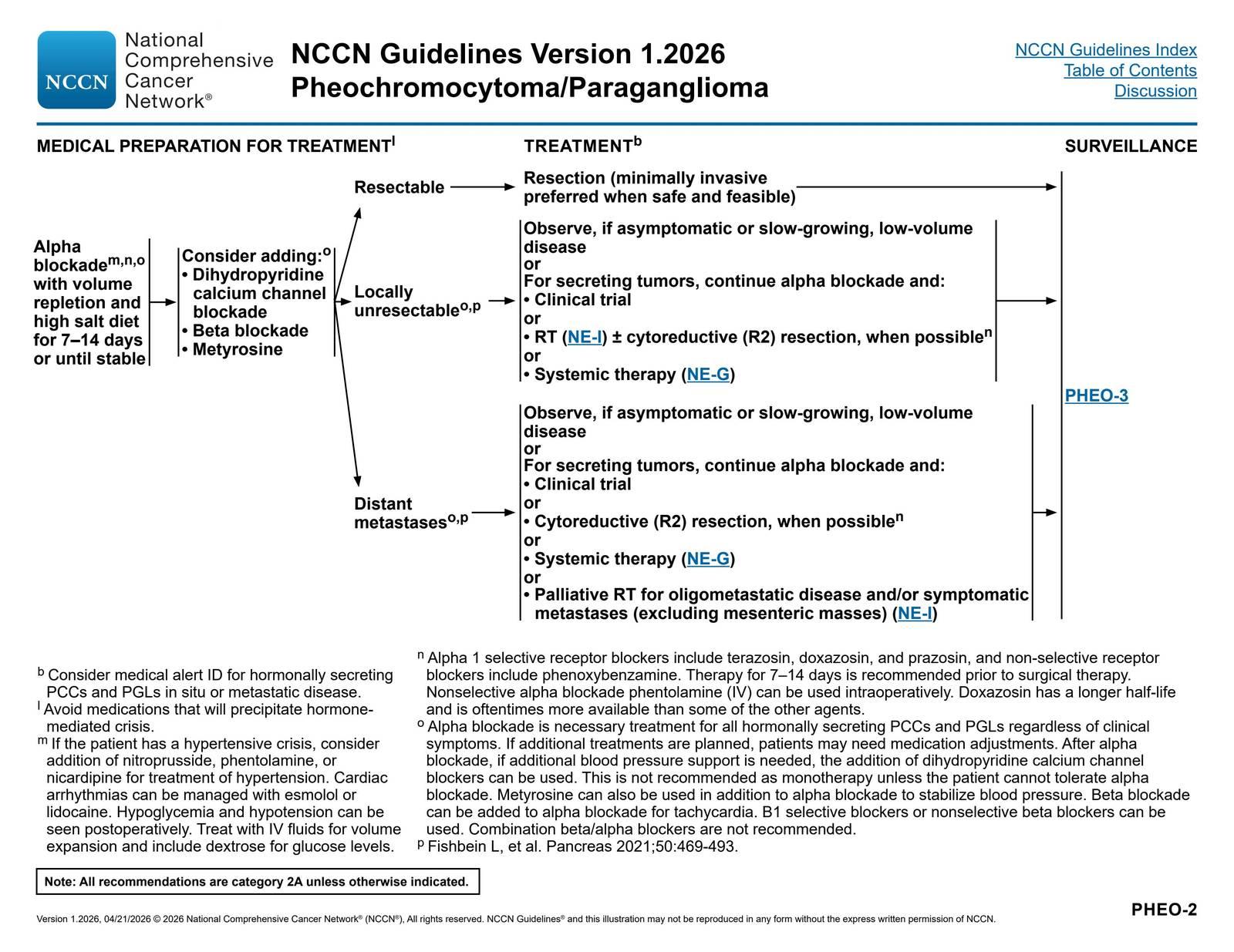
The NCCN algorithm for pheochromocytoma management is shown below:
Disposition
- All pheochromocytoma crises require ICU admission [2]
- Admission criteria: any hemodynamic instability, end-organ damage, arrhythmia, cardiomyopathy, multi-organ dysfunction
- VA-ECMO should be considered early in refractory cardiogenic shock [2][26]
- Urgent/emergent adrenalectomy only if medical management fails and patient is deteriorating despite maximal support [18]
- Endocrinology, cardiology, and surgical (endocrine surgery or urology) consultation
- Transfer to a center with ECMO capability if cardiogenic shock is refractory
Follow Up / Return Precautions
- Postoperative monitoring: Watch for hypotension (loss of catecholamine drive) and hypoglycemia (rebound insulin effect) — treat with IV fluids and dextrose [13]
- After resection, repeat plasma/urine metanephrines at 2–6 weeks to confirm biochemical cure
- Long-term surveillance: annual biochemical screening (metanephrines) lifelong, as recurrence can occur years later [13]
- Genetic testing and counseling for all patients — up to 40% have germline mutations [13][17]
- If hereditary syndrome confirmed, initiate syndrome-specific surveillance for patient and at-risk family members (per NCCN) [13]
- Medical alert ID recommended for patients with hormonally secreting tumors in situ or metastatic disease [13]
- Return precautions: recurrent headache, palpitations, diaphoresis, chest pain, severe hypertension, visual changes, or any "spell" — seek immediate evaluation
References
1. Clinical Characteristics and Outcomes of Pheochromocytoma Crisis: A Literature Review of 200 Cases. — Ando Y, Ono Y, Sano A, et al. Journal of Endocrinological Investigation. 2022.
2. Pheochromocytoma Crisis in the ICU: A French Multicenter Cohort Study With Emphasis on Rescue Extracorporeal Membrane Oxygenation. — Sauneuf B, Chudeau N, Champigneulle B, et al. Critical Care Medicine. 2017.
3. Phaeochromocytoma crisis. — Whitelaw BC, Prague JK, Mustafa OG, et al. Clinical Endocrinology. 2014.
4. Catecholamine-Induced Hypertensive Crises: Current Insights and Management. — Nazari MA, Hasan R, Haigney M, et al. The Lancet. Diabetes & Endocrinology. 2023.
5. Management of pheochromocytomas and paragangliomas: Review of current diagnosis and treatment options. — Eid M, Foukal J, Sochorová D, et al. Cancer Medicine. 2023.
6. Diagnosing phaeochromocytoma/paraganglioma in a patient presenting with critical illness: biochemistry versus imaging. — Amar L, Eisenhofer G. Clinical Endocrinology. 2015.
7. Cardiovascular Manifestations and Complications of Pheochromocytomas and Paragangliomas. — Y-Hassan S, Falhammar H. Journal of Clinical Medicine. 2020.
8. Pheochromocytoma Mimicking Acute Coronary Syndrome: A Case Report. — Cheng Y, Qin L, Chen L. Frontiers in Oncology. 2022.
9. Pheochromocytoma Crisis Rescued by Veno-Arterial Extracorporeal Membrane Oxygenation and Continuous Renal Replacement Therapy. — Nakayama T, Ito K, Inagaki F, et al. The American Surgeon. 2023.
10. Approach to the Management of Gastrointestinal Manifestations in Patients With Phaeochromocytoma and Paraganglioma. — Majumder M, Chiang C, Kong G, et al. Clinical Endocrinology. 2025.
11. Malignant Hypertension:A Systemic Cardiovascular Disease: JACC Review Topic of the Week. — Boulestreau R, Śpiewak M, Januszewicz A, et al. Journal of the American College of Cardiology. 2024.
12. Pathophysiology and Acute Management of Tachyarrhythmias in Pheochromocytoma: JACC Review Topic of the Week. — Nazari MA, Rosenblum JS, Haigney MC, Rosing DR, Pacak K. Journal of the American College of Cardiology. 2020.
13. Neuroendocrine and Adrenal Tumors. — Updated 2026-04-21. National Comprehensive Cancer Network.
14. 2025 AHA/ACC/AANP/AAPA/ABC/ACCP/ACPM/AGS/AMA/ASPC/NMA/PCNA/SGIM Guideline for the Prevention, Detection, Evaluation, and Management of High Blood Pressure in Adults: A Report of the American College of Cardiology/American Heart Association Joint Committee on Clinical Practice Guidelines. — Jones DW, Ferdinand KC, Taler SJ, et al. Journal of the American College of Cardiology. 2025.
15. Treatment of Hypertensive Crisis. — Calhoun DA, Oparil S. The New England Journal of Medicine. 1990.
16. Pheochromocytoma and Paraganglioma. — Neumann HPH, Young WF, Eng C. The New England Journal of Medicine. 2019.
17. Genetics of Pheochromocytoma and Paraganglioma. — Wachtel H, Fishbein L. Current Opinion in Endocrinology, Diabetes, and Obesity. 2021.
18. Pheochromocytoma Manifested as a Surgical Emergency. — Yiannakopoulou E. European Journal of Surgical Oncology : The Journal of the European Society of Surgical Oncology and the British Association of Surgical Oncology. 2025.
19. Cardiac Phenotypes in Secondary Hypertension: JACC State-of-the-Art Review. — Januszewicz A, Mulatero P, Dobrowolski P, et al. Journal of the American College of Cardiology. 2022.
20. A Rare Initial Presentation of Giant Pheochromocytoma: A Case Report of Acute Pulmonary Embolism Complicated by Catecholamine-Induced Cardiomyopathy. — Zhao L, Zhang Y, Zhou Y, et al. Frontiers in Oncology. 2025.
21. European Association of Nuclear Medicine Practice Guideline/Society of Nuclear Medicine and Molecular Imaging Procedure Standard 2019 for Radionuclide Imaging of Phaeochromocytoma and Paraganglioma. — Taïeb D, Hicks RJ, Hindié E, et al. European Journal of Nuclear Medicine and Molecular Imaging. 2019.
22. Acute Myocardial Injury Induced by Pheochromocytoma: A Case Report. — Li G, Xu S, Shi S, et al. BMC Cardiovascular Disorders. 2025.
23. Pheochromocytoma/Paraganglioma-Associated Cardiomyopathy. — Szatko A, Glinicki P, Gietka-Czernel M. Frontiers in Endocrinology. 2023.
24. Pheochromocytoma Is Characterized By Catecholamine-Mediated Myocarditis, Focal and Diffuse Myocardial Fibrosis, And Myocardial Dysfunction. — Ferreira VM, Marcelino M, Piechnik SK, et al. Journal of the American College of Cardiology. 2016.
25. Cardiovascular Complications in Pheochromocytoma and Paraganglioma: Does Phenotype Matter?. — Petrák O, Krátká Z, Holaj R, et al. Hypertension. 2024.
26. Pheochromocytoma Multisystem Crisis Requiring Temporary Mechanical Circulatory Support: A Narrative Review. — Giraud R, Glauser A, Looyens C, et al. Journal of Clinical Medicine. 2025.
Figure
More from Deep Dive
Reviewed by Dr. Lucas Mastropaolo. SymptomDx is an educational tool for medical professionals — it does not replace clinical judgment. Verify all clinical data and drug dosages with authoritative sources.