Pilonidal Abscess
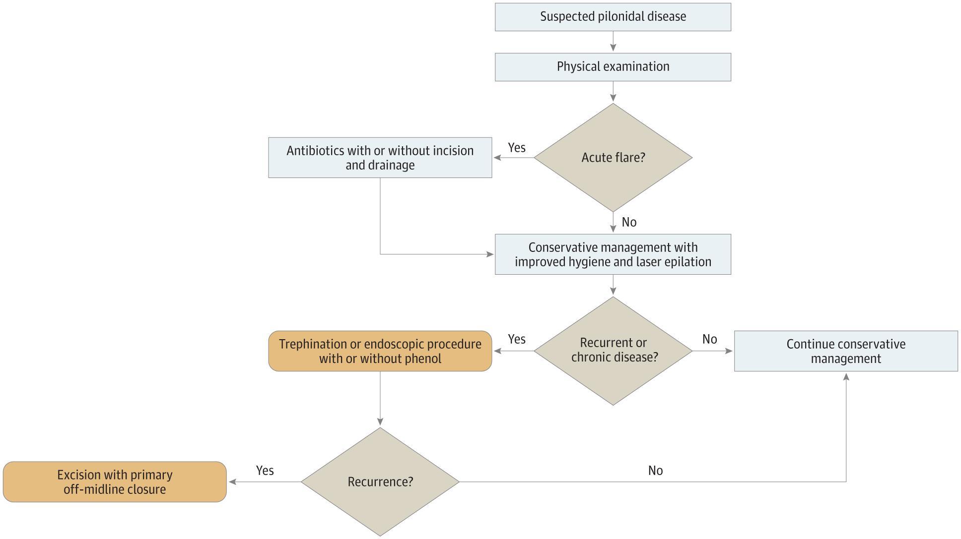
Pilonidal abscess is an acute infection of the sacrococcygeal region resulting from hair follicle obstruction and foreign-body reaction in the natal cleft, predominantly affecting young adults (peak incidence ~19–21 years), with a male predominance of at least 2:1. [1-2] The following figure outlines a hierarchical management algorithm for pilonidal disease:
History
- Onset and progression: Rapid onset of pain in the gluteal cleft, often over hours to days; ask about prior episodes or chronic intermittent drainage [1]
- Symptom characterization: Throbbing sacrococcygeal pain worsened by sitting, walking, or pressure; may report spontaneous purulent or serosanguinous drainage [1]
- Triggers: Prolonged sitting (long drives, desk work), recent trauma to the area, increased sweating
- Associated symptoms: Foul-smelling discharge, bleeding from the site, fever/chills (suggests systemic involvement)
- Important negatives: Perianal symptoms (fistula vs. pilonidal), groin involvement (hidradenitis), GI symptoms (Crohn's disease), weight loss
Alarm Features
- Fever, tachycardia, hypotension → concern for sepsis/necrotizing soft tissue infection [3]
- Rapidly expanding cellulitis, crepitus, or skin necrosis → necrotizing fasciitis until proven otherwise
- SIRS criteria (temp >38°C or <36°C, HR >90, RR >24, WBC >12,000 or <4,000) → indication for antibiotics and possible admission [3]
- Immunocompromised patients (diabetes, HIV, chemotherapy) → higher risk of complicated infection
- Suspicious skin lesions overlying chronic disease → biopsy to rule out rare malignant degeneration (squamous cell carcinoma) [4]
Medications
- Antibiotics are NOT routinely required after uncomplicated I&D of a pilonidal abscess. The ASCRS guidelines emphasize drainage as the mainstay [4]
- Indications for antibiotics: SIRS, significant surrounding cellulitis, immunocompromised host, failed initial treatment [3]
- TMP-SMX (1 DS tab BID × 7–10 days) or clindamycin (300–450 mg TID × 7–10 days) for MRSA coverage [5]
- Amoxicillin-clavulanate if mixed flora suspected (anaerobic coverage)
- Needle aspiration + empirical antibiotics (covering aerobes and anaerobes) is an emerging alternative to I&D in select non-septic patients, with 95% success in one prospective cohort [6]
- Perioperative prophylaxis for elective excision: mixed evidence; single-dose cefazolin + metronidazole has not shown clear benefit [4]
- Avoid: No specific contraindicated medications, but be cautious with clindamycin in patients with C. difficile history
Diet
- No specific acute dietary modifications are required
- Weight management is important for long-term recurrence prevention, as obesity is a significant risk factor [1][7]
- Adequate hydration and nutrition support wound healing post-I&D
Review of Systems
- Constitutional: Fever, chills, malaise (systemic infection)
- GI: Perianal drainage, diarrhea, bloody stools (Crohn's disease with perianal fistula)
- Skin: Other abscesses, axillary/groin nodules (hidradenitis suppurativa), chronic wounds
- Musculoskeletal: Sacral/coccygeal pain (osteomyelitis in chronic/neglected cases)
- Endocrine: Polyuria, polydipsia (undiagnosed diabetes increasing infection risk)
Collateral History and Family History
- Family history of pilonidal disease is associated with increased overall incidence and earlier onset [1]
- Family history of hidradenitis suppurativa or inflammatory bowel disease
- Occupational history: Sedentary occupation (truck drivers, office workers) is a well-established risk factor [2]
- Social context: Impact on school/work attendance, sports participation, and social activities — significant psychosocial burden especially in adolescents [1]
Risk Factors
- Male sex (≥2:1 male-to-female ratio) [1]
- Young adulthood (peak incidence late teens to early 20s) [1]
- Obesity [2][7]
- Hirsutism / excessive body hair [1][7]
- Sedentary lifestyle / prolonged sitting [2]
- Deep natal cleft anatomy [1]
- Local trauma or chronic irritation to the sacrococcygeal area [7]
- Family history of pilonidal disease [1]
- Prior pilonidal disease — recurrence rate 15–40% after simple I&D [4]
Differential Diagnosis
- Perianal/anorectal abscess or fistula — more caudal location, closer to the anal verge; perform thorough anorectal exam [4]
- Hidradenitis suppurativa — recurrent abscesses in apocrine gland-bearing areas (axillae, groin, gluteal); look for bilateral/multifocal involvement [4][8]
- Infected skin furuncle/carbuncle — no midline pits, no sinus tracts [4]
- Dermoid cyst — congenital, midline, may be deeper [8]
- Crohn's disease with perianal fistula — GI symptoms, complex fistulous disease [4]
- Infectious etiologies (rare): Tuberculosis, syphilis, actinomycosis [4]
- Malignant degeneration (squamous cell carcinoma) — extremely rare, consider in chronic/recurrent disease, especially immunosuppressed [4]
Past Medical History
- Prior pilonidal episodes — number, treatments received, time to healing, recurrence pattern
- Previous I&D or surgical excision — type of procedure, wound closure method, complications
- Diabetes mellitus — impaired wound healing, increased infection risk
- Immunosuppression (medications, HIV) — higher complication risk
- Inflammatory bowel disease — particularly Crohn's with perianal manifestations
- Smoking — impairs wound healing
Physical Exam
- Vital signs: Temperature, heart rate, blood pressure (assess for SIRS/sepsis)
- Inspection of natal cleft:
- Palpation: Fluctuance (abscess ready for drainage), tenderness, induration extent, crepitus (concerning for necrotizing infection)
- Anorectal exam: Essential to rule out concomitant fistulous disease, perianal abscess, or Crohn's-related pathology [4]
- Assess surrounding skin: Extent of cellulitis, skin necrosis, satellite lesions
Lab Studies
- Routine labs are generally unnecessary for uncomplicated pilonidal abscess
- Obtain if systemic illness suspected:
- CBC with differential (leukocytosis)
- BMP (renal function if considering antibiotics)
- Blood cultures (if septic)
- Lactate (if concern for sepsis)
- Blood glucose / HbA1c (if diabetes suspected)
- Wound culture: Gram stain and culture of purulent drainage are recommended for abscesses, though treatment without these studies is reasonable in typical cases [3]
- Staphylococcus aureus
Imaging
- Imaging is generally unnecessary — diagnosis is clinical based on history and physical exam [4]
- Ultrasound (point-of-care): Useful when diagnosis is uncertain, to confirm fluctuance, delineate abscess cavity size, or guide drainage [9]
- MRI: Reserved for complex or recurrent disease to map sinus tracts, rule out deep extension, or differentiate from perianal fistula
- CT: Rarely needed; consider if concern for deep space infection or necrotizing soft tissue infection
Special Tests
- No validated scoring systems specific to pilonidal abscess severity; no universally accepted classification system exists [8]
- Point-of-care ultrasound (POCUS): Valuable for confirming abscess vs. cellulitis and guiding I&D [9]
- Biopsy: Indicated if skin lesions have a suspicious appearance in chronic/recurrent disease to rule out malignancy [4]
- Fistulography or MRI fistula protocol: For complex recurrent disease with suspected extensive sinus tracts
ECG
- Not routinely indicated for pilonidal abscess
- Obtain if:
- Sepsis or SIRS present (tachycardia evaluation, septic cardiomyopathy)
- Pre-procedural assessment for patients requiring operative management under general anesthesia
- Significant comorbidities warranting cardiac evaluation
Assessment
Pilonidal abscess is a clinical diagnosis characterized by a painful, fluctuant mass in the sacrococcygeal region with characteristic midline pits in the natal cleft. [4] Approximately 50% of patients with pilonidal disease present with an acute infection. [1] The condition is acquired, resulting from hair follicle obstruction and foreign-body reaction, and is a chronic disease process with intermittent flares and quiescent periods. [1]
Severity stratification
- Uncomplicated: Localized abscess, no SIRS, immunocompetent → ED I&D and discharge
- Moderate: Significant cellulitis, SIRS criteria met → I&D + antibiotics, close follow-up
- Severe: Sepsis, necrotizing features, immunocompromised → emergent drainage, IV antibiotics, admission
Recurrence is the hallmark challenge: simple I&D carries a 15–40% recurrence rate, and overall recurrence across all treatments ranges from 0–66% with ~80% of recurrences occurring within the first year. [1][4]
Treatment Plan
Initial Stabilization (ED)
- Incision and drainage is the definitive ED treatment — strong recommendation (ASCRS Grade 1B) [4]
- Incise over the point of maximal fluctuance, lateral to midline pits
- Express pus, break up loculations, remove hair and debris
- Unroofing with curettage (lay open technique) is a one-step option that significantly reduces recurrence (10% vs. 54%) and improves healing at 10 weeks (96% vs. 79%) compared to simple I&D alone [4]
- Needle aspiration + oral antibiotics is an alternative in select non-septic, non-immunocompromised patients without skin necrosis — 95% success rate in converting to elective procedure [6]
Wound Care
- Routine packing is likely unnecessary — contemporary evidence for cutaneous abscesses shows no outcome advantage with packing, and it increases pain [10]
- Simple dressing changes; wound heals by secondary intention
- Sitz baths for comfort and hygiene
Antibiotics
- Not routinely needed post-I&D for uncomplicated abscess [3-4]
- If indicated (SIRS, cellulitis, immunocompromised): TMP-SMX DS BID or clindamycin 300–450 mg TID × 7–10 days [5]
Recurrence Prevention
- Improved hygiene: Regular cleaning of natal cleft, chlorhexidine washes [1]
- Hair removal: Weekly shaving or laser epilation of the gluteal cleft area [1][7]
- Weight loss if obese
- Avoid prolonged sitting; use cushions if sedentary occupation
Disposition
Discharge criteria (majority of patients)
- Uncomplicated abscess successfully drained
- No SIRS/sepsis
- Tolerating PO, adequate pain control
- Reliable follow-up available
Admission criteria
- Sepsis or hemodynamic instability
- Necrotizing soft tissue infection
- Failed outpatient management
- Significant immunocompromise with complicated infection
- Need for operative drainage under general anesthesia (large/complex abscess)
Observation
Specialist consultation
- General surgery or colorectal surgery referral for all patients post-I&D to discuss definitive management and recurrence prevention [4]
- Urgent surgical consultation if concern for necrotizing infection or complex disease
Follow Up / Return Precautions
Follow-up timing
- 48–72 hours for wound check (primary care or surgery)
- Surgical consultation within 2–4 weeks for discussion of definitive management if recurrent or chronic disease [6]
Return precautions — instruct patients to return immediately for:
- Fever >38°C (100.4°F)
- Worsening pain, swelling, or redness despite drainage
- Red streaking from the wound
- Foul-smelling drainage or inability to care for wound
- Dizziness, lightheadedness, or feeling generally unwell
Patient counseling
- Pilonidal disease is chronic with high recurrence rates — this is not a one-time problem [1]
- Emphasize hygiene, hair removal, and weight management as modifiable factors [1]
- Expected recovery: wound healing by secondary intention takes 4–10 weeks depending on cavity size
- Avoid prolonged sitting and strenuous activity until wound healed
- Definitive surgical options (Karydakis flap, Bascom cleft lift, excision with off-midline closure) should be discussed with a surgeon if disease recurs, as these offer lower long-term recurrence rates [4][7][11]
References
1. Management of Pilonidal Disease: A Review. — Gil LA, Deans KJ, Minneci PC. JAMA Surgery. 2023.
2. Dressings and Topical Agents for the Management of Open Wounds After Surgical Treatment for Sacrococcygeal Pilonidal Sinus. — Herrod PJ, Doleman B, Hardy EJ, et al. The Cochrane Database of Systematic Reviews. 2022.
3. Practice Guidelines for the Diagnosis and Management of Skin and Soft Tissue Infections: 2014 Update by the Infectious Diseases Society of America. — Stevens DL, Bisno AL, Chambers HF, et al. Clinical Infectious Diseases : An Official Publication of the Infectious Diseases Society of America. 2014.
4. The American Society of Colon and Rectal Surgeons' Clinical Practice Guidelines for the Management of Pilonidal Disease. — Johnson EK, Vogel JD, Cowan ML, Feingold DL, Steele SR. Diseases of the Colon and Rectum. 2019.
5. A Placebo-Controlled Trial of Antibiotics for Smaller Skin Abscesses. — Daum RS, Miller LG, Immergluck L, et al. The New England Journal of Medicine. 2017.
6. Converting Emergency Pilonidal Abscess Into an Elective Procedure. — Hussain ZI, Aghahoseini A, Alexander D. Diseases of the Colon and Rectum. 2012.
7. Pilonidal Disease. — O'Guinn ML, Nguyen ML, Raman S, Lin M. The Surgical Clinics of North America. 2026.
8. International Society of Laser Proctology Position Paper on SiLaC and EPSiT/SiLaC in the Management of Pilonidal Sinus Disease. — Ouali M, Martin-Martin GP, Avdicausevic S, et al. Lasers in Surgery and Medicine. 2025.
9. Abscess Management: An Evidence-Based Review for Emergency Medicine Clinicians. — Menegas S, Moayedi S, Torres M. The Journal of Emergency Medicine. 2021.
10. Beyond the Knife: A Contemporary Review of Subcutaneous Abscesses. — Mersal M, Embaby O, Ayyad M, et al. ANZ Journal of Surgery. 2025.
11. Midline and Off-Midline Wound Closure Methods After Surgical Treatment for Pilonidal Sinus. — Cai Z, Zhao Z, Ma Q, et al. The Cochrane Database of Systematic Reviews. 2024.
Figure
More from Deep Dive
Reviewed by Dr. Lucas Mastropaolo. SymptomDx is an educational tool for medical professionals — it does not replace clinical judgment. Verify all clinical data and drug dosages with authoritative sources.