Pleurisy
Pleurisy (pleuritis) is inflammation of the pleural membranes causing sharp, stabbing chest pain that worsens with inspiration and expiration. It is a clinical diagnosis of exclusion — life-threatening causes of pleuritic chest pain (PE, MI, pneumothorax, aortic dissection) must be ruled out first. [1-2] Viral pleurisy is the most common benign etiology, and treatment centers on NSAIDs for pain control plus management of the underlying cause. [1-2]
History
- Pain characterization: Sudden-onset, sharp/stabbing/burning chest pain; worsened by deep breathing, coughing, sneezing, and trunk movement; often well-localized [2]
- Timing: Acute onset; ask about preceding URI symptoms (viral prodrome in 1–2 weeks prior), recent travel, immobilization, surgery [1-2]
- Associated symptoms: Dyspnea, cough (dry or productive), fever, malaise, pleuritic shoulder pain (diaphragmatic irritation via phrenic nerve) [1]
- Important negatives to elicit: Hemoptysis, unilateral leg swelling, syncope, positional component (worse supine → think pericarditis), tearing/ripping quality (aortic dissection), exertional component [2-3]
Alarm Features
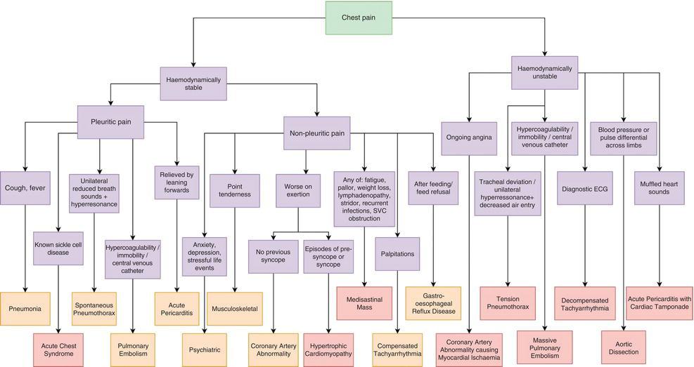
- Hemodynamic instability (tachycardia, hypotension, hypoxia) → PE, tension pneumothorax, tamponade [3]
- Sudden severe "tearing" pain radiating to back → aortic dissection [3]
- Unilateral absent breath sounds → pneumothorax [3]
- Fever + productive cough + hypoxia → pneumonia with possible empyema [4]
- Hemoptysis, unilateral leg swelling, recent immobilization → PE (found in 5–21% of ED patients with pleuritic chest pain) [2]
- Pain worse supine, relieved leaning forward → pericarditis [5]
- Subcutaneous emphysema → esophageal rupture or pneumomediastinum [3]
Medications
- First-line treatment: NSAIDs (ibuprofen 400–800 mg TID or naproxen 500 mg BID) — mainstay of viral/idiopathic pleurisy [1-2]
- Adjunctive analgesia: Acetaminophen if NSAIDs contraindicated; short-course opioids for severe pain refractory to NSAIDs
- Medications that can cause pleurisy/effusions: Nitrofurantoin, dantrolene, methotrexate, dasatinib, amiodarone, procarbazine, clozapine, phenytoin, β-blockers [6]
- Contraindicated: Avoid NSAIDs if renal insufficiency, active GI bleeding, or anticoagulation for PE; aminoglycosides have poor pleural penetration and should be avoided in pleural infections [7-8]
- If infectious etiology: Antibiotics targeted to cause; empiric coverage for parapneumonic effusion includes ceftriaxone + metronidazole or ampicillin-sulbactam [9]
- If autoimmune (SLE/RA): Corticosteroids for moderate-severe cases; mild lupus pleuritis may respond to NSAIDs alone [10]
Diet
- No specific dietary triggers for pleurisy
- Hydration: Encourage adequate oral hydration, especially if febrile
- Long-term: If pleurisy secondary to autoimmune disease, anti-inflammatory dietary patterns may be supportive but are not primary therapy
Review of Systems
- Pulmonary: Cough, dyspnea, hemoptysis, wheezing
- Cardiovascular: Palpitations, syncope, leg swelling, orthopnea, PND
- GI: Dysphagia, vomiting (esophageal rupture), abdominal pain (pancreatitis, subdiaphragmatic abscess)
- MSK: Joint pain, swelling, morning stiffness (rheumatologic causes) [11-12]
- Dermatologic: Rash (SLE malar rash, dermatomyositis, herpes zoster) [3]
- Constitutional: Fever, weight loss, night sweats (TB, malignancy)
Collateral History and Family History
- Collateral: Recent sick contacts, travel history (TB-endemic areas), occupational exposures (asbestos), substance use, IV drug use
- Family history: VTE/hypercoagulable states, autoimmune diseases (SLE, RA, familial Mediterranean fever), malignancy [6]
- Social context: Smoking history (malignancy risk), recent immobilization/long travel (PE risk), hormone use (OCP/HRT)
Risk Factors
- Viral infection (most common cause): Coxsackievirus, RSV, influenza, parainfluenza, EBV, CMV, adenovirus [2][13]
- PE risk factors: Immobilization, recent surgery, malignancy, OCP/HRT use, prior VTE, obesity, pregnancy [2]
- Pneumonia: Aspiration risk, COPD, immunosuppression [14]
- Autoimmune: SLE (pleuritis in up to 50%), RA, Sjögren syndrome [10-11]
- Malignancy: Smoking, age >50, asbestos exposure, known cancer [6]
- TB: Endemic area residence, immunosuppression, incarceration, homelessness [14]
Differential Diagnosis
Cannot-miss diagnoses
- Pulmonary embolism — most common life-threatening cause (5–21% of ED pleuritic chest pain presentations) [2]
- Pneumothorax — sudden dyspnea, absent breath sounds unilaterally [3]
- Acute coronary syndrome — atypical MI can present with pleuritic pain [2]
- Aortic dissection — tearing pain, pulse differential [3]
- Esophageal rupture — post-emesis, subcutaneous emphysema [3]
Common/important diagnoses
- Pneumonia (parapneumonic pleuritis) [4]
- Pericarditis — positional pain, friction rub, diffuse ST elevation [5][15]
- Viral pleurisy — diagnosis of exclusion after ruling out above [1]
- Autoimmune serositis (SLE, RA) [11-12]
- Malignant pleural disease [6]
- Costochondritis — reproducible tenderness at costochondral junctions [3]
- Musculoskeletal chest wall pain [16]
Past Medical History
- Prior episodes of pleurisy or pleuritic chest pain
- History of VTE, hypercoagulable states
- Known autoimmune disease (SLE, RA)
- Malignancy (lung, breast, lymphoma, mesothelioma)
- Recent thoracic surgery or cardiac procedures (Dressler syndrome)
- Chronic liver, kidney, or heart disease (transudative effusions) [6]
- TB exposure or prior treatment
Physical Exam
- Vital signs: Tachycardia, tachypnea, hypoxia (concerning for PE, pneumonia, pneumothorax); fever (infection, autoimmune) [3]
- Auscultation: Pleural friction rub — coarse, grating sound heard in both inspiration and expiration, best at lower anterolateral chest wall; distinguishes from pericardial rub (persists with breath-holding) [3]
- Percussion: Dullness suggests effusion; hyperresonance suggests pneumothorax [9]
- Decreased breath sounds over affected area [9]
- Chest wall palpation: Reproducible tenderness at costochondral junctions suggests costochondritis (LR+ for non-ACS) [3]
- Extremities: Unilateral leg swelling (DVT/PE), joint swelling (autoimmune)
- Skin: Malar rash (SLE), dermatomal vesicles (herpes zoster) [3]
Lab Studies
- Baseline for all: CBC with differential, CMP, CRP/ESR [1]
- Rule out PE: D-dimer (if low pretest probability by Wells/PERC) [2]
- Rule out ACS: Troponin [2]
- If effusion present and thoracentesis performed: Cell count with differential, pH, glucose, LDH, total protein, Gram stain, culture, cytology [9][14]
- If autoimmune suspected: ANA, anti-dsDNA, RF, anti-CCP, complement levels [10][17]
- If TB suspected: Pleural fluid ADA (sensitivity ≥79%, specificity ≥83%), AFB culture, interferon-gamma [14]
- Procalcitonin: May support bacterial infection but not routinely recommended for pleural infection diagnosis [7]
Imaging
- First-line: Chest X-ray (PA and lateral) — recommended for all patients with pleuritic chest pain; detects effusions >75 mL (lateral) or >175 mL (frontal), pneumothorax, pneumonia, widened mediastinum [1][18]
- Point-of-care ultrasound (POCUS): Detects effusions as small as 20 mL; superior to CXR for small/occult effusions; identifies septations, guides thoracentesis, reduces procedural complications [6][16][18]
- CT chest: Gold standard for imaging (detects >10 mL fluid); indicated if PE suspected (CTA), malignancy suspected, or CXR inconclusive [18]
- When imaging is unnecessary: Repeat CXR post-thoracentesis not required unless symptomatic, air aspirated, or multiple attempts [9]
- Lung ultrasound showed sensitivity 97% and specificity 96% for detecting radio-occult pleural-pulmonary lesions in patients with pleuritic pain and normal CXR [16]
Special Tests
- Wells Score / PERC Rule — validated clinical decision rules for PE risk stratification [2][4]
- HEART Score — for ACS risk stratification if cardiac etiology considered
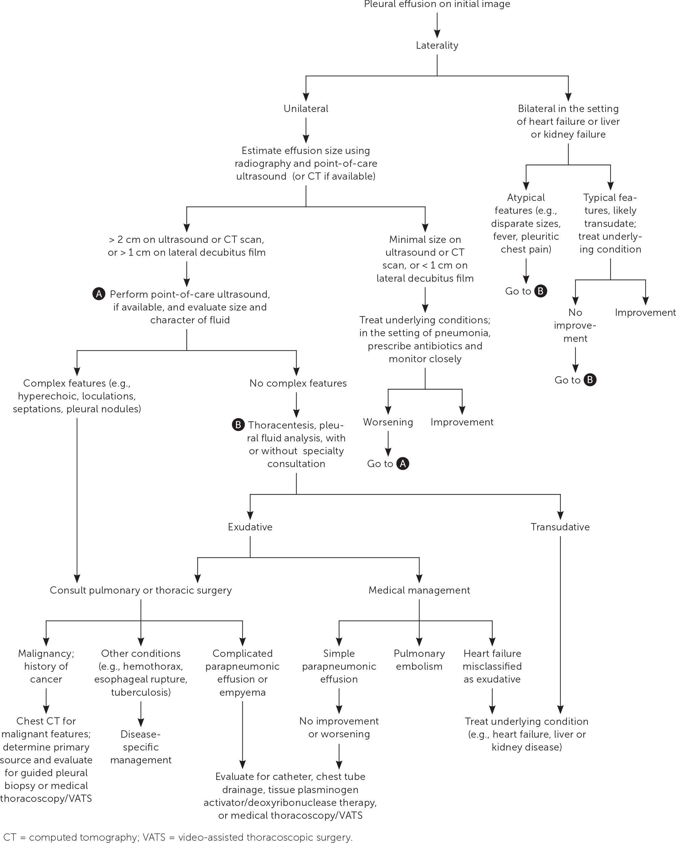
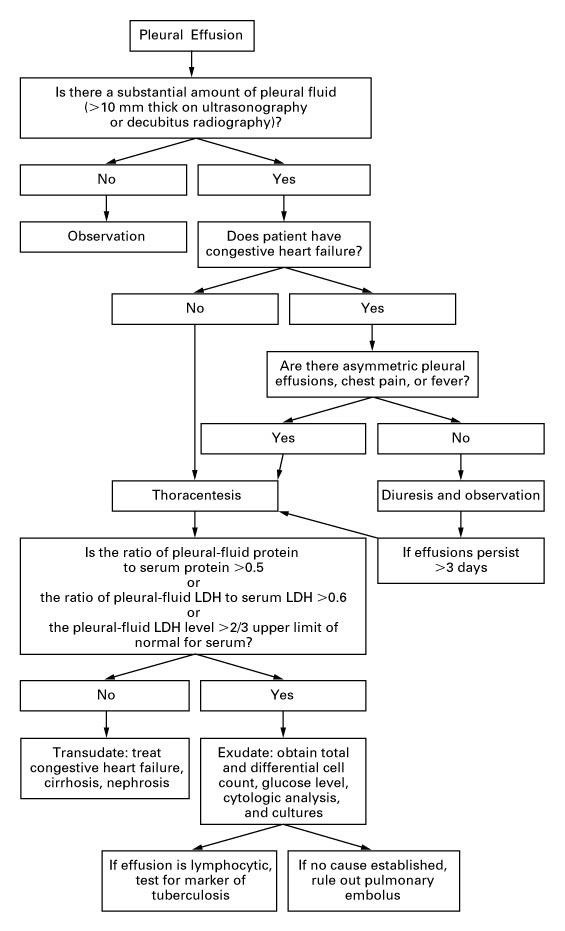
- Light's Criteria — pleural fluid protein ratio >0.5, LDH ratio >0.6, or pleural LDH >2/3 upper limit of normal serum LDH → exudate [9]
- Thoracentesis — indicated for new-onset unilateral effusions >1 cm on decubitus film or >2 cm on ultrasound/CT, not explained by heart failure/cirrhosis/renal failure [9]
- Pleural biopsy — if TB or malignancy suspected and fluid analysis nondiagnostic [14]
ECG
ECG is recommended for all patients with pleuritic chest pain to rule out life-threatening mimics: [1-2]
- Pericarditis: Diffuse concave ST-segment elevation, PR-segment depression (especially leads I, II, aVL, V3–V5); no reciprocal changes; ST/T ratio >0.25 in V6 [5][15]
- Pulmonary embolism: Sinus tachycardia (31%), S1Q3T3 pattern (15%), T-wave inversions V1–V3 (18%), right axis deviation, RBBB (14%) [15][19]
- ACS: ST elevation/depression in coronary distribution with reciprocal changes [15]
- Isolated pleurisy: ECG is typically normal [1]
Assessment
- Pleurisy is a clinical syndrome, not a final diagnosis — the underlying etiology must be identified [1]
- Viral pleurisy is the most common benign cause and is a diagnosis of exclusion after ruling out PE, ACS, pneumothorax, pneumonia, and pericarditis [1-2]
- Severity stratification depends on the underlying cause: uncomplicated viral pleurisy is self-limited (1–2 weeks), while PE, empyema, or malignant pleuritis carry significant morbidity/mortality [2]
- Atypical presentations: Elderly patients may present with dyspnea rather than pain; immunocompromised patients may lack fever despite infection [9]
- Complications: Pleural effusion, empyema, fibrothorax, chronic pain, trapped lung [6-7]
Treatment Plan
Initial stabilization
- ABCs; supplemental O2 if hypoxic; IV access if hemodynamically unstable
- Treat underlying life-threatening cause immediately (anticoagulation for PE, needle decompression for tension pneumothorax, etc.)
Viral/idiopathic pleurisy
- NSAIDs (ibuprofen 400–800 mg TID or indomethacin 25–50 mg TID) for 1–2 weeks with GI prophylaxis as needed [1-2]
- Encourage incentive spirometry and splinting to prevent atelectasis
Infectious pleurisy
- Antibiotics targeted to cause; empiric coverage for community-acquired parapneumonic effusion: ceftriaxone + metronidazole or ampicillin-sulbactam [8-9]
- Complicated parapneumonic effusion (pH <7.2, glucose <40 mg/dL, positive Gram stain/culture, or pus): chest tube drainage ± intrapleural tPA/DNase [7][9]
Autoimmune pleurisy
Malignant pleurisy
Disposition
Admission criteria
- Hemodynamic instability, hypoxia, or respiratory distress
- Confirmed PE (submassive/massive), pneumothorax requiring intervention, empyema
- Large pleural effusion requiring drainage
- Suspected serious underlying etiology (malignancy, aortic dissection)
- Inability to control pain or maintain oral intake
Discharge criteria
- Life-threatening causes excluded (normal CXR, ECG, troponin, low PE probability)
- Hemodynamically stable, adequate oxygenation
- Pain controlled with oral NSAIDs
- Reliable follow-up arranged [1-2]
Observation indications
- Borderline vital signs, pending workup results (D-dimer, serial troponins)
- Small effusion being monitored for progression
Specialist consultation triggers
- Pulmonology: significant or recurrent effusion, need for thoracentesis/chest tube, suspected malignancy [6]
- Cardiology: pericarditis with hemodynamic compromise or myocardial involvement
- Rheumatology: suspected autoimmune serositis [12][20]
- Thoracic surgery: empyema, loculated effusion, fibrothorax [8]
Follow Up / Return Precautions
- Follow-up timing: Primary care within 1–2 weeks for uncomplicated viral pleurisy; sooner if symptoms worsen [2]
- Repeat CXR at 6 weeks in patients >50 years, smokers, or those with pneumonia to document radiographic resolution and exclude underlying malignancy [2]
- Return immediately for: Worsening dyspnea, chest pain at rest, hemoptysis, fever >101°F, syncope, unilateral leg swelling, inability to take oral medications
- Expected recovery: Viral pleurisy typically resolves in 1–2 weeks; persistent symptoms beyond 2–4 weeks warrant further workup for autoimmune, malignant, or TB etiologies [1-2]
- Patient counseling: Pleurisy pain may transiently worsen with deep breathing and coughing; splinting the chest with a pillow during coughs can help; avoid smoking; complete full NSAID course
References
1. Pleurisy. — Kass SM, Williams PM, Reamy BV. American Family Physician. 2007.
2. Pleuritic Chest Pain: Sorting Through the Differential Diagnosis. — Reamy BV, Williams PM, Odom MR. American Family Physician. 2017.
3. 2021 AHA/ACC/ASE/CHEST/SAEM/SCCT/SCMR Guideline for the Evaluation and Diagnosis of Chest Pain: A Report of the American College of Cardiology/American Heart Association Joint Committee on Clinical Practice Guidelines. — Gulati M, Levy PD, Mukherjee D, et al. Journal of the American College of Cardiology. 2021.
4. Acute Chest Pain in Adults: Outpatient Evaluation. — McConaghy JR, Sharma M, Patel H. American Family Physician. 2020.
5. Diagnosis, Risk Stratification, and Treatment of Pericarditis: A Review. — Cremer PC, Klein AL, Imazio M. The Journal of the American Medical Association. 2024.
6. Pleural Disease. — Feller-Kopman D, Light R. The New England Journal of Medicine. 2018.
7. ERS/ESTS Statement on the Management of Pleural Infection in Adults. — Bedawi EO, Ricciardi S, Hassan M, et al. The European Respiratory Journal. 2023.
8. The American Association for Thoracic Surgery Consensus Guidelines for the Management of Empyema. — Shen KR, Bribriesco A, Crabtree T, et al. The Journal of Thoracic and Cardiovascular Surgery. 2017.
9. Pleural Effusion: Diagnostic Approach in Adults. — Shen-Wagner J, Gamble C, MacGilvray P. American Family Physician. 2023.
10. Lung Involvements in Rheumatic Diseases: Update on the Epidemiology, Pathogenesis, Clinical Features, and Treatment. — Ha YJ, Lee YJ, Kang EH. BioMed Research International. 2017.
11. Systemic Lupus Erythematosus and Lung Involvement: A Comprehensive Review. — Shin JI, Lee KH, Park S, et al. Journal of Clinical Medicine. 2022.
12. Thoracic Involvement in Systemic Autoimmune Rheumatic Diseases: Pathogenesis and Management. — De Zorzi E, Spagnolo P, Cocconcelli E, et al. Clinical Reviews in Allergy & Immunology. 2022.
13. Viral Diseases Affecting the Pleura. — Nestor J, Huggins T, Kummerfeldt C, et al. Journal of Clinical Virology : The Official Publication of the Pan American Society for Clinical Virology. 2013.
14. Guide to Utilization of the Microbiology Laboratory for Diagnosis of Infectious Diseases: 2024 Update by the Infectious Diseases Society of America (IDSA) and the American Society for Microbiology (ASM). — Miller JM, Binnicker MJ, Campbell S, et al. Clinical Infectious Diseases : An Official Publication of the Infectious Diseases Society of America. 2024.
15. Acute Pericarditis: Rapid Evidence Review. — Peterson TA, Turner SP, Dolezal KA. American Family Physician. 2024.
16. A Comparison of Different Diagnostic Tests in the Bedside Evaluation of Pleuritic Pain in the ED. — Volpicelli G, Cardinale L, Berchialla P, et al. The American Journal of Emergency Medicine. 2012.
17. Immune Complexes and Other Laboratory Features of Pleural Effusions: A Comparison of Rheumatoid Arthritis, Systemic Lupus Erythematosus, and Other Diseases. — Halla JT, Schrohenloher RE, Volanakis JE. Annals of Internal Medicine. 1980.
18. ACR Appropriateness Criteria® Workup of Pleural Effusion or Pleural Disease. — Morris MF, Henry TS, Raptis CA, et al. Journal of the American College of Radiology : JACR. 2024.
19. Prevalence of Electrocardiographic Abnormalities in Patients With Acute Pulmonary Embolism: A Systematic Review and Meta-Analysis. — Krintratun S, Srichuachom W, Wongtanasarasin W. Journal of Clinical Medicine. 2025.
20. Pleural Diseases in Connective Tissue Diseases. — Guo H, Iqbal B, Rahman NM. Seminars in Respiratory and Critical Care Medicine. 2024.
Figures
More from Deep Dive
Reviewed by Dr. Lucas Mastropaolo. SymptomDx is an educational tool for medical professionals — it does not replace clinical judgment. Verify all clinical data and drug dosages with authoritative sources.