Pneumomediastinum
Pneumomediastinum — the presence of free air within the mediastinal tissues — is classified as primary/spontaneous (no obvious cause such as trauma or instrumentation) or secondary (trauma, iatrogenic, esophageal perforation). Primary spontaneous pneumomediastinum (SPM) is a benign, self-limited condition predominantly affecting young males (mean age ~23 years, 3:1 male predominance), caused by the Macklin phenomenon: alveolar rupture with air dissecting along bronchovascular sheaths into the mediastinum. [1-3] The critical ED challenge is distinguishing this benign entity from Boerhaave syndrome (esophageal perforation), which requires urgent intervention. [3-4]
History
- Onset and character of chest pain: Acute retrosternal or pleuritic chest pain is the most common symptom (~71–89%), often radiating to the neck, back, or shoulders [1][5]
- Dyspnea (43–57%), neck pain (32–55%), sore throat, odynophagia (14%), dysphagia, and voice change [1][6-7]
- Precipitating events: Forceful coughing (27–34%), retching/emesis (13–24%), physical exertion (11–21%), Valsalva maneuvers (straining, weightlifting, childbirth), inhaled drug use (marijuana, cocaine, vaping) [1][3][8]
- No identifiable trigger in 21–53% of cases [6][9]
- Important negatives: Absence of trauma, recent instrumentation/endoscopy, mechanical ventilation, foreign body ingestion
Alarm Features
- Fever — rare in SPM; raises concern for esophageal perforation or mediastinitis [3]
- Pleural effusion on imaging — 100% sensitivity for esophageal perforation when combined with mediastinal fluid; absence of pleural/mediastinal fluid has a 100% NPV for perforation [4]
- Age >40 — rare in SPM and should raise suspicion for secondary causes [3]
- Respiratory compromise (43% of perforation patients vs. 6% of SPM) [4]
- Dysphagia — significantly more common in perforation [4]
- Hemodynamic instability, signs of tension pneumomediastinum or cardiac tamponade physiology [10]
- Concurrent large pneumothorax (~10% of SPM have concomitant pneumothorax) [11]
Medications
- Relevant contributors: Inhaled corticosteroids (asthma management), inhaled recreational drugs (cocaine, marijuana, vaping/e-cigarettes) can precipitate alveolar rupture [3][8]
- Treatment medications:
- Prophylactic antibiotics: Generally not required; no benefit demonstrated in SPM [14]
- Cautions: Avoid positive-pressure ventilation if possible (may worsen air leak); avoid medications that provoke emesis
Diet
- NPO consideration: Only if esophageal perforation is being actively ruled out; otherwise, diet as tolerated
- Soft diet may be considered if odynophagia is present
- No specific long-term dietary restrictions for SPM
Review of Systems
- Pulmonary: Cough, wheezing, dyspnea, history of asthma or reactive airway disease
- GI: Emesis, retching, dysphagia, odynophagia, hematemesis (suggests Boerhaave)
- ENT: Sore throat, voice change, neck swelling/crepitus
- Cardiac: Palpitations (tachycardia common in 31% of SPM) [3]
- Substance use: Inhaled drugs (cocaine, marijuana, nitrous oxide), vaping
- Recent procedures: Dental extraction, endoscopy, intubation
Collateral History and Family History
- Collateral: Witnesses to emesis/retching episodes, substance use history (friends/family may provide more accurate drug use history)
- Family history: Asthma, connective tissue disorders (Marfan, Ehlers-Danlos — associated with spontaneous air leaks), alpha-1 antitrypsin deficiency
- Social context: Occupation (diving, wind instrument players), recreational drug use, recent strenuous exercise or athletic activity
Risk Factors
- Asthma/reactive airway disease — most common comorbidity (22–27%); only independent risk factor for recurrence (OR ~7–8) [1][5]
- Smoking/tobacco use (~30%) [1]
- Thin body habitus / young male
- Inhaled drug use (cocaine, marijuana, vaping) — rising incidence [8]
- Forceful coughing (any cause: URI, pertussis, asthma exacerbation)
- Valsalva-provoking activities: Weightlifting, straining, childbirth, spirometry
- Diabetic ketoacidosis (Kussmaul breathing) [12]
- Interstitial lung disease (secondary pneumomediastinum) [15]
Differential Diagnosis
Past Medical History
- Asthma — most important predisposing condition; associated with recurrence [5]
- Prior pneumomediastinum — recurrence rate ~4.6% [5]
- COPD/emphysema, interstitial lung disease, cystic fibrosis
- Connective tissue disorders
- Prior esophageal or airway surgery
- History of eating disorders (forceful vomiting)
Physical Exam
- Subcutaneous emphysema: Palpable crepitus in the neck, face, and chest — most common sign (33–54%); present in two-thirds at presentation [1][11]
- Hamman's sign (Hamman's crunch): Crunching/crackling sound synchronous with the cardiac cycle on auscultation — pathognomonic [7][10]
- Vital signs: Tachycardia in ~31%; fever is rare and should prompt concern for perforation [3]
- Neck: Swelling, voice change, tracheal deviation (if tension physiology)
- Lungs: May be clear; wheezing if underlying asthma; decreased breath sounds if concurrent pneumothorax
- Abdomen: Assess for peritoneal signs (if concern for hollow viscus perforation)
Lab Studies
- CBC: Leukocytosis is common (~30%) in SPM and does not reliably distinguish from perforation; however, significantly higher WBC is associated with esophageal perforation [3-4][16]
- CRP: Mild elevation common in SPM (mean ~1.0 mg/dL); markedly elevated CRP suggests secondary cause [20]
- BMP: Rule out DKA if clinically suspected
- Lactate: If hemodynamically unstable
- Troponin: If ACS is on the differential
- Blood cultures: Only if febrile or concern for mediastinitis
Imaging
- Chest X-ray (PA and lateral): First-line; diagnostic in ~69–90% of cases [9][11][21]
- CT chest: More sensitive; detects ~30% of cases missed on CXR; indicated when CXR is equivocal or concern for secondary cause [23]
- absence of pleural effusion/mediastinal fluid[4]
- Esophagram (water-soluble contrast): Rarely indicated; reserve for high suspicion of esophageal perforation (older age, pleural effusion, emesis history, respiratory compromise) [3][9]
- CT with oral contrast: Alternative to esophagram if perforation is suspected
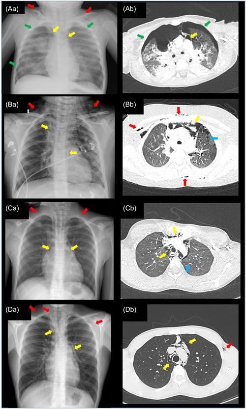
The following figure demonstrates the spectrum of pneumomediastinum findings on both CXR and CT in pediatric patients:
Special Tests
- Esophagogastroduodenoscopy (EGD): Only if high suspicion for esophageal perforation; performed in ~13% historically but invariably negative in true SPM [1][9]
- Bronchoscopy: Rarely needed; consider if tracheobronchial injury suspected [1]
- Pulmonary function testing: After recovery, to evaluate for undiagnosed asthma (especially if no clear precipitant) [13]
- Methacholine challenge: If exercise-induced bronchoconstriction suspected as underlying etiology [13]
ECG
- Indications: Obtain to rule out ACS in patients presenting with acute chest pain
- Expected findings in SPM: Usually normal or sinus tachycardia
- Concerning patterns: ST changes, axis deviation, or low voltage (may suggest tension pneumomediastinum with cardiac compression or concurrent pneumopericardium) [10]
Assessment
SPM is a benign, self-limited condition with no reported mortality across large case series totaling hundreds of patients. [1][6][9] The key clinical decision point is distinguishing SPM from esophageal perforation (Boerhaave syndrome). A practical risk stratification approach:
- Low risk for perforation: Age <40, known precipitant (cough, asthma, exertion), no emesis history, no pleural effusion, no mediastinal fluid on CT → observation alone is appropriate [3-4]
- Higher risk for perforation: Age >40, history of retching/emesis, pleural effusion, elevated WBC, fever, respiratory compromise, dysphagia → further workup with esophagram or CT with oral contrast [3-4][16]
Recurrence rate is low (~4.6%), with asthma being the only independent risk factor for recurrence. [5]
Treatment Plan
- Conservative management is the standard of care for SPM: [6][9][14]
- Rest and activity restriction (avoid Valsalva maneuvers)
- Analgesics: NSAIDs or acetaminophen
- Supplemental oxygen (high-flow/100%): Promotes nitrogen washout and accelerates air reabsorption [6][12]
- Treat underlying trigger: Bronchodilators and inhaled corticosteroids for asthma; antiemetics if vomiting [13]
- Prophylactic antibiotics are NOT routinely indicated [14]
- NPO status: Only if esophageal perforation workup is ongoing
- Surgical intervention: Extremely rare; reserved for tension pneumomediastinum with hemodynamic compromise or confirmed esophageal/airway perforation [25]
Disposition
- ED discharge is safe for well-appearing patients with confirmed SPM, stable vitals, no hypoxia, no pleural effusion, and no concern for secondary cause [14][26]
- no significant difference in outcomes[26]
- Admission/observation criteria:
- Significant dyspnea or hypoxia
- Hemodynamic instability
- Concern for esophageal perforation (pending workup)
- Large concurrent pneumothorax
- Inability to tolerate oral intake
- Significant comorbidities or diagnostic uncertainty
- Median length of stay when admitted: ~27 hours (pediatric) to 1.8–2.3 days (adult) [3][9][21]
- Specialist consultation: Thoracic surgery if concern for esophageal/airway perforation or tension physiology; pulmonology for undiagnosed underlying lung disease
Follow Up / Return Precautions
- Follow-up: Outpatient visit in 1–2 weeks; repeat CXR only if symptoms persist or worsen [2][21]
- Pulmonary follow-up: If asthma is suspected as the underlying trigger, arrange PFTs and asthma management [5][13]
- Return precautions — instruct patients to return immediately for:
- Worsening chest pain or new-onset severe pain
- Increasing shortness of breath or difficulty breathing
- Fever
- Difficulty swallowing or inability to tolerate fluids
- Neck swelling that is worsening
- Expected recovery: Symptoms typically resolve within 1–2 days; radiographic resolution within 1–2 weeks [7][20]
- Counseling: Avoid Valsalva maneuvers, heavy lifting, and strenuous exercise until symptoms resolve; smoking cessation; avoid inhaled recreational drugs; inform asthma patients of small recurrence risk (~5%) [5]
References
1. A Narrative Review of Primary Spontaneous Pneumomediastinum: A Poorly Understood and Resource-Intensive Problem. — Morgan CT, Maloney JD, Decamp MM, McCarthy DP. Journal of Thoracic Disease. 2021.
2. Recurrent spontaneous pneumomediastinum in a child with tracheomalacia. — Dewulf J, Van Daele S, De Baets F. Pediatric Pulmonology. 2017.
3. One Hundred Cases of Primary Spontaneous Pneumomediastinum: Leukocytosis Is Common, Pleural Effusions and Age Over 40 Are Rare. — Morgan CT, Kanne JP, Lewis EE, et al. Journal of Thoracic Disease. 2023.
4. Diagnostic Utility of Computed Tomography in Patients With Spontaneous Pneumomediastinum Evaluated for Suspected Esophageal Perforation. — Keogan AG, Rybachok A, Patel S, et al. World Journal of Surgery. 2025.
5. Primary Spontaneous Pneumomediastinum: 237 Cases in a Single-Center Experience Over a 10-Year Period and Assessment of Factors Related With Recurrence. — Yu MH, Kim JK, Kim T, Lee HS, Kim DK. PloS One. 2023.
6. Clinical Experience of Spontaneous Pneumomediastinum: Diagnosis and Treatment. — Kim KS, Jeon HW, Moon Y, et al. Journal of Thoracic Disease. 2015.
7. Hamman's Sign and Syndrome: A Reminder of Important Clinical Clues. — Mendes J, Boas NV, Gomes C, Santos SD. Pediatric Pulmonology. 2025.
8. Current Assessment and Management of Spontaneous Pneumomediastinum: Experience in 24 Adult Patients. — Koullias GJ, Korkolis DP, Wang XJ, Hammond GL. European Journal of Cardio-Thoracic Surgery : Official Journal of the European Association for Cardio-Thoracic Surgery. 2004.
9. Spontaneous Pneumomediastinum: An Extensive Workup Is Not Required. — Bakhos CT, Pupovac SS, Ata A, Fantauzzi JP, Fabian T. Journal of the American College of Surgeons. 2014.
10. Pneumomediastinum. — Kouritas VK, Papagiannopoulos K, Lazaridis G, et al. Journal of Thoracic Disease. 2015.
11. ACR Appropriateness Criteria® Chest Pain-Child. — Expert Panel on Pediatric Imaging, Dorfman SR, Chan SS, et al. Journal of the American College of Radiology : JACR. 2026.
12. Spontaneous Pneumomediastinum With Subcutaneous Emphysema in a Young Healthy Female, a Case Report. — Molla YD, Abegaz SH, Desita ZT. Heliyon. 2023.
13. Spontaneous Pneumomediastinum Secondary to Undiagnosed Asthma In Military Adult. — Klamfoth J, Koroscil M. Military Medicine. 2024.
14. Management of Spontaneous Pneumomediastinum: Are Hospitalization and Prophylactic Antibiotics Needed?. — Ebina M, Inoue A, Takaba A, Ariyoshi K. The American Journal of Emergency Medicine. 2017.
15. Image Findings and Background Characteristics of Extrapleural Air Collection: A Pneumothorax Mimicker. — Nishinaka H, Oi M, Hiroi T, et al. Journal of Computer Assisted Tomography. 2025.
16. Clinical Significance of Spontaneous Pneumomediastinum. — Potz BA, Chao LH, Ng TT, Okereke IC. The Annals of Thoracic Surgery. 2017.
17. Spontaneous Pneumomediastinum: A Comparative Study and Review of the Literature. — Caceres M, Ali SZ, Braud R, Weiman D, Garrett HE. The Annals of Thoracic Surgery. 2008.
18. Pneumomediastinum: Old Signs and New Signs. — Bejvan SM, Godwin JD. AJR. American Journal of Roentgenology. 1996.
19. Computed Tomographic Findings and Mortality in Patients With Pneumomediastinum From Blunt Trauma. — Lee WS, Chong VE, Victorino GP. JAMA Surgery. 2015.
20. Management of Spontaneous Pneumomediastinum Based on Clinical Experience of 25 Cases. — Takada K, Matsumoto S, Hiramatsu T, et al. Respiratory Medicine. 2008.
21. Management and Outcomes of Spontaneous Pneumomediastinum in Children. — Noorbakhsh KA, Williams AE, Langham JJW, et al. Pediatric Emergency Care. 2021.
22. Pneumomediastinum Revisited. — Zylak CM, Standen JR, Barnes GR, Zylak CJ. Radiographics : A Review Publication of the Radiological Society of North America, Inc. 2000.
23. Spontaneous Pneumomediastinum in 33 Patients: Yield of Chest Computed Tomography for the Diagnosis of the Mild Type. — Kaneki T, Kubo K, Kawashima A, et al. Respiration; International Review of Thoracic Diseases. 2000.
24. Spontaneous pneumomediastinum: A complication of SARS‐CoV‐2 variant delta infection in children. — Prevost B, Leger PL, Sileo C, et al. Pediatric Pulmonology. 2024.
25. Surgical Drainage of Tension Pneumomediastinum in a Newborn: A Rare Lifesaving Intervention. — Yamamichi T, Ibuka S, Yamashita S, et al. Pediatric Pulmonology. 2025.
26. The Unnecessary Workups and Admissions of Adolescents and Young Adults With Spontaneous Pneumomediastinum. — Wald L, Yergin C, Petroze R, Larson S, Islam S. Scientific Reports. 2024.
Figure
More from Deep Dive
Reviewed by Dr. Lucas Mastropaolo. SymptomDx is an educational tool for medical professionals — it does not replace clinical judgment. Verify all clinical data and drug dosages with authoritative sources.