Pyelonephritis
Acute pyelonephritis is a bacterial infection of the renal pelvis and kidney, most commonly caused by ascending Escherichia coli (>90% in young women), presenting with flank pain, fever, and urinalysis evidence of UTI. [1] Clinical presentations range from mild flank pain with low-grade fever to septic shock, with bacteremia rates varying from <10% to >50% depending on host factors. [1] The estimated annual incidence in the United States is 459,000–1,138,000 cases. [1]
History
- Onset and progression: Sudden onset of flank pain (unilateral or bilateral), fever, chills, malaise — ask about timing and rapidity of symptom development [1]
- Lower urinary tract symptoms: Dysuria, frequency, urgency, hematuria — present in ~80% but absent in up to 20% of patients [1-2]
- Systemic symptoms: Fever (may be absent early), rigors, nausea, vomiting, anorexia [3-4]
- Prior UTI history: Recent cystitis, prior pyelonephritis episodes, recurrent UTIs
- Recent antibiotic use or hospitalization: Increases risk of resistant organisms [1]
- Recent travel: Particularly to areas endemic for ESBL-producing organisms [1]
- Urologic history: Known stones, structural abnormalities, catheterization, recent urologic procedures [5]
- Pregnancy status: Must be assessed in all women of reproductive age [3]
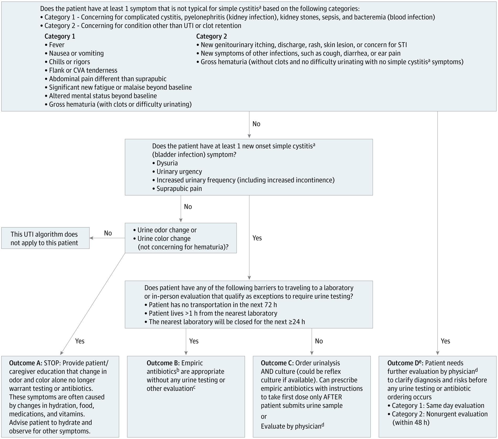
- Important negatives: Vaginal discharge (suggests alternative diagnosis), diarrhea, cough, genital discharge [6]
Alarm Features
- Sepsis signs: Hypotension, tachycardia, tachypnea, altered mental status, lactate elevation [7]
- Inability to tolerate oral intake (persistent vomiting) [1]
- Urine pH ≥7.0 (suggests urease-producing organism, e.g., Proteus; associated with struvite stones) [1]
- New decrease in GFR to ≤40 mL/min (suggestive of obstruction) [1]
- Known or suspected urolithiasis with concurrent infection (obstructed infected system = urologic emergency) [1][8]
- Failure to improve within 48–72 hours of appropriate antibiotics [4][8]
- Abnormalities in multiple CBC cell lines (leukocytosis + thrombocytopenia + anemia) — highly correlated with ICU admission [3]
- Immunocompromised state, diabetes, pregnancy, advanced age — higher risk for complications [9]
Medications
- Empiric outpatient oral options:
- Fluoroquinolones (ciprofloxacin 500 mg BID or levofloxacin 750 mg daily × 5–7 days) — if local resistance <10% [1][10]
- TMP-SMX (160/800 mg BID × 7–14 days) — if susceptibility known or local resistance <10% [10]
- Oral 3rd-generation cephalosporins (cefpodoxime) — limited evidence but used in practice [11]
- Supplemental parenteral dose at discharge: Ceftriaxone 1 g IV, gentamicin, amikacin, or ertapenem — recommended when anticipated resistance to the oral agent exceeds 10% [1][11]
- Inpatient parenteral options: Ceftriaxone, fluoroquinolones IV, piperacillin-tazobactam, carbapenems (for ESBL concern), aminoglycosides [8][12]
- Contraindicated/avoid:
- IV-to-oral transition: The 2025 IDSA guideline suggests transitioning to oral therapy once clinically improving, able to take PO, and an effective oral option is available — even in the setting of gram-negative bacteremia [13]
- Duration: 7 days for fluoroquinolones; 10–14 days for TMP-SMX or beta-lactams [1][10]
Diet
- Aggressive hydration is essential — oral if tolerated, IV if not [1]
- No specific dietary triggers for pyelonephritis
- Encourage adequate fluid intake during recovery to maintain urine output
- Cranberry products have limited evidence for prevention of recurrent UTI and no role in acute treatment
Review of Systems
- GU: Dysuria, frequency, urgency, hematuria, flank pain, suprapubic pain
- GI: Nausea, vomiting, diarrhea, abdominal pain (may mimic intra-abdominal pathology)
- Constitutional: Fever, chills, rigors, malaise, fatigue
- Respiratory: Dyspnea (consider ARDS in severe cases, especially pregnancy) [3]
- Neurologic: Altered mental status (sepsis indicator) [7]
- OB/GYN: Vaginal discharge (alternative diagnosis), contractions (in pregnancy) [3]
Collateral History and Family History
- Collateral: Medication compliance, recent antibiotic courses, recent healthcare exposures, functional status in elderly patients
- Family history: Vesicoureteral reflux (familial predisposition), polycystic kidney disease, recurrent UTIs
- Social context: Ability to take oral medications at home, reliable follow-up, stable home situation — critical for disposition decisions [1]
Risk Factors
- Female sex (short urethra, proximity to GI flora) [1]
- Sexual intercourse (strongest behavioral risk factor in young women) [9]
- Recent spermicide use
- Prior UTI or pyelonephritis [9]
- Urinary tract obstruction (stones, BPH, strictures) [5]
- Indwelling urinary catheter or recent instrumentation [5]
- Diabetes mellitus [9]
- Immunosuppression (transplant, HIV, chemotherapy) [9]
- Pregnancy (physiologic hydronephrosis, ureteral dilation) [3]
- Advanced age (higher hospitalization rates) [1]
- Anatomic/congenital urinary tract abnormalities [9]
- Recent antibiotic use or hospitalization (risk for resistant organisms) [1]
Differential Diagnosis
- Nephrolithiasis/ureterolithiasis — colicky pain, hematuria, may coexist with infection (pyonephrosis = emergency) [1]
- Renal abscess/perinephric abscess — failure to improve on antibiotics, CT needed [1]
- Acute cholecystitis — right-sided pain, Murphy's sign, may overlap with right-sided pyelonephritis [1]
- Appendicitis — right lower quadrant/flank pain, especially retrocecal appendix [1]
- Pelvic inflammatory disease — lower abdominal pain, cervical motion tenderness, vaginal discharge [1]
- Acute prostatitis (in men) — fever + pyuria without flank pain [1]
- Renal vein thrombosis — flank pain, hematuria, proteinuria [1]
- Paraspinous muscle disorders — musculoskeletal flank pain without systemic signs [1]
- Emphysematous pyelonephritis — diabetic patients, gas on imaging, high mortality [9]
- Lower lobe pneumonia — may present with upper abdominal/flank pain and fever
Past Medical History
- Prior UTIs or pyelonephritis episodes (recurrence risk)
- Known urolithiasis or urologic surgery
- Diabetes mellitus (risk for emphysematous pyelonephritis, complicated course)
- Immunocompromising conditions
- Chronic kidney disease (baseline GFR important for comparison)
- Structural urinary tract abnormalities (vesicoureteral reflux, neurogenic bladder)
- Pregnancy history
- Drug allergies — particularly beta-lactams and fluoroquinolones
- Recent antibiotic use (resistance risk) [1]
Physical Exam
- Vital signs: Fever (≥38°C), tachycardia, hypotension (sepsis), tachypnea [2-3]
- CVA tenderness — the only physical exam finding that increases the probability of upper tract UTI; nearly pathognomonic when combined with fever and pyuria [2][4]
- Abdominal exam: Flank tenderness, suprapubic tenderness; assess for peritoneal signs (alternative diagnosis)
- Pelvic exam (if indicated): Rule out PID, vaginitis, cervicitis
- Skin: Assess for mottling, delayed capillary refill (sepsis)
- Mental status: Confusion or altered sensorium suggests sepsis [7]
- Absence of flank pain should raise suspicion for an alternative diagnosis [4]
Lab Studies
- Urinalysis with microscopy: Pyuria (WBC), bacteriuria, nitrites, leukocyte esterase — dipstick sensitivity ~75%, specificity ~82% for combined LE/nitrite [2]
- Urine culture with susceptibilities: Mandatory in all suspected pyelonephritis — the cardinal confirmatory test; typically ≥10⁴ CFU/mL of a uropathogen [1-2][8]
- Blood cultures: Not needed in uncomplicated cases; obtain if sepsis suspected, immunocompromised, or diagnostic uncertainty [8][14]
- CBC: Leukocytosis expected; multi-cell-line abnormalities predict adverse outcomes [3]
- BMP/CMP: Creatinine (assess for obstruction if GFR ≤40), electrolytes, glucose
- Lactate: If sepsis suspected [7]
- CRP/procalcitonin: Not routinely needed in uncomplicated cases; procalcitonin ≥0.50 ng/mL has ~82% sensitivity for bacteremia in febrile UTI [15]
- Pregnancy test: All women of reproductive age
Imaging
- Uncomplicated pyelonephritis: Imaging is NOT indicated [4][8]
- Indications for imaging: [1][9]
- Sepsis or septic shock
- Known or suspected urolithiasis
- Urine pH ≥7.0
- New GFR decrease to ≤40 mL/min
- Failure to improve within 48–72 hours
- Complicated patient (diabetes, immunocompromised, recurrent pyelonephritis)
- First-line: CT abdomen/pelvis with IV contrast — detection rate ~84% vs ~40% for ultrasound [9]
- CT findings: Focal/multifocal decreased parenchymal enhancement, renal swelling, perinephric stranding, abscess, gas (emphysematous pyelonephritis) [9]
- Ultrasound: Reasonable first-line in pregnancy and when CT is contraindicated; good for detecting hydronephrosis and stones but poor sensitivity for parenchymal changes [9]
- Non-contrast CT: Adequate if primary concern is urolithiasis [9]
Special Tests
- qSOFA score: ≥2 of altered mentation, SBP ≤100, RR ≥22 — identifies patients at risk for poor outcomes from sepsis [16]
- SOFA score: For ICU-level assessment of organ dysfunction [16]
- Point-of-care ultrasound (POCUS): Assess for hydronephrosis at bedside in the ED
- Bedside bladder scan: Evaluate for urinary retention
- Post-void residual: If obstruction suspected
ECG
- Not routinely indicated for uncomplicated pyelonephritis
- Obtain ECG if:
- Sepsis or hemodynamic instability (to evaluate for sepsis-related cardiac dysfunction)
- Elderly patients or those with cardiac comorbidities
- Pre-existing cardiac disease with new tachycardia
- ECG findings in sepsis-associated pyelonephritis: Sinus tachycardia (most common), QT prolongation (54% of sepsis patients), new atrial fibrillation/flutter (associated with poor outcomes, OR 2.19), ST-T wave changes [17]
- QT prolongation is particularly relevant given that fluoroquinolones (commonly used for pyelonephritis) also prolong QT — exercise caution with concurrent QT-prolonging medications [17]
Assessment
Typical presentation: Young woman with acute onset of fever, unilateral flank pain, CVA tenderness, dysuria/frequency, and pyuria/bacteriuria on UA. [1]
Atypical presentations to recognize
- Absence of lower urinary tract symptoms (~20%) [1]
- Absence of fever early in the course [4]
- Elderly patients may present with altered mental status, generalized weakness, or GI symptoms without classic localizing signs
- Bilateral pyelonephritis (less common, more severe)
Severity stratification: [1]
- Mild: Low-grade fever, minimal nausea, no vomiting, stable vitals → outpatient
- Moderate: Higher fever, vomiting, unable to tolerate PO initially → ED observation/short stay
- Severe: Sepsis, septic shock, hemodynamic instability, obstruction → admission
Complications: Renal/perinephric abscess, emphysematous pyelonephritis, urosepsis, septic shock, ARDS, renal insufficiency, preterm labor (in pregnancy) [1][3]
Treatment Plan
Initial stabilization
- IV fluid resuscitation if dehydrated or hemodynamically unstable [1]
- Antipyretics/analgesics (acetaminophen, NSAIDs if no renal contraindication)
- Antiemetics (ondansetron) if vomiting
Outpatient management (uncomplicated, tolerating PO, stable): [1][8]
- Fluoroquinolone (ciprofloxacin 500 mg PO BID × 7 days or levofloxacin 750 mg PO daily × 5 days) if local resistance <10%
- TMP-SMX DS PO BID × 14 days (or 7 days if susceptible E. coli) [10]
- Consider single dose of ceftriaxone 1 g IV or IM before discharge if resistance concern >10% [1][11]
Inpatient management (severe illness, unable to tolerate PO, sepsis, pregnancy, obstruction): [1][3][8]
- IV antibiotics: Ceftriaxone 1–2 g IV daily, fluoroquinolone IV, or piperacillin-tazobactam
- If ESBL concern: Carbapenem (ertapenem or meropenem) [12]
- Transition to oral therapy once clinically improving, afebrile, tolerating PO [13]
Source control: Urgent urologic decompression (percutaneous nephrostomy or ureteral stent) if obstructed infected system [1][8]
Pregnancy: Admit all patients; IV ampicillin + gentamicin or ceftriaxone; complete 14 days total therapy [3]
Disposition
Specialist consultation triggers
- Urology: Obstructing stone with infection, abscess requiring drainage, emphysematous pyelonephritis
- Infectious disease: MDR organisms, ESBL-producing pathogens, treatment failure, immunocompromised host [1]
Follow Up / Return Precautions
- Follow-up timing: Primary care or urology within 48–72 hours if discharged from ED; sooner if culture results require antibiotic change [8]
- Expected course: Most patients improve within 48–72 hours of appropriate antibiotics [4][8]
- Return immediately for:
- Worsening or persistent fever >48–72 hours on antibiotics
- Inability to keep down oral medications or fluids
- Worsening flank pain
- Signs of sepsis: confusion, lightheadedness, rapid heart rate, difficulty breathing
- Decreased urine output
- No routine follow-up testing needed if clinical course is uneventful [1]
- If no improvement at 48–72 hours: Repeat urine culture, obtain imaging (CT with contrast), and reconsider diagnosis [1][4][8]
- Patient counseling: Complete the full antibiotic course; maintain hydration; avoid nephrotoxic agents (NSAIDs with caution if renal function borderline)
References
1. Acute Pyelonephritis in Adults. — Johnson JR, Russo TA. The New England Journal of Medicine. 2018.
2. Uncomplicated Urinary Tract Infection. — Hooton TM. The New England Journal of Medicine. 2012.
3. Urinary Tract Infections in Pregnant Individuals. — Committee on Clinical Consensus—Obstetrics Obstetrics and Gynecology. 2023.
4. Diagnosis and Treatment of Acute Pyelonephritis in Women. — Colgan R, Williams M, Johnson JR. American Family Physician. 2011.
5. Clinical Practice Guideline by Infectious Diseases Society of America (IDSA): 2025 Guideline on Management and Treatment of Complicated Urinary Tract Infections: Introduction and Methods. — Barbara W. Trautner, Nicolás W. Cortés-Penfield, Kalpana Gupta, et al Infectious Diseases Society of America. 2025.
6. Ann Arbor Guide to Triaging Adults With Suspected Urinary Tract Infection for In-Person and Telehealth Settings. — Meddings J, Chrouser K, Fowler KE, et al. JAMA Network Open. 2026.
7. Sepsis and Septic Shock. — Meyer NJ, Prescott HC. The New England Journal of Medicine. 2024.
8. Acute Pyelonephritis in Adults: Rapid Evidence Review. — Herness J, Buttolph A, Hammer NC. American Family Physician. 2020.
9. ACR Appropriateness Criteria® Acute Pyelonephritis: 2022 Update. — Expert Panel on Urological Imaging, Smith AD, Nikolaidis P, et al. Journal of the American College of Radiology : JACR. 2022.
10. Appropriate Use of Short-Course Antibiotics in Common Infections: Best Practice Advice From the American College of Physicians. — Lee RA, Centor RM, Humphrey LL, et al. Annals of Internal Medicine. 2021.
11. Clinical Practice Guideline by Infectious Diseases Society of America (IDSA): 2025 Guideline on Management and Treatment of Complicated Urinary Tract Infections: Selection of Antibiotic Therapy for Complicated UTI. — Barbara W. Trautner, Nicolás W. Cortés-Penfield, Kalpana Gupta, et al Infectious Diseases Society of America. 2025.
12. Infectious Diseases Society of America 2024 Guidance on the Treatment of Antimicrobial-Resistant Gram-Negative Infections. — Tamma PD, Heil EL, Justo JA, et al. Clinical Infectious Diseases : An Official Publication of the Infectious Diseases Society of America. 2024.
13. Clinical Practice Guideline by Infectious Diseases Society of America (IDSA): 2025 Guideline on Management and Treatment of Complicated Urinary Tract Infections: Timing of Intravenous to Oral Antibiotics Transition for Complicated UTI. — Barbara W. Trautner, Nicolás W. Cortés-Penfield, Kalpana Gupta, et al Infectious Diseases Society of America. 2025.
14. Early Care of Adults With Suspected Sepsis in The Emergency Department and Out-of-Hospital Environment: A Consensus-Based Task Force Report. — Yealy DM, Mohr NM, Shapiro NI, et al. Annals of Emergency Medicine. 2021.
15. A Reference Standard for Urinary Tract Infection Research: A Multidisciplinary Delphi Consensus Study. — Bilsen MP, Conroy SP, Schneeberger C, et al. The Lancet. Infectious Diseases. 2024.
16. Surviving Sepsis Campaign: International Guidelines for Management of Sepsis and Septic Shock 2021. — Evans L, Rhodes A, Alhazzani W, et al. Critical Care Medicine. 2021.
17. Association of Electrocardiogram Abnormalities With Clinical Outcomes in Emergency Department Sepsis Patients. — Kotruchin P, Chuehongthong M, Tangpaisarn T, et al. The Western Journal of Emergency Medicine. 2026.
Figures
More from Deep Dive
Reviewed by Dr. Lucas Mastropaolo. SymptomDx is an educational tool for medical professionals — it does not replace clinical judgment. Verify all clinical data and drug dosages with authoritative sources.