Rabies (Encephalitic)
Encephalitic (furious) rabies is an almost invariably fatal acute viral encephalitis caused by rabies virus (RABV), accounting for ~80% of clinical rabies cases. [1] It is characterized by fluctuating consciousness, hydrophobia, aerophobia, inspiratory spasms, and autonomic dysfunction, progressing to coma and death typically within 5–7 days of symptom onset without intensive care. [2] Approximately 59,000 human rabies deaths occur annually worldwide, with 98% linked to canine exposures. [3-4]
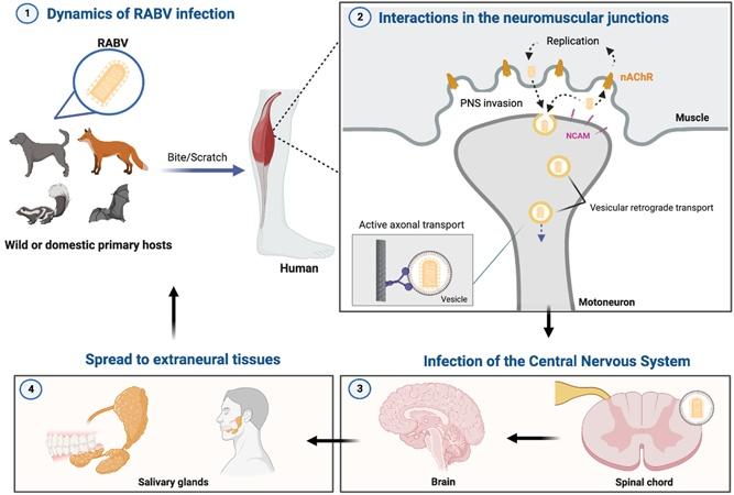
The following figure illustrates the pathogenic journey of RABV from initial bite inoculation through neuromuscular junction entry, CNS invasion, and centrifugal spread to salivary glands:
History
- Exposure history is the single most critical element: animal bite (dog, bat, raccoon, skunk, fox), scratch, or mucous membrane contact with saliva — may have occurred weeks to months prior [6]
- Ask about geographic location, travel to rabies-endemic regions (Asia, Africa), occupation (veterinarian, animal handler, cave explorer), and outdoor/recreational activities [4][7]
- Prodromal symptoms (2–10 days): fever, malaise, headache, nausea, sore throat — nonspecific and easily missed [2][8]
- Pain, paresthesia, or pruritus at the bite site is often the earliest neurological symptom and is highly suggestive [2][8]
- Progression to agitation, anxiety, confusion, hallucinations, hypersalivation, difficulty swallowing
- Hydrophobia: pharyngeal spasms triggered by sight, sound, or perception of water [6][8]
- Aerophobia: spasms triggered by air drafts
- Periods of agitation alternating with lucidity — a key feature of furious rabies [8]
- In the US, rabies diagnosis is almost always missed at the first clinical encounter because exposure history may be remote or unrecognized, particularly with bat exposures [6][9]
Alarm Features
- Hydrophobia or aerophobia — virtually pathognomonic for rabies [2][10]
- Inspiratory muscle spasms (spontaneous or provoked)
- Rapidly progressive encephalitis with autonomic instability (tachycardia, hypersalivation, hyperpyrexia, piloerection)
- Fluctuating consciousness with episodes of extreme agitation
- Cardiac arrhythmias — supraventricular arrhythmias, heart block, myocarditis mimicking STEMI [11-13]
- Seizures, delirium, or coma
- Any acute encephalitis in a patient with a history of animal bite in an endemic area
Medications
- No effective antiviral therapy exists once clinical symptoms develop [1][14-15]
- Milwaukee Protocol (therapeutic coma + ketamine + ribavirin + amantadine): now considered ineffective with at least 64 documented failures; should be abandoned [14-16]
- Ribavirin and interferon-α have been disappointing [15]
- Minocycline and corticosteroids should NOT be used — may aggravate disease [15]
- Ketamine and midazolam are used for symptom palliation (sedation, dysautonomia control) rather than as curative therapy [2]
- Nimodipine has been added in some protocols for vasospasm but carries risk of severe hypotension; use with extreme caution [2]
- Post-exposure prophylaxis (PEP) is the only effective intervention and must be given before symptom onset:
Diet
- Dysphagia is a cardinal feature — patients cannot swallow due to pharyngeal spasms; oral intake is impossible once hydrophobia develops [8][10]
- IV hydration and nutritional support are required
- No specific dietary triggers or long-term dietary management apply
Review of Systems
- Neurological: altered mental status, agitation, hallucinations, seizures, focal deficits, cranial nerve palsies (especially in bat-acquired cases), myoclonus, tremor [2][19]
- Respiratory: inspiratory spasms, periodic/ataxic breathing, respiratory arrest [10][13]
- Cardiovascular: palpitations, chest pain (myocarditis), arrhythmias [11][13]
- GI: nausea, vomiting, hypersalivation, GI bleeding [10]
- Autonomic: diaphoresis, piloerection, priapism, lacrimation, pupillary abnormalities
- Endocrine: diabetes insipidus, SIADH, hypothalamic-pituitary dysfunction [20-21]
- Musculoskeletal: fasciculations, weakness at bitten extremity
Collateral History and Family History
- Collateral history is essential: witnesses to animal exposure, travel companions, family members who may recall a bite the patient has forgotten or dismissed
- Children may not report bat contact or minor bites [6]
- Bat exposure may be unrecognized (e.g., bat found in room of sleeping person) [17]
- No hereditary predisposition; family history is not contributory
- Organ/tissue transplant history — rare cases of transmission via transplanted corneas and organs [17]
Risk Factors
- Dog bite in Asia or Africa — accounts for ~98% of global human rabies [4][22]
- Bat exposure in the Americas — most common source in the US (70% of indigenously acquired cases) [9]
- Bites to highly innervated areas (face, hands) or deep muscle bites increase transmission efficiency [2][6]
- Proximity of bite to CNS (head, neck) may shorten incubation period [6]
- Children — higher risk due to inquisitive behavior, smaller stature (more head/face bites), and failure to report exposures [6]
- Failure to receive or complete PEP [3]
- Occupational exposure: veterinarians, animal control workers, laboratory workers, cave explorers [4]
- Travel to endemic regions without pre-exposure prophylaxis [22]
Differential Diagnosis
- Tetanus — trismus (lockjaw), opisthotonus, sustained muscle rigidity (vs. intermittent spasms in rabies); no altered consciousness until late [10]
- Herpes simplex encephalitis (HSV-1) — temporal lobe predilection on MRI, hemorrhagic changes; no hydrophobia [7][23]
- Other viral encephalitides (arboviral, enteroviral, VZV) — distinguished by epidemiology, CSF findings, MRI patterns [7][24]
- Autoimmune encephalitis (anti-NMDAR, LGI1) — more subacute onset, psychiatric features, seizures; antibody testing differentiates [23][25]
- Guillain-Barré syndrome — mimics paralytic rabies; ascending weakness without altered consciousness [2]
- Delirium tremens — alcohol withdrawal history, tremor, hallucinations; no hydrophobia [10]
- Hysterical pseudohydrophobia — psychogenic; no autonomic dysfunction or progression [10]
- Postvaccinal encephalomyelitis — history of nervous-tissue rabies vaccine (older formulations) [10]
- Cerebral malaria, bacterial meningitis — in endemic settings, consider concurrent infections
Past Medical History
- Prior rabies vaccination status — critical for determining PEP regimen and prognosis; most survivors had received at least partial vaccination before symptom onset [8][16]
- Previous animal bites or PEP courses
- Immunocompromised states (HIV, chemotherapy) — may alter immune response and PEP efficacy; immunocompromised patients should receive 5-dose PEP schedule [18]
- History of organ transplantation (rare transmission route) [17]
- No specific chronic illnesses predispose to rabies, but immunosuppression may impair viral clearance
Physical Exam
- Vital signs: tachycardia, hypertension or hypotension, hyperpyrexia, irregular respirations [10][13]
- Inspection of bite wound: look for healing bite marks, especially on extremities, face, or hands; may show local inflammation
- Neurological exam:
- Fluctuating level of consciousness with periods of lucidity and agitation [2]
- Hydrophobia test: offering water provokes visible pharyngeal spasms and terror [8]
- Aerophobia test: blowing air on face triggers spasms
- Hypersalivation ("foaming at the mouth")
- Cranial nerve palsies (more common in bat-acquired cases) [19]
- Myoclonus, tremor, fasciculations
- Late: flaccid paralysis, areflexia, coma [2]
- Autonomic signs: piloerection, diaphoresis, pupillary dilation, priapism, lacrimation
- Cardiac: irregular rhythm, signs of heart failure (myocarditis) [11][13]
- Myoedema (mounding of muscle on percussion) — seen in paralytic form [2]
Lab Studies
- Routine labs: plasma neutrophil leukocytosis; otherwise often unremarkable [26]
- CSF: mild lymphocytic pleocytosis in only ~60% of patients in the first week; mildly elevated protein (more pronounced in bat-acquired cases); normal glucose [19][26]
- Antemortem diagnostic panel (contact CDC or public health lab): [6][27]
- Saliva: RT-PCR for RABV RNA (sensitivity ~86%) [28]
- Nuchal skin biopsy (nape of neck, hair follicle-containing): immunohistochemistry for rabies antigen in cutaneous nerves [6][27]
- Serum and CSF: rabies virus neutralizing antibody (RFFIT) — in unvaccinated patients, CSF antibody confirms diagnosis [6]
- CSF: RT-PCR for RABV RNA (sensitivity ~45% antemortem, higher postmortem) [28]
- Sequential sampling is indicated if initial tests are negative but clinical suspicion remains high [6]
- Neurofilament light chain (NfL): emerging biomarker — significantly elevated in rabies vs. other encephalitides [29]
- No single test is sufficient for antemortem diagnosis; multiple specimen types are required [27]
Imaging
- MRI brain (preferred over CT): [2][30]
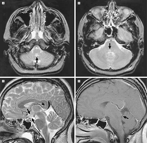
- Non-comatose phase: subtle, ill-defined T2/FLAIR hyperintensities in brainstem, hippocampi, hypothalami, deep/subcortical white matter — without gadolinium enhancement (a distinguishing feature from other viral encephalitides) [2][30]
- Comatose phase: more widespread T2 hyperintensities; gadolinium enhancement may appear along brainstem and midline structures [2]
- Brachial plexus enhancement on the bitten side may be seen even during prodrome [2]
- Novel findings include dentate nuclei involvement, cranial nerve enhancement, "hot cross bun" sign in pons [31]
- CT head: low sensitivity; may show only diffuse edema in late stages
- The lack of contrast enhancement in a noncomatose encephalitis patient should raise suspicion for rabies over other viral encephalitides [30]
The following MRI demonstrates characteristic brainstem T2 hyperintensities in a confirmed case of rabies encephalitis:
Special Tests
- Direct fluorescent antibody (DFA) test: gold standard for postmortem diagnosis on brain tissue; detects rabies antigen [27]
- Negri bodies: eosinophilic cytoplasmic inclusions in hippocampal and cerebellar Purkinje neurons — pathognomonic on histopathology [32]
- Virus isolation from saliva [27]
- WHO clinical case definition: acute neurologic syndrome dominated by hyperactivity (furious) or paralysis, progressing to coma and death within 7–10 days [6]
- Animal testing: if the biting animal is available, DFA testing of brain tissue; 10-day observation period for dogs, cats, and ferrets [8][17]
ECG
- Cardiac involvement is common and clinically significant [1][11][13]
- Supraventricular arrhythmias (most common cardiac finding) [13]
- ST-segment elevation mimicking STEMI — due to interstitial myocarditis, not coronary occlusion [11]
- Heart block (including complete heart block) — may be the terminal event [12]
- Autonomic denervation manifests as loss of heart rate variability [33]
- ECG should be obtained in all suspected cases; continuous telemetry monitoring is essential
- Vagus nerve neuritis found at autopsy explains many of the cardiac manifestations [12]
Assessment
- Encephalitic rabies is a clinical diagnosis supported by exposure history and confirmed by specialized laboratory testing [6]
- Average survival from symptom onset to death: 5.7 days (furious form) without intensive care [2]
- Only ~34 well-documented survivors exist; most had received partial vaccination and/or were infected with bat RABV variants; many had severe neurological sequelae [8][14]
- The disease is almost 100% fatal once clinical signs appear [1][5]
- Atypical presentations (focal brainstem signs, myoclonus, hemichorea, Horner syndrome) are more common with bat RABV variants and contribute to frequent misdiagnosis [2][34]
- Complications in ICU-managed patients include ARDS, diabetes insipidus, myocarditis, GI bleeding, and multi-organ failure [1][20]
Treatment Plan
Initial stabilization
- Airway management — early intubation may be needed for inspiratory spasms and airway protection
- IV access, continuous telemetry, ICU admission
- Aggressive sedation with benzodiazepines (midazolam) and ketamine for agitation, spasms, and dysautonomia [2]
- Avoid neuromuscular paralysis if possible (masks clinical progression)
Definitive management
- No proven curative therapy exists [1][14-15]
- Decision between aggressive critical care vs. palliative approach should be made early with the patient's family:
- Favorable factors for aggressive approach: prior partial vaccination, young age, immunocompetent, bat RABV variant, early neutralizing antibody detection, mild neurological disease at presentation [8]
- Palliative approach: liberal sedation (midazolam, opioids) to relieve suffering; appropriate for most cases, especially canine RABV variant [2][8]
- Milwaukee Protocol should no longer be used — at least 64 failures documented [14]
- Manage complications: vasopressors for hemodynamic instability, antiarrhythmics, correction of DI/SIADH, ventilatory support
PEP (pre-symptom only)
- Wound cleansing with soap/water for ≥15 minutes + povidone-iodine [3][8]
- HRIG 20 IU/kg infiltrated into and around wound [17]
- Rabies vaccine IM: days 0, 3, 7, 14 (4-dose schedule per ACIP) [18]
Disposition
- All suspected rabies cases require ICU admission once neurological symptoms develop [1][8]
- Strict contact and droplet precautions for healthcare workers; PEP for any staff with mucous membrane or open wound exposure to patient saliva [17]
- Rabies is a nationally notifiable disease — immediate notification to local/state public health department and CDC [6]
- Infectious disease and neurology consultation
- Palliative care consultation when aggressive treatment is not pursued or deemed futile
- No role for observation unit or outpatient management once symptomatic
Follow Up / Return Precautions
For exposed but asymptomatic patients receiving PEP
- Complete the full vaccine series on schedule (days 0, 3, 7, 14) [18]
- Return immediately for any neurological symptoms: paresthesia at bite site, anxiety, agitation, difficulty swallowing, fever with behavioral changes
- Wound care follow-up; monitor for secondary bacterial infection
For symptomatic patients
- Prognosis is nearly uniformly fatal; family counseling and goals-of-care discussions are paramount [1][14]
- Rare survivors require prolonged neurorehabilitation; most have significant neurological sequelae [8][14]
- Healthcare workers exposed to patient secretions should receive PEP evaluation
Public health follow-up
- Ensure the biting animal is captured for testing or observation if possible [8]
- Contact tracing for other potential exposures from the same animal source
References
1. Rabies: A Medical Perspective. — Jackson AC. Revue Scientifique Et Technique. 2018.
2. Human Rabies: Neuropathogenesis, Diagnosis, and Management. — Hemachudha T, Ugolini G, Wacharapluesadee S, et al. The Lancet. Neurology. 2013.
3. Human Rabies Despite Post-Exposure Prophylaxis: A Systematic Review of Fatal Breakthrough Infections After Zoonotic Exposures. — Whitehouse ER, Mandra A, Bonwitt J, et al. The Lancet. Infectious Diseases. 2023.
4. Use of a Modified Preexposure Prophylaxis Vaccination Schedule to Prevent Human Rabies: Recommendations of the Advisory Committee on Immunization Practices - United States, 2022. — Rao AK, Briggs D, Moore SM, et al. MMWR. Morbidity and Mortality Weekly Report. 2022.
5. Neuroimmunology of rabies: New insights into an ancient disease. — Bastos V, Pacheco V, Rodrigues ÉDL, et al. Journal of Medical Virology. 2023.
6. Rabies. — Ryan M. Wallace and David R. Shlim CDC Yellow Book. 2025.
7. Acute Viral Encephalitis. — Tyler KL. The New England Journal of Medicine. 2018.
8. Current Status of Rabies and Prospects for Elimination. — Fooks AR, Banyard AC, Horton DL, et al. Lancet. 2014.
9. Vital Signs: Trends in Human Rabies Deaths and Exposures - United States, 1938-2018. — Pieracci EG, Pearson CM, Wallace RM, et al. MMWR. Morbidity and Mortality Weekly Report. 2019.
10. The Clinical Picture of Rabies in Man. — Warrell DA. Transactions of the Royal Society of Tropical Medicine and Hygiene. 1976.
11. Rabies Encephalitis With Myocarditis Mimicking ST-Elevation Myocardial Infarction. — Park SC, Crane IM, Pal K, Cagnina RE. Open Forum Infectious Diseases. 2019.
12. Diagnosis, Management and Post-Mortem Findings of a Human Case of Rabies Imported Into the United Kingdom From India: A Case Report. — Pathak S, Horton DL, Lucas S, et al. Virology Journal. 2014.
13. Pathophysiologic Studies in Human Rabies. — Warrell DA, Davidson NM, Pope HM, et al. The American Journal of Medicine. 1976.
14. Demise of the Milwaukee Protocol for Rabies. — Jackson AC. Clinical Infectious Diseases : An Official Publication of the Infectious Diseases Society of America. 2025.
15. Antiviral Therapy for Human Rabies. — Appolinario CM, Jackson AC. Antiviral Therapy. 2014.
16. Current and Future Approaches to the Therapy of Human Rabies. — Jackson AC. Antiviral Research. 2013.
17. FDA Drug Label. — Food and Drug Administration (DailyMed)..
18. Use of a Reduced (4-Dose) Vaccine Schedule for Postexposure Prophylaxis to Prevent Human Rabies: Recommendations of the Advisory Committee on Immunization Practices. — Rupprecht CE, Briggs D, Brown CM, et al. MMWR. Recommendations and Reports : Morbidity and Mortality Weekly Report. Recommendations and Reports. 2010.
19. Clinical Features of Dog- And Bat-Acquired Rabies in Humans. — Udow SJ, Marrie RA, Jackson AC. Clinical Infectious Diseases : An Official Publication of the Infectious Diseases Society of America. 2013.
20. Human Rabies: Clinical Features, Diagnosis, Complications, and Management. — Udwadia ZF, Udwadia FE, Katrak SM, et al. Critical Care Medicine. 1989.
21. Correlation of Clinical and Neuroimaging Findings in a Case of Rabies Encephalitis. — Pleasure SJ, Fischbein NJ. Archives of Neurology. 2000.
22. Boostability After Single-Visit Pre-Exposure Prophylaxis With Rabies Vaccine: A Randomised Controlled Non-Inferiority Trial. — Overduin LA, Koopman JPR, Prins C, et al. The Lancet. Infectious Diseases. 2024.
23. Encephalitis. — Binks SNM, Saylor D, Easton A, Thakur KT, Irani SR. Lancet. 2026.
24. Viral Encephalitis. — Venkatesan A, Murphy OC. Neurologic Clinics. 2018.
25. Acute Encephalitis in Immunocompetent Adults. — Venkatesan A, Michael BD, Probasco JC, Geocadin RG, Solomon T. Lancet. 2019.
26. Rabies and Other Lyssavirus Diseases. — Warrell MJ, Warrell DA. Lancet. 2004.
27. Guide to Utilization of the Microbiology Laboratory for Diagnosis of Infectious Diseases: 2024 Update by the Infectious Diseases Society of America (IDSA) and the American Society for Microbiology (ASM). — Miller JM, Binnicker MJ, Campbell S, et al. Clinical Infectious Diseases : An Official Publication of the Infectious Diseases Society of America. 2024.
28. Utility of real‐time Taqman PCR for antemortem and postmortem diagnosis of human rabies. — Mani RS, Madhusudana SN, Mahadevan A, et al. Journal of Medical Virology. 2014.
29. Neurofilament Light Chain: A Potential Diagnostic Biomarker for Rabies. — Siddalingaiah N, Lodha L, Ashwini MA, et al. Microbiology and Immunology. 2025.
30. MR Imaging in Human Rabies. — Laothamatas J, Hemachudha T, Mitrabhakdi E, Wannakrairot P, Tulayadaechanont S. AJNR. American Journal of Neuroradiology. 2003.
31. Neuroimaging Findings in Rabies Encephalitis. — Bhat MD, Priyadarshini P, Prasad C, Kulanthaivelu K. Journal of Neuroimaging : Official Journal of the American Society of Neuroimaging. 2021.
32. Histopathology of vaccine‐preventable diseases. — Solomon IH, Milner DA. Histopathology. 2017.
33. Survival after Treatment of Rabies with Induction of Coma. — Willoughby RE, Tieves KS, Hoffman GM, et al. The New England Journal of Medicine. 2005.
34. Comparative Pathogenesis of Rabies in Bats and Carnivores, and Implications for Spillover to Humans. — Begeman L, GeurtsvanKessel C, Finke S, et al. The Lancet. Infectious Diseases. 2018.
Figures
More from Deep Dive
Reviewed by Dr. Lucas Mastropaolo. SymptomDx is an educational tool for medical professionals — it does not replace clinical judgment. Verify all clinical data and drug dosages with authoritative sources.