Raynaud's Disease
Raynaud's phenomenon (RP) is an exaggerated vasospastic response to cold or emotional stress causing episodic digital ischemia with characteristic triphasic color changes (white → blue → red). [1-2] It affects 3–5% of the general population, with a strong female predominance (F:M ratio ~4:1). [3-4] The critical clinical task is distinguishing primary RP (idiopathic, benign) from secondary RP (associated with underlying disease, particularly connective tissue diseases). [1][5]
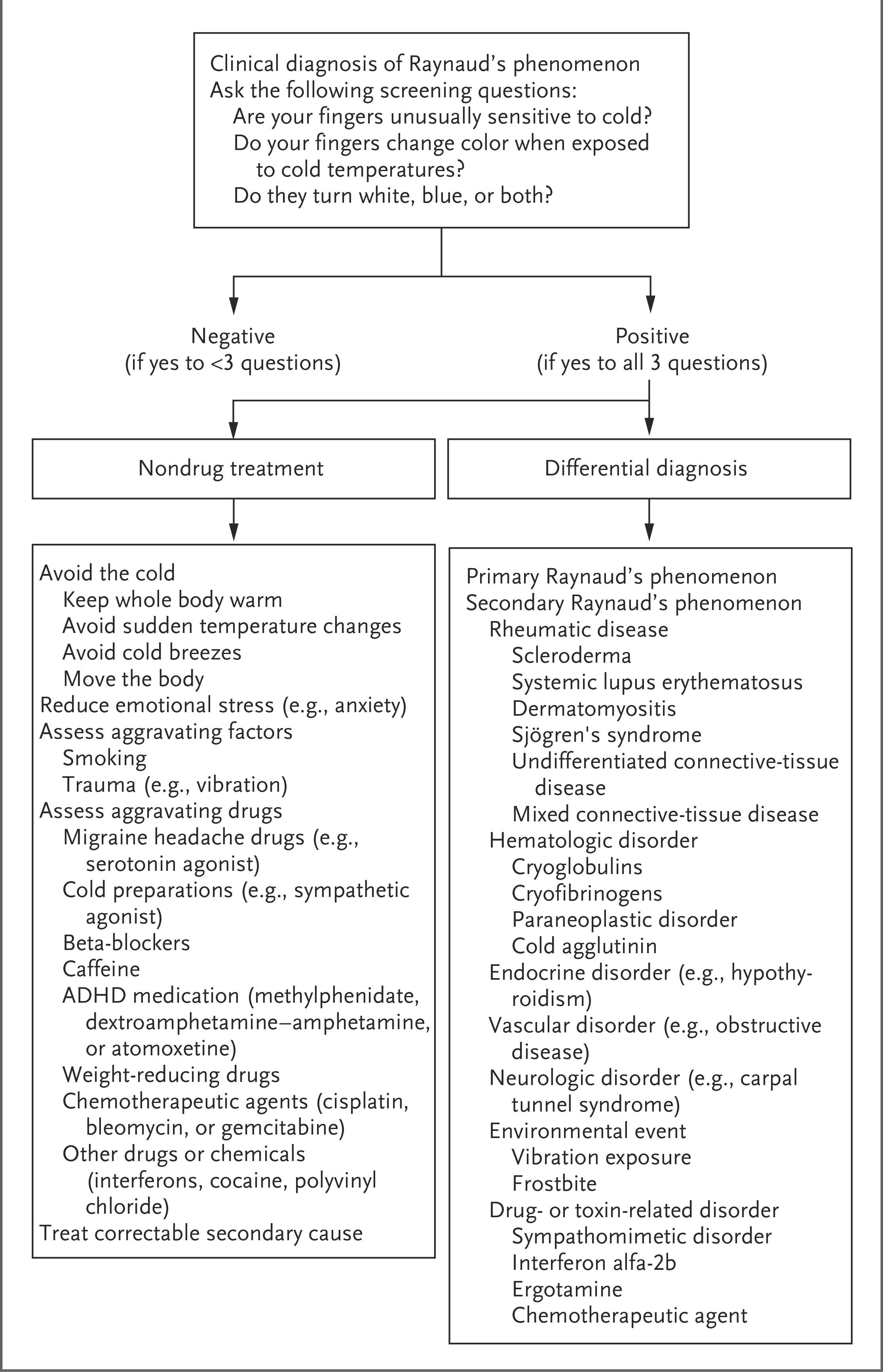
The following NEJM algorithm outlines the diagnostic and nondrug management approach:
History
- Triphasic color change: Ask specifically about well-demarcated pallor (white), cyanosis (blue), and rubor (red) of digits — at least biphasic change (white + blue) is needed for diagnosis [1]
- Triggers: Cold exposure (>80% of cases), emotional stress, rapid temperature shifts (e.g., entering air-conditioned spaces) [1-2]
- Timing and duration: Typical episode lasts ~15–20 minutes after rewarming; ask about frequency, seasonality, and progression over time [1][6]
- Digit involvement: Which fingers/toes? Symmetric vs. asymmetric? Thumb involvement? (Thumb is generally spared in primary RP) [1]
- Associated symptoms: Numbness, tingling, pain during attacks; stinging/throbbing during reperfusion [7]
- Ischemic complications: Digital ulcers, pitting scars, gangrene (strongly suggest secondary RP) [4][8]
- Connective tissue disease symptoms: Arthralgia, skin tightening, dysphagia, dry eyes/mouth, rash, myalgia, fever [5]
- Occupational exposure: Vibratory tools (hand-arm vibration syndrome), repetitive hand trauma [5][8]
- Age of onset: Primary RP typically 15–30 years; secondary RP usually >30–40 years [1][9]
Alarm Features
- Digital ulceration, pitting scars, or gangrene — indicates severe ischemia and secondary RP [4][8]
- Acute persistent digital ischemia (prolonged pallor/cyanosis not resolving with rewarming) — constitutes an ischemic crisis requiring urgent intervention [5]
- Asymmetric attacks or involvement of a single digit — raises concern for embolic or structural vascular disease [5]
- Late onset (>40 years) with no prior history — consider secondary causes including malignancy (tumor screening warranted if B symptoms present) [3][5]
- New onset in childhood — rare and points to secondary origin [3]
- Signs of connective tissue disease: Sclerodactyly, skin thickening, telangiectasias, calcinosis, pulmonary hypertension symptoms [1][5]
- Abnormal nailfold capillaries — highly predictive of progression to systemic sclerosis [10-11]
Medications
Medications that can trigger/worsen RP (avoid or reassess)
- Beta-blockers (nonselective > selective)
- Ergot alkaloids and triptans (migraine drugs)
- Sympathomimetics, amphetamines, ADHD medications
- Chemotherapeutics (bleomycin, cisplatin, vinblastine)
- Clonidine, cocaine, decongestants
- Caffeine (evidence not solid but commonly listed) [1-2][8]
First-line treatment: Dihydropyridine calcium channel blockers [5][10][12]
- Nifedipine SR 30 mg/day (titrate up; maintenance can exceed 60 mg/day) or amlodipine 5 mg/day
- Cochrane review: ~1.7 fewer attacks/week vs. placebo for primary RP [1][13]
- Side effects: headache, flushing, peripheral edema, hypotension [14]
Second-line options: [1][12][15]
- PDE-5 inhibitors (sildenafil, tadalafil) — especially for refractory RP or digital ulcers
- Topical nitroglycerin 2% ointment (¼–½ inch daily)
- ARBs (losartan 50 mg/day) — some evidence of benefit
- SSRIs (fluoxetine 20 mg/day) — open-label data suggest efficacy
- Prazosin (alpha-1 blocker)
Severe/refractory or digital ulcers: [5][12][15]
- IV iloprost (prostacyclin analogue) — for acute ischemic crisis or refractory ulcers
- Bosentan (endothelin receptor antagonist) — reduces new digital ulcers in scleroderma
- Botulinum toxin injections — emerging evidence for refractory cases
- Aspirin — recommended during acute digital ischemic crisis [5]
Contraindicated/caution: Oral prostaglandins are ineffective for RP. [12] Avoid combining nitrates with PDE-5 inhibitors (hypotension risk).
Diet
- Caffeine: Commonly recommended to limit, though evidence is not definitive [1][8]
- Alcohol: Limit intake; while acutely vasodilatory, chronic use and rebound vasoconstriction may worsen symptoms [16]
- Smoking cessation: Critically important — smoking reduces digital blood flow and worsens vascular damage [1][5][8]
- Beetroot juice: One study showed improved peripheral blood flow, endothelial function, and anti-inflammatory status in RP patients following nitrate-rich beetroot supplementation, though this is preliminary [17]
- Hydration: Maintain adequate hydration to support peripheral perfusion
- Omega-3 fatty acids: Sometimes recommended for anti-inflammatory properties, though evidence specific to RP is limited
Review of Systems
- Musculoskeletal: Arthralgia, arthritis, myalgia (connective tissue disease)
- Dermatologic: Skin tightening/thickening, telangiectasias, calcinosis, rash (malar, discoid), photosensitivity
- GI: Dysphagia, reflux, bloating (scleroderma esophageal dysmotility)
- Pulmonary: Dyspnea on exertion, dry cough (ILD, pulmonary hypertension)
- Ophthalmologic/Oral: Dry eyes, dry mouth (Sjögren syndrome)
- Constitutional: Fever, weight loss, fatigue (CTD or malignancy)
- Neurologic: Carpal tunnel symptoms, neuropathy
- Vascular: Livedo reticularis, claudication [5][18]
Collateral History and Family History
- Family history: 30–50% of patients with primary RP have a first-degree relative with the condition, suggesting genetic susceptibility [1][9]
- Family history of autoimmune disease: Scleroderma, SLE, Sjögren syndrome, rheumatoid arthritis
- Occupational history: Vibratory tool use, cold workplace exposure, repetitive hand trauma [5]
- Social history: Smoking status (critical), recreational drug use (cocaine, amphetamines)
- Medication reconciliation: Confirm all current medications including OTC decongestants, supplements, and migraine treatments [1]
Risk Factors
- Female sex (prevalence 2–20% in women vs. 1–12% in men) [2][9]
- Cold climate (geographically variable prevalence) [9]
- Family history of RP or autoimmune disease [1][9]
- Smoking [5][8]
- Occupational vibration exposure (hand-arm vibration syndrome) [5]
- Connective tissue disease (>90% of systemic sclerosis patients have RP) [9]
- Positive ANA or disease-specific autoantibodies (anticentromere, anti-Scl-70) [5][8]
- Abnormal nailfold capillaroscopy [10-11]
- Medications: Beta-blockers, ergotamines, chemotherapy agents [2]
- Hematologic disorders: Polycythemia vera, cryoglobulinemia, cold agglutinin disease [15-16]
- Recent data suggest RP is associated with increased cardiovascular disease and VTE risk independent of traditional risk factors [19]
Differential Diagnosis
- Acrocyanosis: Persistent (not episodic) symmetric cyanosis of hands/feet; no pallor phase; benign [15]
- Pernio (chilblains): Inflammatory lesions (erythematous/violaceous papules/nodules) after cold/damp exposure; distinct from vasospasm [15]
- Peripheral arterial disease: Asymmetric, associated with diminished pulses, claudication; older patients with atherosclerotic risk factors
- Thoracic outlet syndrome: Positional symptoms, unilateral, provoked by arm elevation [5]
- Carpal tunnel syndrome: Numbness/tingling in median nerve distribution; may coexist
- Achenbach syndrome: Spontaneous venous hemorrhage of digits; self-limiting, often mistaken for RP [15]
- Complex regional pain syndrome: Pain, swelling, color/temperature changes; usually post-traumatic [15]
- Small fiber neuropathy with vasomotor symptoms [15]
- Embolic disease: Acute unilateral digital ischemia; consider cardiac source, proximal aneurysm
- Cryoglobulinemia / cold agglutinin disease: Hematologic causes of cold-induced digital ischemia
- Erythromelalgia: Episodic burning pain with erythema and warmth (opposite of RP)
- Buerger disease (thromboangiitis obliterans): Young male smokers with distal extremity ischemia
Past Medical History
- Connective tissue disease: Systemic sclerosis, SLE, Sjögren syndrome, rheumatoid arthritis, dermatomyositis, mixed CTD [1][4]
- Hypothyroidism: Endocrine cause of secondary RP [3]
- Hematologic disorders: Polycythemia, cryoglobulinemia, paraproteinemia [3]
- Prior digital ulcers or amputations
- Carpal tunnel syndrome (may coexist or mimic)
- Atherosclerotic disease / PAD
- Previous frostbite or hand trauma
- Malignancy (paraneoplastic RP, especially with late-onset) [3]
- Pulmonary hypertension [20]
Physical Exam
- Vital signs: Blood pressure in both arms (asymmetry suggests vascular pathology); heart rate
- Hands/feet: Inspect for color changes, digital pitting scars, ulcers, gangrene, calcinosis, sclerodactyly, skin thickening [1][8]
- Nailfold capillaroscopy: Performed with ophthalmoscope or dermatoscope at nail base with drop of oil — look for dilated capillary loops, avascular areas, hemorrhages (scleroderma pattern) [1][10-11]
- Allen test: Assess radial and ulnar artery patency
- Peripheral pulses: Radial, ulnar, dorsalis pedis, posterior tibial — diminished pulses suggest structural vascular disease
- Skin exam: Telangiectasias, malar rash, livedo reticularis, skin tightening, Raynaud-related changes on ears/nose/nipples [6][21]
- Musculoskeletal: Joint swelling, synovitis, tendon friction rubs (scleroderma)
- Thoracic outlet maneuvers: Adson test, costoclavicular maneuver if positional symptoms [5]
- Oral exam: Dry mucous membranes, microstomia (scleroderma)
Lab Studies
Initial workup (to distinguish primary from secondary RP): [1][5][16]
- ANA — negative or low-titer (≤1:40) supports primary RP; positive warrants further workup
- Anti-centromere antibody — associated with limited scleroderma (CREST)
- Anti-Scl-70 (anti-topoisomerase I) — associated with diffuse scleroderma
- Anti-RNP — mixed connective tissue disease
- ESR / CRP — elevated suggests secondary cause (though normal ESR no longer required to diagnose primary RP) [1]
- CBC — polycythemia, thrombocytosis, anemia
- Comprehensive metabolic panel — renal function (scleroderma renal crisis)
- TSH — hypothyroidism as secondary cause
- Rheumatoid factor
- Complement levels (C3, C4) — if SLE suspected
- Cryoglobulins — if hematologic cause suspected
- Urinalysis — proteinuria (renal involvement in CTD)
A connective tissue disease developed in only 2% of patients who were seronegative for ANA and RF, versus ~15–20% of those with positive autoantibodies or abnormal capillaroscopy. [5]
Imaging
- Nailfold capillaroscopy: First-line and most important imaging tool — gold standard for microvascular assessment; abnormal patterns (giant capillaries, hemorrhages, avascular areas) are highly predictive of SSc [1][10-11]
- Arterial Doppler / duplex ultrasound: If concern for proximal arterial occlusion, thoracic outlet syndrome, or embolic disease
- CT angiography or MR angiography: For suspected large-vessel disease or thoracic outlet syndrome
- Chest X-ray / CT chest: If pulmonary symptoms suggest ILD or pulmonary hypertension in secondary RP
- Echocardiography: If pulmonary hypertension suspected (elevated RVSP)
- Imaging is generally unnecessary for straightforward primary RP in young patients with normal exam and negative serologies [1][5]
Special Tests
- Nailfold capillaroscopy: Key risk-stratification tool; scleroderma pattern predicts CTD transition [10-11]
- Cold stimulation test: Hands immersed in ice water; recovery time >20 minutes suggests RP [16]
- Raynaud's Condition Score (RCS): Patient-reported 0–10 scale assessing daily difficulty; used to guide treatment escalation [1]
- Laser Doppler flowmetry / thermography: Research tools for quantifying digital perfusion; not widely used clinically [22]
- Digital plethysmography: Assesses digital artery pressures and waveforms
- Botulinum toxin injection: Both diagnostic (response confirms vasospastic component) and therapeutic in refractory cases [15]
ECG
- ECG is not routinely indicated for isolated primary RP
- In secondary RP with systemic sclerosis, ECG may reveal:
- Conduction abnormalities (bundle branch blocks, AV blocks)
- Arrhythmias (atrial and ventricular)
- ST-T wave changes during cold-induced "cardiac Raynaud's" (transient myocardial ischemia) [23-24]
- Cold-induced segmental myocardial dysfunction has been demonstrated in SSc patients with long-standing RP (>5 years), even with normal coronary arteries [24-25]
- Consider ECG if chest pain, palpitations, or syncope in the setting of secondary RP
- RP patients have higher rates of ischemic heart disease (22.9% vs. 19.5%) and heart failure (19.2% vs. 14.5%) compared to matched controls [20]
Assessment
Primary RP (80–90% of cases): [9]
- Onset age 15–30, female predominance, symmetric, thumb spared
- Mild attacks, no digital ulcers, normal nailfold capillaries, negative/low-titer ANA
- Benign course; however, ~12.6% may develop a secondary cause over time (almost all CTD) [5]
Secondary RP (10–20% of cases)
- Onset >30–40 years, more severe/painful attacks, asymmetric involvement possible
- Digital ulcers, pitting scars, gangrene may occur [4][8]
- Most commonly associated with systemic sclerosis (>90% of SSc patients have RP) [9]
- Other associations: SLE, Sjögren, RA, dermatomyositis, MCTD, hematologic disorders [6]
Complications: Digital ulceration, critical digital ischemia, tissue necrosis, amputation (rare, almost exclusively secondary RP), and increased cardiovascular/VTE risk. [14][19]
Treatment Plan
All patients — nonpharmacologic (foundation of therapy): [1][5][8]
- Cold avoidance: layered clothing, gloves, hats, heated gloves/footwear, chemical hand warmers
- Avoid rapid temperature shifts
- Smoking cessation (strongly recommended)
- Stress reduction techniques
- Avoid offending medications (beta-blockers, ergotamines, sympathomimetics)
- Regular exercise to improve peripheral circulation
Pharmacologic — stepwise escalation: [1][5][12]
Acute digital ischemic crisis: Immediate rewarming, vasodilator therapy (nifedipine or IV iloprost), aspirin, and urgent evaluation for underlying cause. [5] Early intervention is key before irreversible tissue injury.
Disposition
Discharge criteria (majority of RP presentations)
- Primary RP with mild-moderate symptoms responding to rewarming
- No digital ulcers, gangrene, or signs of critical ischemia
- Able to follow up with PCP or rheumatology
- Appropriate prescriptions and lifestyle counseling provided
Observation / Admission criteria
- Acute digital ischemic crisis with persistent ischemia despite rewarming and oral vasodilators [5]
- Digital gangrene or threatened tissue loss requiring IV prostanoid therapy
- Severe pain uncontrolled with oral medications
- New diagnosis of secondary RP with concerning systemic features (e.g., scleroderma renal crisis, pulmonary hypertension)
Specialist consultation triggers: [10][12]
- Rheumatology: Suspected secondary RP, positive autoantibodies, abnormal capillaroscopy, digital ulcers
- Vascular surgery: Refractory ischemia, consideration for sympathectomy, suspected large-vessel disease
- Hand surgery: Digital ulcers requiring debridement or threatened amputation
- Cardiology: Suspected pulmonary hypertension or cardiac involvement
- Pulmonology: ILD screening in SSc-associated RP
Follow Up / Return Precautions
Follow-up timing
- Primary RP: PCP follow-up in 2–4 weeks if starting medication; annual reassessment for evolution to secondary RP [3]
- Secondary RP: Rheumatology follow-up within 2–4 weeks; ongoing monitoring for organ involvement
- Patients with positive ANA or abnormal capillaroscopy but no definite CTD: ~15–20% will develop a defined CTD, usually within 2 years — close surveillance warranted [5]
Return precautions — instruct patients to seek immediate care for:
- Persistent color change (white or blue) in a digit lasting >30 minutes despite rewarming
- New digital ulcers, open sores, or blackened skin on fingertips
- Severe unrelenting pain in digits
- Fever, new joint swelling, skin changes, or difficulty swallowing (suggesting CTD progression)
- Chest pain or shortness of breath
Patient counseling
- RP is a chronic condition; primary RP is generally benign with excellent prognosis
- Lifestyle modifications are the cornerstone and often sufficient for primary RP [1]
- Medication side effects (headache, flushing, edema with CCBs) are common but manageable
- Expected recovery: attacks typically resolve within 15–20 minutes of rewarming [6]
- Long-term specialist care is necessary for patients with risk factors for secondary RP [3]
References
1. Raynaud’s Phenomenon. — Wigley FM, Flavahan NA. The New England Journal of Medicine. 2016.
2. Vasodilators for Primary Raynaud's Phenomenon. — Su KY, Sharma M, Kim HJ, et al. The Cochrane Database of Systematic Reviews. 2021.
3. Raynaud's Phenomenon: A Vascular Acrosyndrome That Requires Long-Term Care. — Klein-Weigel P, Sander O, Reinhold S, et al. Deutsches Arzteblatt International. 2021.
4. Raynaud's Phenomenon. — Haque A, Hughes M. Clinical Medicine. 2020.
5. Raynaud's Phenomenon. — Wigley FM. The New England Journal of Medicine. 2002.
6. Raynaud phenomenon. — National Library of Medicine (MedlinePlus) 2018.
7. Clinically Relevant Differences Between Primary Raynaud's Phenomenon and Secondary to Connective Tissue Disease. — Di Donato S, Huang S, Pauling JD, et al. Seminars in Arthritis and Rheumatism. 2024.
8. Raynaud's Phenomenon. — Block JA, Sequeira W. Lancet. 2001.
9. Calcium Channel Blockers for Primary and Secondary Raynaud's Phenomenon. — Rirash F, Tingey PC, Harding SE, et al. The Cochrane Database of Systematic Reviews. 2017.
10. Practical Management of Raynaud's Phenomenon - A Primer for Practicing Physicians. — Ramahi A, Hughes M, Khanna D. Current Opinion in Rheumatology. 2022.
11. Causes of Raynaud's Phenomenon and the Predictive Laboratory and Capillaroscopy Features for the Evolution to a Definite Connective Tissue Disease. — Shenavandeh S, Ajri M, Hamidi S. Rheumatology. 2022.
12. Systemic Sclerosis: Evaluation and Treatment. — Frazier W, Goettel C, Chaudhri P. American Family Physician. 2026.
13. Calcium Channel Blockers for Primary Raynaud's Phenomenon. — Ennis H, Hughes M, Anderson ME, Wilkinson J, Herrick AL. The Cochrane Database of Systematic Reviews. 2016.
14. The Diagnosis and Treatment of Raynaud's Phenomenon: A Practical Approach. — Pope JE. Drugs. 2007.
15. Cold Hands or Feet: Is It Raynaud's or Not?. — Kadian-Dodov D. The Medical Clinics of North America. 2023.
16. Cold Stimulation Test. — National Library of Medicine (MedlinePlus) 2021.
17. "Beet" the Cold: Beetroot Juice Supplementation Improves Peripheral Blood Flow, Endothelial Function, and Anti-Inflammatory Status in Individuals With Raynaud's Phenomenon. — Shepherd AI, Costello JT, Bailey SJ, et al. Journal of Applied Physiology. 2019.
18. Raynaud's Phenomenon-an Update on Diagnosis, Classification and Management. — Pauling JD, Hughes M, Pope JE. Clinical Rheumatology. 2019.
19. Raynaud's Phenomenon Is Associated With an Increased Risk of Cardiovascular Disease and Venous Thromboembolism. — Hughes M, Ruaro B, McMahan ZH, et al. Seminars in Arthritis and Rheumatism. 2025.
20. Cardiovascular Disease and Inpatient Complications in Raynaud's Syndrome: Propensity Score Analysis. — Alzahrani T. The American Journal of the Medical Sciences. 2025.
21. The Role of the Dermatologist in Raynaud's Phenomenon: A Clinical Challenge. — Matucci-Cerinic C, Nagaraja V, Prignano F, Kahaleh B, Bellando-Randone S. Journal of the European Academy of Dermatology and Venereology : JEADV. 2018.
22. Raynaud Phenomenon and Microvasculopathy in Systemic Sclerosis: Multi-Modality Imaging for Diagnosis and Evaluation. — Markousis-Mavrogenis G, Bournia VK, Sfikakis PP, Mavrogeni SI. Current Opinion in Rheumatology. 2023.
23. The Cardiac Magnetic Resonance in the Diagnosis of Cardiac Raynaud Phenomenon in a Patient With Systemic Sclerosis: Case Report and Review of Literature. — Quarta S, Galea N, Gigante A, et al. Expert Review of Clinical Immunology. 2015.
24. Cold-Induced Coronary Raynaud's Phenomenon in Patients With Systemic Sclerosis. — Lekakis J, Mavrikakis M, Emmanuel M, et al. Clinical and Experimental Rheumatology. 1998.
25. Left Ventricular Dysfunction Induced by Cold Exposure in Patients With Systemic Sclerosis. — Ellis WW, Baer AN, Robertson RM, Pincus T, Kronenberg MW. The American Journal of Medicine. 1986.
Figure
More from Deep Dive
Reviewed by Dr. Lucas Mastropaolo. SymptomDx is an educational tool for medical professionals — it does not replace clinical judgment. Verify all clinical data and drug dosages with authoritative sources.