Respiratory Acidosis
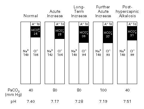
Respiratory acidosis is a primary acid-base disorder resulting from an increase in arterial PaCO₂ (>45 mmHg) due to alveolar hypoventilation, leading to acidemia (pH <7.35 in acute cases). [1-2] It develops from disorders affecting gas exchange, chest wall/respiratory muscles, or inhibition of the medullary respiratory center. [1] The following figure illustrates the pathophysiological progression from acute to chronic respiratory acidosis and the risk of posthypercapnic alkalosis with overly rapid correction.
History
- Key HPI questions: Onset and tempo of dyspnea, orthopnea, sleep quality (snoring, witnessed apneas, morning headaches), medication/substance use (opioids, benzodiazepines, alcohol), recent illness or infection, trauma
- Symptom characterization: Sleepiness, morning headaches, general tiredness, malaise, ankle edema, confusion [4]
- Timing: Acute (hours–days) vs. chronic (weeks–months); acute-on-chronic decompensation is common in COPD [4]
- Triggers: Infection, sedating medications, oxygen over-supplementation in CO₂ retainers, postoperative state, neuromuscular flare
- Important negatives: Absence of chest pain, hemoptysis, fever, focal neurologic deficits, toxic ingestion
Alarm Features
- pH <7.25 — severe acidemia with high risk of cardiovascular collapse and NIV failure [5]
- Rapidly declining mental status — confusion, somnolence, or coma indicating CO₂ narcosis [2][4]
- Respiratory rate <8 or >30 with accessory muscle use, abdominal paradox, or inability to speak in full sentences [6]
- Hemodynamic instability — hypotension unresponsive to fluids [5]
- Coexistent metabolic acidemia (low base excess) — mixed acidosis portends worse outcomes [5]
- Inability to protect airway or clear secretions — absolute contraindication to NIV, mandates intubation [6]
- Late-onset hypercapnia developing after hospital admission despite primary therapy [5]
Medications
Causative medications (CNS/respiratory depression)
- Opioids, benzodiazepines, barbiturates, general anesthetics, muscle relaxants
- Excessive supplemental O₂ in chronic CO₂ retainers (blunts hypoxic drive) [4]
Treatment medications
- Inhaled bronchodilators (albuterol, ipratropium) and systemic corticosteroids for obstructive etiologies [4]
- Naloxone for opioid-induced hypoventilation; flumazenil for benzodiazepine overdose (use with caution — seizure risk)
- Acetazolamide — carbonic anhydrase inhibitor that promotes renal bicarbonate excretion, may augment ventilatory drive in chronic compensated respiratory acidosis (e.g., COPD, OHS) [7]
Contraindicated/caution
- Sodium bicarbonate is not recommended for isolated respiratory acidosis — no RCT evidence of benefit and potential risks including paradoxical intracellular acidosis and volume overload. May be considered only in mixed respiratory + metabolic acidosis or permissive hypercapnia with severe acidemia [8-10]
- Avoid sedatives, opioids, and neuromuscular blockers unless intubation is planned
Diet
- High-carbohydrate diets increase CO₂ production (RQ ~1.0 for carbohydrates vs. ~0.7 for fat); in ventilator-dependent or borderline patients, high-fat/low-carb enteral formulas may reduce CO₂ load [1]
- Adequate hydration to support mucociliary clearance
- In obesity hypoventilation syndrome (OHS), long-term weight management is a cornerstone of therapy
Review of Systems
- Pulmonary: Dyspnea, cough, sputum production, wheezing, orthopnea, exercise intolerance
- Neurologic: Headache (especially morning), somnolence, confusion, tremor, asterixis, seizures [2]
- Cardiovascular: Peripheral edema, palpitations (arrhythmias from acidemia/hypercapnia)
- Psychiatric: Anxiety, panic, insomnia (or hypersomnia)
- Musculoskeletal: Weakness, fatigue (neuromuscular disease screening)
Collateral History and Family History
- Collateral: Witnessed apneas, snoring, baseline functional status, home O₂ or CPAP/BiPAP use, prior intubations, recent medication changes, substance use
- Family history: Neuromuscular diseases (muscular dystrophy, myasthenia gravis), COPD, obesity, obstructive sleep apnea [4]
- Social context: Smoking history, occupational exposures, home environment (ability to use NIV at home), access to follow-up
Risk Factors
- COPD — most common cause of chronic respiratory acidosis [2][11]
- Obesity/OHS — restrictive physiology with blunted ventilatory drive [4]
- Neuromuscular disease — ALS, muscular dystrophy, myasthenia gravis, Guillain-Barré syndrome [4]
- CNS depression — drug overdose (opioids, benzodiazepines), stroke, brainstem lesions [2]
- Chest wall deformity — severe kyphoscoliosis [4]
- Severe asthma — status asthmaticus with air trapping
- Obstructive sleep apnea — especially when combined with obesity
- Metabolic abnormalities — severe hypokalemia, hypothyroidism (myxedema) [4]
Differential Diagnosis
- COPD exacerbation — most common cause; look for wheezing, prolonged expiratory phase, smoking history
- Opioid/sedative overdose — pinpoint pupils, depressed RR, toxicology screen
- Severe asthma (status asthmaticus) — rising PaCO₂ is a late and ominous sign
- Neuromuscular crisis — Guillain-Barré, myasthenic crisis; ascending weakness, bulbar symptoms
- Obesity hypoventilation syndrome — BMI >30, daytime hypercapnia, often with OSA
- Pulmonary edema (cardiogenic or ARDS) — may cause mixed respiratory failure
- Pneumothorax — sudden onset, unilateral absent breath sounds
- CNS event — brainstem stroke, intracranial hemorrhage affecting respiratory centers
- Metabolic acidosis with respiratory fatigue — distinguish from primary respiratory acidosis by ABG pattern [11]
Past Medical History
- Prior episodes of hypercapnic respiratory failure, prior intubations
- Baseline PaCO₂ and HCO₃⁻ (critical for distinguishing acute vs. chronic vs. acute-on-chronic)
- COPD severity (GOLD stage), home O₂ use, home NIV/CPAP
- Neuromuscular diagnoses, chest wall abnormalities
- Cardiac comorbidities (heart failure, pulmonary hypertension)
- Surgical history (thoracic, abdominal — diaphragmatic dysfunction)
Physical Exam
Vital signs
- Tachypnea (>24 in obstructive, >30 in restrictive) or paradoxically low RR in CNS depression [6]
- Tachycardia, hypertension (sympathetic response to hypercapnia), or hypotension in severe cases
- SpO₂ may be normal or low; SpO₂ does not reflect CO₂ levels
Focused exam
- Pulmonary: Wheezing, prolonged expiration, diminished breath sounds, accessory muscle use, abdominal paradox (diaphragm fatigue)
- Neurologic: Altered mental status, asterixis, papilledema (from cerebral vasodilation), tremor [2]
- Cardiovascular: Bounding pulses, flushed skin, peripheral edema, elevated JVP (cor pulmonale) [4]
- Musculoskeletal: Proximal weakness, kyphoscoliosis, obesity habitus
Lab Studies
Essential
- Arterial blood gas (ABG) — gold standard; defines pH, PaCO₂, PaO₂, HCO₃⁻, base excess [4]
- Acute: HCO₃⁻ rises ~1 mEq/L per 10 mmHg rise in PaCO₂
- Chronic: HCO₃⁻ rises ~3.5–4 mEq/L per 10 mmHg rise in PaCO₂ [12]
- Venous blood gas (VBG) — reasonable screen; venous PCO₂ correlates with arterial values and can rule out significant hypercarbia [4]
- Basic metabolic panel — serum HCO₃⁻, electrolytes (K⁺, Cl⁻), BUN/Cr, glucose
- CBC — infection screening
- Lactate — assess tissue perfusion
Situational
- Toxicology screen — if overdose suspected
- TSH — if myxedema suspected
- BNP/NT-proBNP — if heart failure contributing
- Phosphate, magnesium — neuromuscular function
Imaging
- Chest X-ray — first-line; evaluate for pneumonia, pulmonary edema, pneumothorax, pleural effusion, hyperinflation (COPD), kyphoscoliosis [2]
- CT chest — if PE suspected, mass lesion, or CXR inconclusive
- CT head — if CNS etiology suspected (brainstem stroke, hemorrhage)
- Imaging is unnecessary if the etiology is clearly established (e.g., known opioid overdose with response to naloxone)
Special Tests
- A-a gradient — helps distinguish intrinsic lung disease (elevated A-a gradient) from pure hypoventilation (normal A-a gradient) [13]
- Pulmonary function tests (PFTs) — outpatient; assess severity of obstructive/restrictive disease
- Negative inspiratory force (NIF) and vital capacity — bedside assessment of neuromuscular respiratory reserve (NIF worse than −20 cmH₂O or VC <15 mL/kg suggests impending respiratory failure)
- Polysomnography — outpatient for suspected OSA/OHS
- Point-of-care ultrasound — diaphragm excursion, B-lines (pulmonary edema), pleural effusion, cardiac function
ECG
- Indications: All patients with acute respiratory acidosis to assess for arrhythmia and cardiac comorbidity
- Findings to watch for:
- Sinus tachycardia (most common)
- Atrial fibrillation (associated with worse outcomes in COPD exacerbation) [5]
- Right heart strain pattern (P pulmonale, right axis deviation, RV hypertrophy) — suggests chronic cor pulmonale
- Peaked T waves — hyperkalemia from severe acidemia
- Widened QRS or arrhythmias — severe acidemia (pH <7.1)
Assessment
Severity stratification
- Mild: pH 7.30–7.35, PaCO₂ 45–60 mmHg — may respond to medical therapy alone
- Moderate: pH 7.25–7.30, PaCO₂ 60–80 mmHg — strong indication for NIV [5]
- Severe: pH <7.25 — high risk of NIV failure, prepare for intubation [5]
Acute vs. chronic vs. acute-on-chronic
- Acute: SBE = 0 ± 2 mEq/L (no renal compensation yet) [12]
- Chronic: SBE = 0.4 × (PaCO₂ − 40); HCO₃⁻ elevated proportionally [12]
- Acute-on-chronic: Compensation present but insufficient for degree of hypercapnia — most common ED presentation in COPD [4]
Complications: CO₂ narcosis, cardiac arrhythmias, cardiovascular collapse, posthypercapnic metabolic alkalosis (if corrected too rapidly) [3]
Treatment Plan
Initial stabilization
- Airway assessment — if unable to protect airway → intubate
- Controlled oxygen therapy — target SpO₂ 88–92% in known CO₂ retainers; avoid high-flow O₂ which may worsen hypercapnia [14]
Non-invasive ventilation (NIV)
- First-line for acute hypercapnic respiratory failure with pH <7.35 and PaCO₂ >45 mmHg [5-6]
- BiPAP: typical starting settings IPAP 10–12 cmH₂O, EPAP 4–5 cmH₂O; titrate to clinical response
- Repeat ABG at 1–2 hours to assess response; improvement in pH and PaCO₂ expected [4]
- Contraindications to NIV: Cardiac/respiratory arrest, inability to protect airway, copious secretions, hemodynamic instability, facial trauma/surgery [6]
Etiology-directed therapy
- COPD exacerbation: Bronchodilators (albuterol/ipratropium), systemic corticosteroids, antibiotics if indicated [4]
- Opioid overdose: Naloxone 0.4–2 mg IV, repeat as needed
- Neuromuscular crisis: Urgent neurology consultation; plasmapheresis/IVIG for myasthenic crisis or GBS
- Status asthmaticus: Aggressive bronchodilators, IV magnesium, consider ketamine for sedation if intubating
Invasive mechanical ventilation
- If NIV fails (worsening pH, rising PaCO₂, declining mental status) or contraindicated
- In chronic hypercapnia, target the patient's baseline PaCO₂, not normal values — rapid correction risks posthypercapnic alkalosis [3]
Sodium bicarbonate: Not recommended for isolated respiratory acidosis; may be considered for mixed respiratory + metabolic acidosis with severe acidemia [8][10]
Acetazolamide: May be used as adjunct in chronic compensated respiratory acidosis to promote bicarbonate excretion and augment ventilatory drive [7]
Disposition
- ICU admission: pH <7.25, hemodynamic instability, altered mental status, need for intubation, NIV failure, rapidly worsening trajectory [5][15]
- Stepdown/telemetry admission: pH 7.25–7.35 on NIV with improving trend, stable hemodynamics, new supplemental O₂ requirement [15]
- Observation: Mild respiratory acidosis responding to medical therapy, stable on room air or baseline O₂
- Discharge: Rarely appropriate for acute respiratory acidosis; consider only if etiology is fully reversed (e.g., opioid overdose with sustained response to naloxone, no recurrence after observation period), patient returns to baseline, and reliable follow-up is ensured [15]
- Specialist consultation triggers: Pulmonology (COPD, OHS, neuromuscular disease), neurology (GBS, myasthenic crisis), toxicology (complex overdose), palliative care (end-stage disease)
Follow Up / Return Precautions
- Follow-up timing: Pulmonology or PCP within 1–2 weeks of discharge; sooner if on new home NIV [15]
- Return precautions — instruct patients to return immediately for:
- Worsening shortness of breath or inability to speak in full sentences
- Increasing drowsiness, confusion, or difficulty waking
- New or worsening swelling in legs
- Fever or worsening cough with colored sputum
- Patient counseling:
- Smoking cessation (if applicable) [15]
- Medication adherence (inhalers, home NIV)
- Avoid sedating medications unless prescribed
- Weight management in OHS
- Expected recovery: Acute respiratory acidosis from reversible causes (e.g., COPD exacerbation) typically improves within 24–72 hours with appropriate therapy; chronic respiratory acidosis requires long-term management of the underlying condition
References
1. Respiratory Acidosis and Respiratory Alkalosis: Core Curriculum 2023. — Palmer BF, Clegg DJ. American Journal of Kidney Diseases : The Official Journal of the National Kidney Foundation. 2023.
2. Respiratory Acidosis. — Epstein SK, Singh N. Respiratory Care. 2001.
3. Management of Life-Threatening Acid–Base Disorders. — Adrogué HJ, Madias NE. The New England Journal of Medicine. 1998.
4. Peripheral Venous Blood Gas Analysis for the Diagnosis of Respiratory Failure, Hypercarbia and Metabolic Disturbance in Adults. — Byrne AL, Pace NL, Thomas PS, et al. The Cochrane Database of Systematic Reviews. 2025.
5. Beyond the Guidelines for Non-Invasive Ventilation in Acute Respiratory Failure: Implications for Practice. — Bourke SC, Piraino T, Pisani L, Brochard L, Elliott MW. The Lancet. Respiratory Medicine. 2018.
6. Non-Invasive Ventilation in Acute Respiratory Failure. — Nava S, Hill N. Lancet. 2009.
7. Carbonic Anhydrase Inhibitors for Hypercapnic Ventilatory Failure in Chronic Obstructive Pulmonary Disease. — Jones PW, Greenstone M. The Cochrane Database of Systematic Reviews. 2001.
8. Sodium Bicarbonate Therapy for Acute Respiratory Acidosis. — Chand R, Swenson ER, Goldfarb DS. Current Opinion in Nephrology and Hypertension. 2021.
9. Acid-Base Disorders in the Critically Ill Patient. — Achanti A, Szerlip HM. Clinical Journal of the American Society of Nephrology : CJASN. 2023.
10. Alkali Therapy for Respiratory Acidosis: A Medical Controversy. — Adrogué HJ, Madias NE. American Journal of Kidney Diseases : The Official Journal of the National Kidney Foundation. 2020.
11. Acid-Base Interpretation: A Practical Approach. — Morikawa MJ, Ganesh PR. American Family Physician. 2025.
12. Diagnostic Use of Base Excess in Acid–Base Disorders. — Berend K. The New England Journal of Medicine. 2018.
13. Physiological Approach to Assessment of Acid–Base Disturbances. — Berend K, de Vries AP, Gans RO. The New England Journal of Medicine. 2014.
14. Management of Severe COPD. — Wouters EF. Lancet. 2004.
15. Acute Care of Patients With Moderate Respiratory Distress: Recommendations From an American College of Emergency Physicians Expert Panel. — Baugh CW, Neuenschwander JF, Lenox J, et al. The Western Journal of Emergency Medicine. 2025.
Figure
More from Deep Dive
Reviewed by Dr. Lucas Mastropaolo. SymptomDx is an educational tool for medical professionals — it does not replace clinical judgment. Verify all clinical data and drug dosages with authoritative sources.