Salter-Harris III Fracture
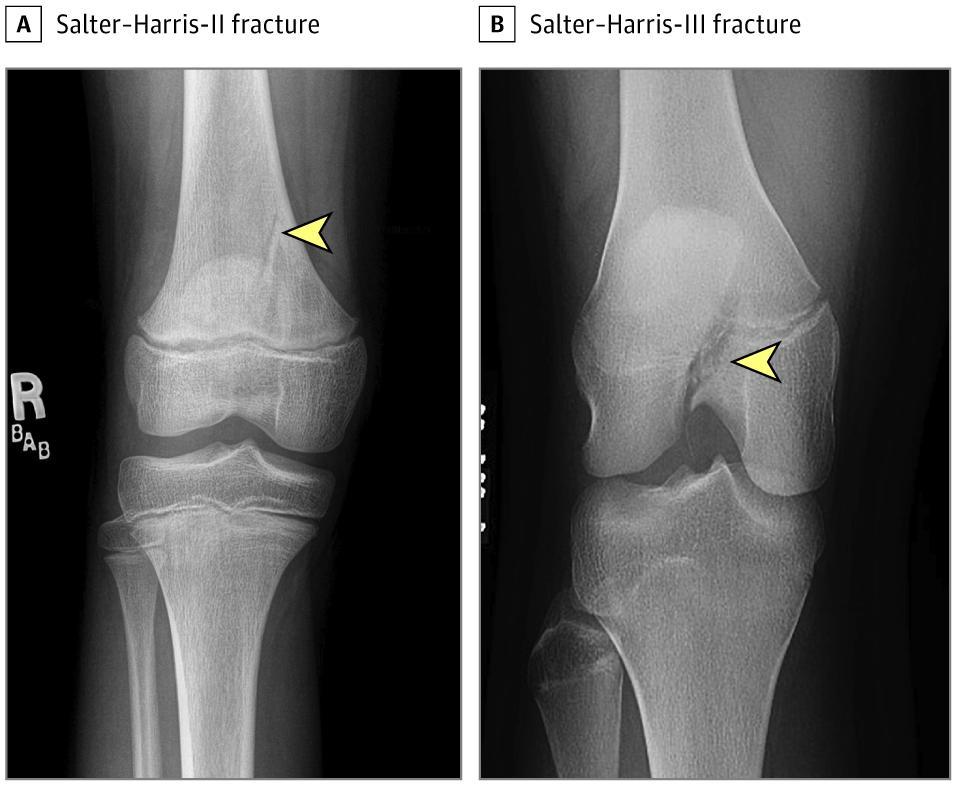
A Salter-Harris (SH) type III fracture is an intra-articular physeal fracture in which the fracture line extends through the growth plate (physis) and into the epiphysis, exiting through the articular surface, without involving the metaphysis. [1-2] This is a relatively uncommon but clinically significant injury in skeletally immature patients, accounting for only ~2% of all physeal fractures in large series. [3] Because it involves the joint surface and the growth plate, it carries a high risk of both growth disturbance and posttraumatic arthritis if not anatomically reduced. [4-6]
The following figure demonstrates the SH classification system, including a Type III fracture of the distal femur:
History
- Mechanism of injury: falls, sports (especially contact/pivoting sports), motor vehicle accidents, or twisting injuries [2-3]
- At the ankle (most common SH III location), the classic mechanism is external rotation of the foot on the leg — this produces the juvenile Tillaux fracture pattern [2][8]
- At the knee, a valgus force (medial femoral condyle SH III) or hyperextension mechanism is typical [7][9]
- Symptom characterization: acute onset of pain, swelling, inability to bear weight, and deformity
- Timing: immediate pain and functional limitation after trauma
- Important negatives: absence of paresthesias, pallor, or pulselessness (to rule out neurovascular compromise); no history of prior fracture or bone disease
Alarm Features
- Neurovascular compromise: diminished or absent distal pulses, pallor, paresthesias, paralysis — particularly with distal femoral and proximal tibial fractures where the popliteal artery and peroneal nerve are at risk [7]
- Compartment syndrome: pain out of proportion, pain with passive stretch, tense swelling
- Open fracture: skin breach overlying the fracture site
- Significant displacement on radiographs: displaced SH III fractures have a 4-fold increased risk of growth arrest compared to nondisplaced fractures [7]
- Physeal gapping >3 mm after reduction raises suspicion for entrapped periosteum, increasing risk of premature physeal closure [4]
Medications
- Acute pain management: weight-based ibuprofen (10 mg/kg PO q6h) and/or acetaminophen (15 mg/kg PO q4-6h); intranasal fentanyl (1.5–2 mcg/kg) for severe pain in the ED
- Procedural sedation agents (if closed reduction attempted): ketamine, propofol, or midazolam/fentanyl per institutional protocol [10]
- Avoid: repeated manipulation attempts — multiple reduction attempts should be avoided as they increase physeal damage [4]
- NSAIDs are generally safe short-term for pediatric fracture pain; concerns about impaired bone healing are not well-supported in the pediatric literature
Diet
- No specific acute dietary restrictions unless procedural sedation is planned (NPO guidelines apply)
- Adequate calcium and vitamin D intake during healing
- Ensure adequate caloric intake for growing children during immobilization
Review of Systems
- MSK: pain localization (over the physis vs. joint line vs. metaphysis), range of motion, ability to bear weight
- Neurologic: numbness, tingling, weakness distal to injury
- Vascular: color changes, temperature changes in the extremity
- Constitutional: fever (consider infection if atraumatic or subacute presentation)
- Skin: open wounds, abrasions overlying the fracture
Collateral History and Family History
- Witnessed mechanism of injury (especially in younger children or non-verbal patients)
- In non-accidental trauma (NAT) considerations: inconsistency between history and injury pattern, delay in presentation, prior fractures
- Skeletal maturity status: age, pubertal stage, prior growth plate injuries
- Family history of bone disorders (osteogenesis imperfecta, metabolic bone disease) if the mechanism seems trivial
Risk Factors
- Age: peak incidence in adolescents; physeal fractures peak during the adolescent growth spurt [3]
- Sex: male predominance (~2:1) [3]
- Sports participation: contact sports, basketball, football, gymnastics [3]
- Transitional period (ages 12–16): the distal tibial physis closes asymmetrically over ~18 months, creating vulnerability to Tillaux (SH III) and triplane fractures [2][11]
- Falls are the most common cause overall (64% of physeal fractures) [3]
Differential Diagnosis
- Salter-Harris Type IV fracture: fracture crosses the physis involving both epiphysis and metaphysis — distinguished by metaphyseal fragment on imaging [1]
- Ligamentous sprain: in skeletally immature patients, the physis is weaker than ligaments, so what appears to be a "sprain" may actually be a physeal fracture. A high index of suspicion is critical [7]
- Osteochondral fracture/osteochondritis dissecans: intra-articular fragment without physeal involvement
- Juvenile Tillaux fracture: a specific SH III pattern of the anterolateral distal tibial epiphysis — must be distinguished from triplane fractures [2][12]
- Triplane fracture: a complex transitional fracture with components in 3 planes (sagittal through epiphysis, axial through physis, coronal through metaphysis) — may appear as SH III on AP view but has a metaphyseal component on lateral [12]
- Non-accidental trauma: always consider in young children with inconsistent history
Past Medical History
- Prior physeal injuries or fractures (increases risk of growth disturbance)
- Metabolic bone disease, renal osteodystrophy, rickets
- Chronic steroid use
- Previous surgical hardware near the physis
- Underlying connective tissue disorders
Physical Exam
- Vital signs: assess for tachycardia/hypotension (hemorrhage from long-bone fractures)
- Inspection: swelling, ecchymosis, deformity, skin integrity (open vs. closed)
- Palpation: point tenderness directly over the physis is the hallmark finding and helps distinguish physeal fractures from ligament injuries [11]
- Neurovascular exam: mandatory — assess distal pulses (dorsalis pedis, posterior tibial for ankle; popliteal for knee), capillary refill, sensation, and motor function [7]
- Joint effusion: a lipohemarthrosis (fat globules in aspirated joint fluid) confirms intra-articular fracture
- Range of motion: typically markedly limited by pain
- Compartment assessment: palpate compartments for tenseness; pain with passive stretch
Lab Studies
- Labs are generally not required for isolated SH III fractures
- If surgical intervention is planned: CBC, BMP, type and screen per institutional protocol
- If concern for NAT: skeletal survey, vitamin D, calcium, phosphorus, alkaline phosphatase, PTH
- If open fracture: CBC, blood cultures if febrile
Imaging
- First-line: AP, lateral, and oblique plain radiographs of the affected joint [11][13]
- SH III fractures show a fracture line through the epiphysis extending to the articular surface, with the physis involved but the metaphysis intact
- Plain films may significantly underestimate displacement — one study found mean displacement of 0.42 mm on plain films vs. 2.70 mm on CT/MRI (p = 0.005) [13]
- CT scan: recommended for all displaced SH III fractures, especially at the ankle (Tillaux, triplane) and distal femur, to accurately assess articular step-off and guide surgical planning [11-13]
- MRI: most sensitive for detecting occult physeal injury, physeal bar formation, and associated soft tissue injuries; recommended when plain films are negative but clinical suspicion is high [7][14]
- When imaging may be unnecessary: if the patient has a clearly nondisplaced SH III on adequate orthogonal views and is near skeletal maturity with minimal growth remaining
Special Tests
- Salter-Harris Classification (mnemonic: SALTR — Straight across/Slip, Above, Lower/beLow, Through, Rammed/cRush):
- Fat pad sign / lipohemarthrosis on cross-table lateral radiograph confirms intra-articular fracture [9]
- Stress views under anesthesia may be needed for occult SH III fractures of the medial femoral condyle [9]
- Point-of-care ultrasound: can identify joint effusion and cortical irregularity at the bedside
ECG
- Not routinely indicated for isolated SH III fractures
- Consider if procedural sedation is planned (per institutional sedation protocols)
- Obtain if polytrauma or hemodynamic instability
Assessment
A Salter-Harris type III fracture is an intra-articular physeal fracture that demands anatomic reduction to restore joint congruity and minimize growth plate damage. Key clinical considerations:
- Growth arrest occurs in approximately 49% of SH III fractures of the distal femur; rates vary by location but are substantial across all sites [7]
- Displaced fractures carry a 4-fold increased risk of growth disturbance compared to nondisplaced fractures [7]
- The juvenile Tillaux fracture (SH III of the anterolateral distal tibial epiphysis) is the classic SH III pattern at the ankle, occurring during the transitional period of physeal closure [2][15]
- Complications include: premature physeal closure, angular deformity (valgus/varus), limb-length discrepancy, posttraumatic arthritis, and joint stiffness [6-7]
- Poor outcomes have been reported in 29–32% of SH III and IV fractures of the distal femur [5]
Treatment Plan
Initial stabilization (ED)
- Immobilize in a well-padded posterior splint in the position of comfort
- Ice, elevation, strict non-weight-bearing
- Adequate analgesia
Nondisplaced SH III fractures
- May be treated nonoperatively with cast immobilization and strict non-weight-bearing [4][16]
- Close orthopedic follow-up within 5–7 days with repeat imaging
Displaced SH III fractures (≥2 mm articular step-off)
- Open reduction and internal fixation (ORIF) is the standard of care to restore articular congruity and minimize physeal damage [4-6]
- Fixation is typically with smooth K-wires or cannulated screws placed parallel to the physis (avoiding crossing the growth plate when possible)
- At the ankle (Tillaux fractures), closed reduction may be attempted first; if articular gap remains ≥2 mm, ORIF is indicated [15]
- Avoid multiple closed reduction attempts — these increase the risk of further physeal injury [4]
Postoperative/post-reduction
- Non-weight-bearing in a short or long leg cast (depending on location) for 4–6 weeks
- Serial radiographs to monitor alignment and healing
Disposition
Admission criteria
- Displaced SH III fractures requiring operative fixation
- Neurovascular compromise or compartment syndrome
- Open fractures
- Polytrauma
- Concern for non-accidental trauma
Discharge criteria
- Nondisplaced SH III fracture with adequate splinting, pain control, and reliable follow-up
- Intact neurovascular exam
- Appropriate caregiver education
Orthopedic consultation triggers (from the ED)
- Any displaced SH III fracture — emergent orthopedic consultation is warranted [4-5]
- Neurovascular compromise
- Open fracture
- Failed closed reduction
- Uncertainty about fracture classification (consider CT before disposition) [12-13]
Follow Up / Return Precautions
- Orthopedic follow-up within 5–7 days for all SH III fractures, with repeat radiographs
- Long-term monitoring for 6–12 months minimum to detect growth arrest and angular deformity [7]
- MRI is the most sensitive tool for early detection of physeal bar formation if growth disturbance is suspected [7]
- Physeal bars developed in 30% of SH III/IV medial malleolus fractures at a mean of 8.4 months, with 47% of those requiring secondary surgery [17]
Return precautions (counsel caregivers)
- Return immediately for increasing pain, numbness/tingling, color change (blue/white) of fingers or toes, inability to move digits, worsening swelling not relieved by elevation, fever, or foul odor from the cast
- No weight-bearing until cleared by orthopedics
- Expected recovery: fracture healing typically 4–6 weeks; return to full activity 2–4 months depending on location and severity; growth monitoring continues for at least 1 year
References
1. Growth Plate Injuries: Salter-Harris Classification. — Brown JH, DeLuca SA. American Family Physician. 1992.
2. Interventions for Treating Ankle Fractures in Children. — Yeung DE, Jia X, Miller CA, Barker SL. The Cochrane Database of Systematic Reviews. 2016.
3. Clinical Characteristics of 1124 Children With Epiphyseal Fractures. — Deng H, Zhao Z, Xiong Z, et al. BMC Musculoskeletal Disorders. 2023.
4. Physeal Fractures of the Distal Tibia and Fibula (Salter-Harris Type I, II, III, and IV Fractures). — Podeszwa DA, Mubarak SJ. Journal of Pediatric Orthopedics. 2012.
5. Growth Plate Fractures of the Distal Femur. — Wall EJ, May MM. Journal of Pediatric Orthopedics. 2012.
6. Distal Tibial Physeal Fractures in Children That May Require Open Reduction. — Kling TF, Bright RW, Hensinger RN. The Journal of Bone and Joint Surgery. American Volume. 1984.
7. Acute Knee Injuries in Children and Adolescents: A Review. — MacDonald J, Rodenberg R, Sweeney E. JAMA Pediatrics. 2021.
8. Isolated Adult Tillaux Fracture: A Report of Two Cases. — Oak NR, Sabb BJ, Kadakia AR, Irwin TA. The Journal of Foot and Ankle Surgery : Official Publication of the American College of Foot and Ankle Surgeons. 2014.
9. Salter-Harris Type-Iii Fracture of the Medial Femoral Condyle Occurring in the Adolescent Athlete. — Torg JS, Pavlov H, Morris VB. The Journal of Bone and Joint Surgery. American Volume. 1981.
10. Pediatric Fracture Reduction in the Emergency Department. — Bin K, Rony L, Henric N, Moukoko D. Orthopaedics & Traumatology, Surgery & Research : OTSR. 2022.
11. Pediatric Physeal Ankle Fracture. — Wuerz TH, Gurd DP. The Journal of the American Academy of Orthopaedic Surgeons. 2013.
12. Review of Distal Tibial Epiphyseal Transitional Fractures. — Rosenbaum AJ, DiPreta JA, Uhl RL. Orthopedics. 2012.
13. Salter-Harris Type III Fractures of the Distal Femur: Plain Radiographs Can Be Deceptive. — Lippert WC, Owens RF, Wall EJ. Journal of Pediatric Orthopedics. 2010.
14. Imaging of Epiphyseal Injuries. — Rogers LF, Poznanski AK. Radiology. 1994.
15. Recognition and Management of Tillaux Fractures in Adolescents. — Koury SI, Stone CK, Harrell G, La Charité DD. Pediatric Emergency Care. 1999.
16. Diagnosis and Management of McFarland Fractures. — Birt M, Vopat B, Schroeppel P, et al. The American Journal of Emergency Medicine. 2018.
17. Risk Factors and Surgical Sequelae of Physeal Arrest in Pediatric Salter-Harris III and IV Medial Malleolus Fractures. — Roth OS, Gupta A, Adebayo T, Tretiakov M. Journal of Pediatric Orthopedics. 2025.
Figure
More from Deep Dive
Reviewed by Dr. Lucas Mastropaolo. SymptomDx is an educational tool for medical professionals — it does not replace clinical judgment. Verify all clinical data and drug dosages with authoritative sources.