Testicular Torsion
Testicular torsion is a urological emergency caused by twisting of the spermatic cord, leading to venous congestion, reduced arterial blood flow, and eventual ischemic necrosis of the testicle. The salvage rate is ~90% within 6 hours, dropping to ~50% at 12 hours and ~10% at 24 hours.[1-2] It should be treated with the same urgency as acute stroke or STEMI.[3]
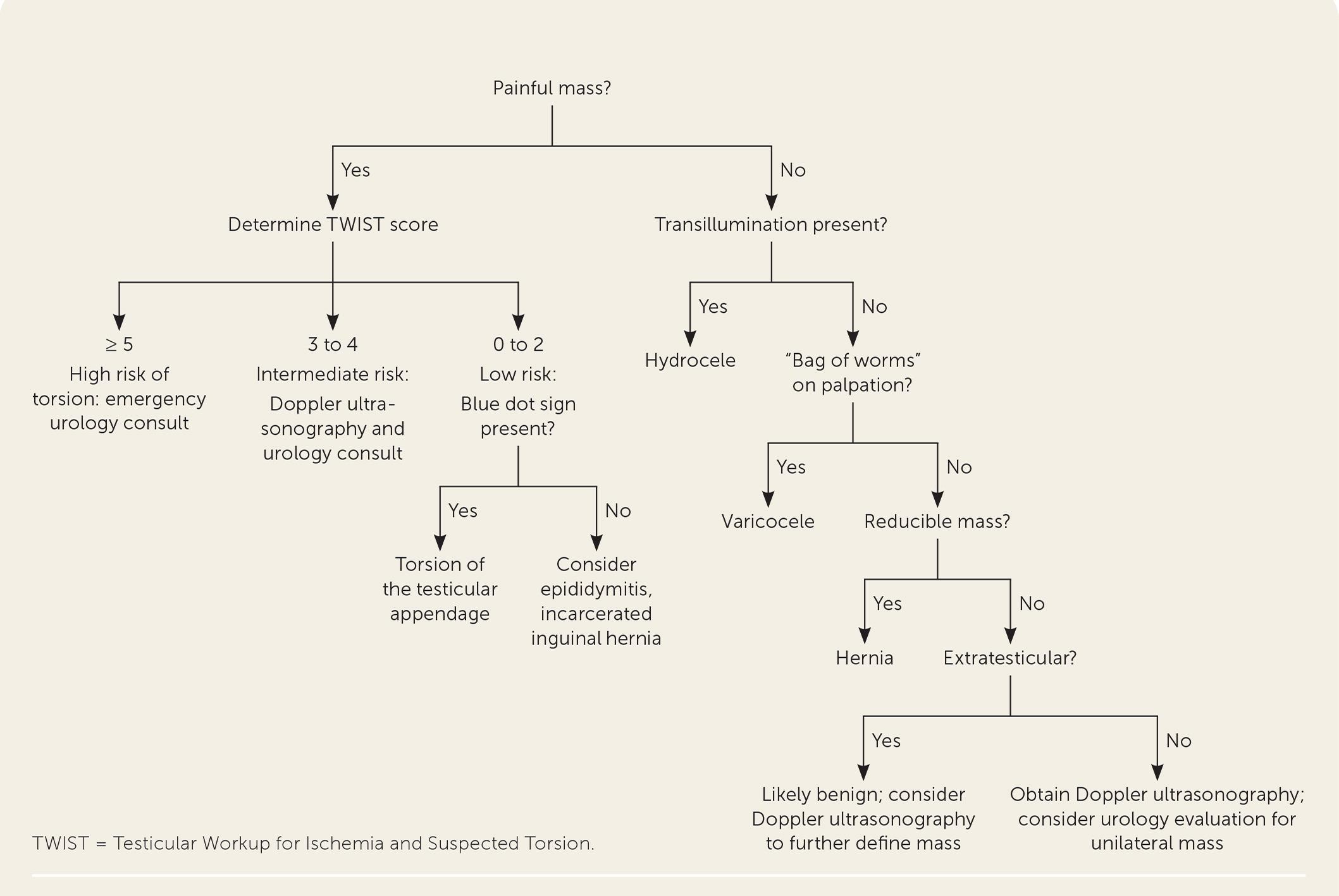
The following algorithm outlines the approach to evaluating scrotal masses, including the TWIST score risk stratification for torsion:
History
- Onset and character: Sudden, severe, unilateral scrotal/testicular pain — the hallmark symptom. Ask exact time of onset (critical for salvage window)[4-5]
- Associated symptoms: Nausea and vomiting are common and strongly predictive (present in majority of torsion cases)[6-7]
- Abdominal pain: Some patients (especially children) present with abdominal pain only — always ask about testicular pain in boys with abdominal pain[8]
- Prior episodes: Intermittent torsion-detorsion may present as recurrent episodes of self-resolving testicular pain[8]
- Activity/triggers: Antecedent trauma, physical activity, cold weather exposure[1]
- Timing: Symptom duration is the single most important prognostic factor — document precisely[2]
- Important negatives: Absence of dysuria, urethral discharge, fever (these suggest epididymitis instead)[1][9]
Alarm Features
- Sudden onset of severe unilateral scrotal pain with nausea/vomiting
- High-riding testicle with transverse lie[1][6]
- Absent cremasteric reflex (OR 47.6 for torsion in one study)[1][7]
- Hard, swollen, exquisitely tender testicle[6][10]
- Symptom duration approaching or exceeding 6 hours — escalate immediately[2][5]
- Any boy with acute abdominal pain and scrotal findings[8]
- Bilateral scrotal pain (rare but catastrophic if bilateral torsion)
Medications
- ED pain management: IV/IM ketorolac, opioids (morphine, fentanyl) for analgesia; do not delay surgical consultation for pain control[4]
- Procedural sedation: Consider for manual detorsion if patient cannot tolerate the procedure due to pain[11]
- Spermatic cord block: Lidocaine injection at the external inguinal ring can facilitate manual detorsion[4]
- No role for antibiotics unless epididymitis is the confirmed diagnosis
- Contraindicated: Do not give antibiotics empirically and discharge if torsion has not been excluded
Diet
- NPO status: Keep patient NPO once torsion is suspected, as emergent surgical exploration is likely[5]
- No specific dietary triggers or long-term dietary considerations
Review of Systems
- GU: Dysuria, urethral discharge, urinary frequency (suggests epididymitis/UTI rather than torsion)[9]
- GI: Nausea, vomiting, abdominal pain (common in torsion; abdominal pain may be the sole complaint in children)[8]
- Constitutional: Fever (more suggestive of infectious etiology — epididymo-orchitis)[9]
- MSK/Derm: Scrotal erythema, edema, warmth
- Heme: Prior history of bleeding disorders (relevant for surgical planning)
Collateral History and Family History
- Family history of torsion: A recognized risk factor; the bell-clapper deformity is often bilateral and may be familial[1]
- Prior urological history: Undescended testis, prior orchiopexy, inguinal hernia repair
- Collateral from parents/caregivers: Essential in pediatric patients — exact time of symptom onset, prior similar episodes, and whether the child has been reluctant to report genital pain[8]
Risk Factors
- Bell-clapper deformity: The most important anatomical risk factor — excessive testicular mobility due to abnormal anchoring of the tunica vaginalis[1][4]
- Age: Bimodal distribution — peak incidence in neonates (first year of life) and adolescents aged 12–18 years[4-5]
- Family history of testicular torsion[1]
- Cold weather / hyperactive cremasteric reflex[1]
- Antecedent trauma[1][6]
- Prior episodes of intermittent testicular pain (intermittent torsion-detorsion)[8][12]
- Incidence: ~1 in 4,000 males younger than 25 years; 3.8 per 100,000 males <18 years annually[5-6]
Differential Diagnosis
- Torsion of the testicular appendage (appendix testis): Gradual onset of superior pole pain, blue dot sign pathognomonic but uncommon, cremasteric reflex preserved, treated conservatively[1][9]
- Epididymitis/epididymo-orchitis: More gradual onset, dysuria, urethral discharge, tender epididymis, positive Prehn sign, increased blood flow on Doppler; in sexually active males think Chlamydia/Gonorrhea, in prepubertal boys think enteric organisms[1][9]
- Incarcerated inguinal hernia: Inguinal swelling extending into scrotum, bowel sounds in scrotum, may have obstructive symptoms[13]
- Testicular trauma/rupture: History of direct trauma, scrotal hematoma
- Idiopathic scrotal edema: Bilateral scrotal wall edema without testicular tenderness, self-limited[13]
- Testicular tumor: Usually painless, firm mass; ~10% present with acute pain from hemorrhage into tumor
- Fournier's gangrene: Rapidly progressive scrotal erythema, crepitus, systemic toxicity — surgical emergency
- Henoch-Schönlein purpura: Scrotal involvement with characteristic purpuric rash
Past Medical History
- Prior episodes of testicular pain (intermittent torsion-detorsion)[8]
- Previous orchiopexy or inguinal surgery
- Undescended testis (cryptorchidism)
- Known bell-clapper deformity
- Urogenital anomalies
- Bleeding disorders or anticoagulant use (surgical planning)
Physical Exam
- Vital signs: Usually normal; tachycardia from pain; fever suggests infectious etiology
- Inspection: Scrotal erythema, edema, high-riding testicle, transverse lie[1][6]
- Palpation: Exquisitely tender, swollen, hard testicle[6][10]
- Cremasteric reflex: Stroke medial thigh → observe for ipsilateral testicular retraction. Absent reflex is the most sensitive physical finding (100% sensitivity in one series). However, its presence does not reliably exclude torsion[4][7]
- Prehn sign: Elevation of the scrotum — classically relieves pain in epididymitis but not torsion. Low reliability; should not be used to rule out torsion[11]
- Blue dot sign: Bluish discoloration at the superior pole — pathognomonic for torsion of the appendix testis but uncommonly seen[1]
- Inguinal exam: Assess for hernia, cord tenderness
- Abdominal exam: Rule out referred pain sources
Lab Studies
- Labs are not required and should not delay surgical consultation or imaging[5]
- Urinalysis: May help differentiate from epididymitis (pyuria, bacteriuria suggest infection); typically normal in torsion
- CBC: Leukocytosis is nonspecific; may be mildly elevated in torsion from stress response
- STI testing: Urine NAAT for Chlamydia/Gonorrhea if epididymitis is suspected in sexually active patients
- Platelet-to-lymphocyte ratio (PLR): Emerging as a predictor of testicular necrosis in some studies, but not yet standard practice[14]
Imaging
- First-line: High-resolution color Doppler ultrasonography — sensitivity ~95–100%, specificity ~94–98% for torsion[1][15]
Key US findings
- Decreased or absent intratesticular blood flow compared to contralateral side[13]
- Whirlpool sign of the spermatic cord — the most specific US sign, pathognomonic in adults[15-16]
- Enlarged, heterogeneous testis with associated hydrocele[13]
- Spermatic cord twisting (target-like concentric rings on cross-section)[13]
- Critical caveat: A normal ultrasound does not exclude torsion — preserved blood flow can occur with partial or early torsion. If clinical suspicion is high, proceed to surgical exploration regardless of US findings[4-5][17]
- Do not delay surgery for imaging when clinical suspicion is high (TWIST ≥5)[1][10]
- ACR Appropriateness Criteria (2024): Scrotal US with Doppler is the recommended initial imaging for acute scrotal pain without trauma[16]
- CT/MRI: Not indicated in the acute setting
Special Tests
- TWIST Score (Testicular Workup for Ischemia and Suspected Torsion):[6][10]
- Export Component Points References Testicular swelling 2[1-2]
- Hard testicle 2[1-2]
- Absent cremasteric reflex 1[1-2]
- Nausea/vomiting 1[1-2]
- High-riding testicle 1[1-2]
- Score 0–2 (Low risk): ~1–2% torsion rate → US may be sufficient to rule out[6][10]
- Score 3–4 (Intermediate risk): ~22% torsion rate → Obtain urgent Doppler US + urology consult[6]
- Score ≥5 (High risk): ~87% torsion rate → Proceed directly to surgical exploration; do not delay for imaging[6][10]
- The TWIST score has been validated when performed by non-urologists including EMTs, with NPV of 100% for low-risk and PPV of 93.5% for high-risk groups.[10] However, it should be used for risk stratification, not as an exclusionary tool, given a non-negligible false-negative rate.[18]
- POCUS by EPs: Sensitivity 96%, specificity 95% when assessing for impaired blood flow or whirlpool sign[18]
- Manual detorsion (see Treatment Plan): Can be performed as a bedside procedure, ideally with US guidance[11][19]
ECG
- Not routinely indicated for testicular torsion
- Consider ECG only if procedural sedation is planned for manual detorsion or if the patient has cardiac comorbidities
Assessment
- Testicular torsion is a time-critical surgical emergency. Key assessment points:
Severity stratification by symptom duration
- 0–6 hours: ~97% salvage rate
- 7–12 hours: ~79% salvage rate
- 13–24 hours: ~42–54% salvage rate
- 24 hours: ~18% salvage rate (but still worth pursuing surgery)
- Atypical presentations to recognize: Isolated abdominal pain in children, intermittent torsion-detorsion with spontaneous resolution, neonatal torsion (painless scrotal mass with discoloration)[4][8]
- Complications: Testicular necrosis requiring orchiectomy (42% overall orchiectomy rate), testicular atrophy even after successful detorsion (significant risk beyond 6 hours), decreased fertility, contralateral testicular damage from immune-mediated mechanisms[5][20]
- Long-term follow-up shows testicular atrophy risk is higher than previously reported, with heterogeneous echotexture on US even in salvaged testes raising concerns about fertility impact[20]
Treatment Plan
Initial stabilization
- IV access, analgesia (ketorolac 0.5 mg/kg IV, opioids as needed), NPO status
- Immediate urology consultation — do not delay for imaging if clinical suspicion is high[4-5]
Manual detorsion in the ED
- Indicated as a temporizing measure while awaiting surgery — not definitive treatment[1][4][19]
- Classic technique: "Open the book" — rotate the affected testicle medially to laterally (toward the thigh). Most torsion occurs inward, so outward rotation detorses[11]
- Success indicators: Immediate pain relief, testicle descends to normal position, restoration of blood flow on Doppler[19][21]
- US-guided detorsion improves success rates and confirms reperfusion in real time[11][19]
- Success rate: ~26–80% depending on series; most effective when presentation delay is <6 hours and scrotal edema is absent[3][21-22]
- Successful manual detorsion was independently associated with improved surgical salvage (OR 17.38) and reduced orchiectomy from 24.6% to 2.8%[23]
- Manual detorsion is NOT definitive — all patients still require surgical orchiopexy[1][19]
Definitive surgical management
- Emergent scrotal exploration with surgical detorsion and bilateral orchiopexy (fixation of both testes to prevent future torsion)[1][5][8]
- If the testicle is nonviable → orchiectomy with contralateral orchiopexy[5]
- After successful manual detorsion, orchiopexy can be performed semi-electively (within 1–14 days) with no increased risk of re-torsion or orchiectomy[3][21]
Aggressive management even with prolonged symptoms
- Testicular survival is significant even past 24 hours — surgical exploration should be pursued regardless of symptom duration[2]
Disposition
- Admission/OR: All confirmed or high-suspicion torsion cases require emergent surgical exploration[5]
- Transfer: If urology is not available on-site, arrange emergent transfer — inter-facility transfer significantly reduces salvage rates, so attempt manual detorsion before/during transfer[24]
- Observation: Not appropriate for suspected torsion. Intermediate-risk TWIST scores warrant urgent imaging and urology consultation, not observation alone[6]
- Discharge: Only if torsion has been definitively excluded (e.g., confirmed alternative diagnosis on US with low TWIST score). Ensure clear return precautions[4]
- Urology consultation triggers: Any concern for torsion — consult before imaging in high-risk cases[1]
Follow Up / Return Precautions
- Post-orchiopexy follow-up: Urology follow-up within 1–2 weeks; long-term US monitoring for testicular atrophy recommended[20]
- Long-term surveillance: Testicular atrophy can develop months to years after salvage, even when initial surgery appeared successful — serial US monitoring of testicular volume is warranted[20]
- Fertility counseling: Discuss potential impact on fertility, especially with delayed presentation or orchiectomy; semen analysis may be considered in post-pubertal patients at appropriate follow-up[5][20]
Return precautions (counsel patient/family)
- Return immediately for any recurrence of sudden testicular pain, swelling, or nausea/vomiting
- Educate adolescents and parents that testicular pain is always an emergency and should never be ignored or "waited out"[8]
- Contralateral torsion can occur even after bilateral orchiopexy (rare)
- Expected recovery: Scrotal swelling and discomfort typically resolve over 1–2 weeks post-surgery
References
1. Scrotal Masses. — Langan RC, Puente MEE. American Family Physician. 2022.
2. A Systematic Review of Testicle Survival Time After a Torsion Event. — Mellick LB, Sinex JE, Gibson RW, Mears K. Pediatric Emergency Care. 2019.
3. The Role of Manual Detorsion in the Management of Testicular Torsion in the Emergency Department: A Single-Group Observational Study. — Başaran E, Baba D, Taşkiran AT, et al. Medicine. 2026.
4. High Risk and Low Prevalence Diseases: Testicular Torsion. — Lacy A, Smith A, Koyfman A, Long B. The American Journal of Emergency Medicine. 2023.
5. Testicular Torsion: Diagnosis, Evaluation, and Management. — Sharp VJ, Kieran K, Arlen AM. American Family Physician. 2013.
6. Clinical Diagnosis of Testicular Torsion. — Ebell MH. American Family Physician. 2022.
7. A Retrospective Review of Pediatric Patients With Epididymitis, Testicular Torsion, and Torsion of Testicular Appendages. — Kadish HA, Bolte RG. Pediatrics. 1998.
8. What Parents and Teens Need to Know About Testicular Pain. — Angulo MI, Collazo GR, Thompson LA. JAMA Pediatrics. 2025.
9. Differentiation of Epididymitis and Appendix Testis Torsion by Clinical and Ultrasound Signs in Children. — Boettcher M, Bergholz R, Krebs TF, et al. Urology. 2013.
10. Diagnosing Testicular Torsion Before Urological Consultation and Imaging: Validation of the TWIST Score. — Sheth KR, Keays M, Grimsby GM, et al. The Journal of Urology. 2016.
11. Successful Outcome of Manual Testicular Detorsion Using Point-of-Care Ultrasound Guidance: A Clinical Experience. — Hsu CT, Chiu PW. Pediatric Emergency Care. 2023.
12. Ultrasound Diagnostic Features and Management of Torsion-Detorsion. — Olawoyin O, Choksi AU, Press BH, et al. Urology. 2026.
13. Differential Diagnosis of Acute Scrotum in Childhood And Adolescence With High-Resolution Duplex Sonography. — Deeg KH. Ultraschall in Der Medizin. 2021.
14. Predictors of Testicular Salvage Failure in Pediatric Testicular Torsion: A Retrospective Analysis of Clinical and Imaging Risk Factors. — Tian Y, Xing X, Cai H, et al. Pediatric Surgery International. 2025.
15. Ultrasound Guidelines: Emergency, Point-of-Care, and Clinical Ultrasound Guidelines in Medicine. — American College of Emergency Physicians (2023). 2023.
16. ACR Appropriateness Criteria® Acute Onset of Scrotal Pain-Without Trauma, Without Antecedent Mass: 2024 Update. — Expert Panel on Urological Imaging, Gerena M, Allen BC, et al. Journal of the American College of Radiology : JACR. 2024.
17. Detection of Testicular Torsion With Preserved Intratesticular Flow by Point of Care Ultrasound in the Emergency Department: A Case Report. — Lee DY, Lee SH, Yoon JA, et al. The Journal of Emergency Medicine. 2022.
18. Evaluating the TWIST Score and Point-of-Care Ultrasound for Paediatric Testicular Torsion. — Nakamura T, Kinoshita M, Ihara T, et al. Emergency Medicine Journal : EMJ. 2026.
19. Ultrasound-Guided Manual Testicular Detorsion in the Emergency Department. — Manivel V, Mirmiran B. The Journal of Emergency Medicine. 2020.
20. Testicular Atrophy Following Torsion in Pediatric Patients: Results of a Long-Term Follow-Up. — Milivojevic S, Topalovic D, Dasic I, et al. Urology. 2025.
21. Manual Detorsion in Testicular Torsion: 5 years of Experience at a Single Center. — Vasconcelos-Castro S, Flor-de-Lima B, Campos JM, Soares-Oliveira M. Journal of Pediatric Surgery. 2020.
22. The Role of Manual Detorsion in Pediatric Testicular Torsion During the Global COVID-19 Pandemic: Experience From 2 Centres. — Russo T, Cozzi DA, Gaglione G, Ceccanti S. Urology. 2023.
23. Improving Organ Salvage in Testicular Torsion: Comparative Study of Patients Undergoing vs Not Undergoing Preoperative Manual Detorsion. — Dias Filho AC, Oliveira Rodrigues R, Riccetto CL, Oliveira PG. The Journal of Urology. 2017.
24. Multicenter Retrospective Analysis of Acute Scrotum in the Kyushu-Okinawa Region: Diagnostic Practices, Management Patterns, and Predictors of Testicular Salvage. — Tobu S, Kakinoki Y, Konomoto T, et al. International Journal of Urology : Official Journal of the Japanese Urological Association. 2025.
Figure
More from Deep Dive
Reviewed by Dr. Lucas Mastropaolo. SymptomDx is an educational tool for medical professionals — it does not replace clinical judgment. Verify all clinical data and drug dosages with authoritative sources.