Urticaria
Urticaria is a common mast cell–mediated dermatologic condition characterized by pruritic, erythematous, raised wheals that individually resolve within 24 hours. It is classified as acute (<6 weeks) or chronic (≥6 weeks), with 80–90% of chronic cases being idiopathic.[1] The primary mechanism is histamine release from dermal mast cells, most commonly via IgE-mediated type 1 hypersensitivity.[1]
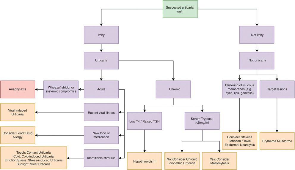
The following diagnostic algorithm provides a useful framework for evaluating urticarial presentations:
History
- Onset and timing: Acute (<6 weeks) vs. chronic (≥6 weeks); continuous vs. episodic; time of day; relationship to activities[1][3]
- Characterization of lesions: Size, shape, distribution, blanching, duration of individual wheals (<24 hours is typical; >24 hours suggests vasculitis)[1][4]
- Triggers: Foods (nuts, shellfish, eggs, dairy), medications, insect stings, infections, physical stimuli (cold, heat, pressure, exercise), stress, latex, aeroallergens[1][3]
- Temporal relationship: Food ingestion within 2 hours, new medication within days to weeks, recent infection[3][5]
- Associated symptoms: Angioedema (lip/tongue/eyelid swelling), pruritus severity, burning vs. itching quality[1]
- Important negatives: Absence of dyspnea, wheezing, stridor, lightheadedness, abdominal pain, vomiting, diarrhea, syncope (rules out anaphylaxis)[3]
- Review patient photographs of lesions if not present at time of evaluation[1][6]
Alarm Features
- Anaphylaxis signs: Hypotension, tachycardia, bronchospasm, stridor, laryngeal edema, abdominal pain, vomiting, diarrhea, dizziness, loss of consciousness[1][3]
- Urticarial vasculitis: Wheals lasting >24 hours, burning pain (not itch), residual ecchymosis/hyperpigmentation, fever, arthralgias[1][4]
- Systemic disease clues: Fevers, night sweats, unintentional weight loss (lymphoma); joint pain, uveitis (autoimmune disease)[1]
- Angioedema without wheals: Consider bradykinin-mediated angioedema (ACE inhibitor–induced or hereditary angioedema) — antihistamines are ineffective in these cases[1][7]
- Epinephrine should be prescribed if anaphylaxis has not been excluded[3]
Medications
Common culprits/exacerbators
- NSAIDs — can induce or exacerbate urticaria via prostaglandin E2 suppression; present in up to 30% of chronic urticaria exacerbations. Recommend acetaminophen as alternative analgesic/antipyretic[1][8-9]
- ACE inhibitors — associated with bradykinin-mediated angioedema (not histamine-mediated); discontinue and observe for 6 weeks[1][3]
- β-lactam antibiotics — most common drug-induced urticaria trigger in children[5]
- Opioids, vancomycin — direct mast cell degranulation (non-IgE mediated)[1]
- Aspirin — cross-reactivity with NSAID-exacerbated urticaria
Treatments
- First-line: Second-generation H1 antihistamines (cetirizine, loratadine, fexofenadine, levocetirizine)[1]
- Refractory acute: Short course oral corticosteroids (prednisone 40–50 mg × 3–5 days), though evidence of benefit is conflicting[3][10]
- Chronic refractory: Omalizumab (Xolair) 300 mg SC q4 weeks for antihistamine-resistant chronic spontaneous urticaria[6][11]
Contraindicated/avoid
- Topical antihistamines and topical corticosteroids — not effective[1]
- Long-term systemic corticosteroids for chronic urticaria[1]
Diet
- Acute urticaria: Nuts, shellfish, eggs, cow's milk, and fish are the most common IgE-mediated food triggers[5][12]
- Histamine-rich foods may exacerbate symptoms: aged cheese, preserved/cured meats, improperly stored fish (scombroid), fermented foods, alcohol[1]
- Pseudoallergens: Food additives (tartrazine dye, benzoates, salicylates) may trigger non-IgE–mediated reactions[1][7]
- Empiric elimination diets are NOT recommended unless guided by specific history and testing[3]
- Adequate hydration is supportive but not specifically evidence-based for urticaria
- Diets rich in omega-3 fatty acids, legumes, nuts, and whole grains may have a protective association[13]
Review of Systems
- Pulmonary: Dyspnea, wheezing, cough, stridor (anaphylaxis, angioedema)
- GI: Abdominal pain, nausea, vomiting, diarrhea (anaphylaxis, food allergy)
- Cardiovascular: Lightheadedness, syncope, palpitations (anaphylaxis)
- ENT: Lip/tongue/throat swelling, voice change, dysphagia (angioedema)
- Musculoskeletal: Joint pain, swelling (autoimmune disease, serum sickness, urticarial vasculitis)
- Constitutional: Fevers, night sweats, weight loss (lymphoma, infection, autoinflammatory syndromes)[1]
- Endocrine: Cold intolerance, weight gain, fatigue (hypothyroidism)[1]
- Dermatologic: Lesion duration, burning vs. itching, residual skin changes[1][4]
Collateral History and Family History
- Family history of urticaria or atopy (asthma, eczema, allergic rhinitis)[1]
- Hereditary angioedema (HAE types 1–3): Family history of recurrent angioedema without urticaria, autosomal dominant[7]
- Cryopyrin-associated periodic syndromes (CAPS): Familial cold autoinflammatory syndrome, Muckle-Wells syndrome[7]
- Social context: Occupational exposures (latex, chemicals), travel history (parasitic infections), sexual history and substance use (hepatitis B/C, HIV risk)[1][3]
- Transfusion history — risk for hepatitis-associated urticaria[3]
Risk Factors
- Female sex — higher prevalence, especially for chronic spontaneous urticaria[6][13]
- Atopic history — asthma, allergic rhinitis, eczema
- Infections: Viral URIs (most common trigger in children, up to 23%), UTIs, GI infections, H. pylori[1][5]
- Autoimmune disease: Autoimmune thyroiditis, SLE, Sjögren syndrome, rheumatoid arthritis, celiac disease[1]
- Medication use: NSAIDs, ACE inhibitors, antibiotics, opioids[1][6]
- Stress — reported trigger in ~14% of chronic urticaria patients[6]
- Environmental: Air pollution (ozone, particulate matter) positively correlated with urticaria incidence[13]
- Younger age groups carry higher disease burden globally[13]
Differential Diagnosis
- Anaphylaxis — urticaria + systemic symptoms (hypotension, bronchospasm, GI symptoms); must be excluded in every ED presentation[3]
- Urticarial vasculitis — wheals >24 hours, burning pain, ecchymosis, residual hyperpigmentation; biopsy shows leukocytoclastic vasculitis[4][8]
- Erythema multiforme — target lesions, fixed (not migratory), often post-infectious (HSV) or drug-related
- Bullous pemphigoid — urticarial prodrome may precede blistering; older adults[8]
- Contact dermatitis — localized, distribution matches exposure
- Drug eruption — morbilliform rash, fixed drug eruption; temporal relationship to medication
- Serum sickness–like reaction — urticaria + fever + arthralgias, typically 1–3 weeks after drug exposure (cefaclor, amoxicillin)
- Hereditary angioedema — recurrent angioedema WITHOUT wheals; does not respond to antihistamines[7]
- Mastocytosis/systemic mastocytosis — urticaria pigmentosa, Darier sign[1]
- Autoinflammatory syndromes: Schnitzler syndrome (urticaria + monoclonal gammopathy), CAPS[7]
- Stevens-Johnson syndrome/TEN — painful, non-pruritic, mucosal involvement (cannot-miss)[2]
Past Medical History
- Prior episodes of urticaria or angioedema and identified triggers
- History of atopy (asthma, allergic rhinitis, eczema)
- Autoimmune thyroid disease — most common autoimmune comorbidity[1][6]
- Prior anaphylaxis and epinephrine auto-injector use
- Chronic infections (hepatitis B/C, HIV, H. pylori)[1]
- Surgical history (implants, transfusions)
- Pregnancy — pruritic urticarial papules and plaques of pregnancy (PUPPP)[1]
Physical Exam
- Vital signs: Tachycardia, hypotension, tachypnea → concern for anaphylaxis[3]
- Skin: Characterize wheals — raised, erythematous, blanching, well-circumscribed; note distribution, size, confluence; check for ecchymosis or residual pigmentation (vasculitis)[1][3]
- Dermatographism testing: Stroke skin with blunt end of tongue blade; positive = linear wheal at site of pressure[1]
- Angioedema: Examine lips, tongue, periorbital area, hands, feet, genitalia for deep tissue swelling[3]
- HEENT: Oropharyngeal edema, stridor, voice changes (airway compromise)[3]
- Lungs: Wheezing, decreased air entry
- Abdomen: Tenderness (visceral angioedema)
- Lymph nodes/thyroid: Lymphadenopathy (lymphoma), thyromegaly (hypothyroidism)[1][3]
- Musculoskeletal: Joint swelling/tenderness (autoimmune, vasculitis)
Lab Studies
- Acute urticaria: Diagnostic testing is typically not required[1]
If clinically indicated (recurrent or atypical)
- CBC with differential (eosinophilia → allergic/parasitic; basopenia → autoimmune CSU)[3][6]
- ESR and/or CRP (elevated → vasculitis, infection, autoimmune)[1][6]
Chronic urticaria workup
- CBC with differential, ESR, CRP[1]
- Total IgE[1]
- TSH and anti-thyroid peroxidase (anti-TPO) IgG[1][6]
- Consider: hepatitis B/C serologies, HIV, H. pylori testing if history suggests[1][6]
- Complement levels (C3, C4, CH50) if urticarial vasculitis or hereditary angioedema suspected[1][4]
- Tryptase — if mastocytosis or anaphylaxis suspected
- Intensive and costly general screening programs are strongly advised against[6-7]
Imaging
- Imaging is generally NOT indicated for uncomplicated urticaria
- Consider chest X-ray if respiratory symptoms present to evaluate for alternative diagnoses
- CT neck/soft tissue if concern for airway-compromising angioedema not responding to treatment
- Imaging for underlying malignancy only if constitutional symptoms (weight loss, night sweats, fevers) are present[1]
Special Tests
- Urticaria Activity Score (UAS7): Patient-reported daily wheal count and itch severity over 7 days; useful for monitoring chronic urticaria[1][6]
- Urticaria Control Test (UCT): 4-item questionnaire to assess disease control; guides step-up therapy decisions[6]
- Skin-prick testing: For suspected IgE-mediated food or venom allergy; perform after acute episode resolves and antihistamines are held[3]
- Serum-specific IgE: Alternative to skin-prick testing, especially with dermatographism[3]
- Autologous serum skin test (ASST): Screens for autoimmune urticaria (functional autoantibodies)[1]
- Basophil activation test / basophil histamine release assay: Identifies autoimmune CSU[6]
- Skin biopsy: Indicated if wheals last >24 hours, are painful/burning, leave ecchymosis, or vasculitis is suspected[1][4]
- Physical provocation testing: Ice cube test (cold urticaria), exercise challenge (cholinergic), pressure test (delayed pressure urticaria)[7]
ECG
- Not routinely indicated for uncomplicated urticaria
Obtain ECG if
- Anaphylaxis is suspected (Kounis syndrome — allergic coronary vasospasm)
- Hemodynamic instability or chest pain
- Pre-existing cardiac disease with epinephrine administration
- Patterns to recognize: Sinus tachycardia (anaphylaxis), ST changes (Kounis syndrome), arrhythmias in setting of epinephrine use
Assessment
- Acute urticaria is self-limited in most cases; the most common cause in children is viral infection[3][5]
- Chronic spontaneous urticaria is idiopathic in 80–90% of cases; remission occurs in ~50% within 1–5 years[1][9]
- Severity stratification: Isolated wheals (mild) → widespread wheals with angioedema (moderate) → systemic symptoms/anaphylaxis (severe)[3]
- Atypical features warranting further workup: wheals >24 hours, burning pain, residual hyperpigmentation, systemic symptoms, isolated angioedema without wheals[1][4][8]
- Complications: Anaphylaxis, airway compromise from angioedema, significant quality-of-life impairment, sleep disruption, psychological distress[1][9]
Treatment Plan
Acute Urticaria — ED/Urgent Care
- First-line: Second-generation H1 antihistamine (e.g., cetirizine 10 mg PO, fexofenadine 180 mg PO)[1][14]
- If IV needed in ED: IV second-generation antihistamine preferred over first-generation (lower return-to-ED rate, fewer adverse events)[14]
- Refractory to antihistamines: Consider short course oral prednisone 40–50 mg/day × 3–5 days, though evidence of benefit is conflicting and one RCT found no benefit of adding IV dexamethasone. Continue antihistamines during and after steroid course[1][3][10]
- If anaphylaxis suspected: IM epinephrine 0.3–0.5 mg (1:1000) into anterolateral thigh; repeat q5–15 min as needed[3]
- Prescribe epinephrine auto-injector if anaphylaxis cannot be excluded[3]
Chronic Urticaria — Stepped Approach
- Step 1: Second-generation H1 antihistamine at standard dose (daily, not PRN)[3][9]
- Step 2: Increase dose up to 4× standard dose (e.g., cetirizine 10 mg QID) — FDA-approved, minimal additional adverse effects[1][6][9]
- Step 3: Add leukotriene receptor antagonist (montelukast) or H2 blocker (evidence is conflicting); consider hydroxyzine or doxepin at bedtime[1][3]
- Step 4: Omalizumab 300 mg SC q4 weeks for antihistamine-refractory CSU[6][11]
- Step 5: Cyclosporine 3 mg/kg/day (off-label) for omalizumab-refractory cases; requires BP and renal monitoring[6]
- Avoid long-term systemic corticosteroids for chronic urticaria[1]
- Non-pharmacologic: Avoid known triggers, NSAIDs, tight clothing, excessive heat, alcohol, stress[3][9]
Disposition
Admission criteria
- Anaphylaxis requiring epinephrine or hemodynamic support
- Airway-compromising angioedema (laryngeal, oropharyngeal)
- Refractory symptoms despite ED treatment with ongoing systemic symptoms
- Concern for serious underlying etiology (vasculitis with systemic involvement, serum sickness)
Observation
- Post-anaphylaxis observation for 4–6 hours minimum (biphasic reaction risk)
- Angioedema involving face/neck with good response to treatment — observe for rebound
Discharge criteria
- Isolated urticaria without systemic symptoms
- Symptoms improving with antihistamines
- Able to tolerate oral medications
- No airway involvement
Specialist consultation triggers
- Wheals lasting >24 hours with residual hyperpigmentation → dermatology/allergy (biopsy for vasculitis)[6]
- Antihistamine-refractory chronic urticaria → allergy/immunology[6]
- Isolated angioedema without wheals → allergy (rule out HAE)[7]
- Extracutaneous symptoms (fever, arthralgias, abdominal pain) → allergy/rheumatology[6]
Follow Up / Return Precautions
Follow-up timing
- Acute urticaria: PCP follow-up within 1–2 weeks if symptoms persist; most cases resolve spontaneously[1][3]
- If approaching 6 weeks of symptoms → initiate chronic urticaria workup[3]
- Chronic urticaria: Regular follow-up q4–8 weeks to titrate therapy and assess disease control (UCT score)[6]
Return precautions — seek immediate care for
- Difficulty breathing, throat tightness, voice changes, tongue/lip swelling
- Lightheadedness, dizziness, fainting
- Abdominal pain, vomiting, diarrhea with hives
- Rapid spread of hives with worsening symptoms despite antihistamines
Patient counseling
- Urticaria is common and usually self-limited; most acute cases resolve within days to weeks[1]
- Take antihistamines daily (not PRN) for best control[9]
- Avoid NSAIDs (use acetaminophen instead), alcohol, and known triggers[6][9]
- Photograph lesions when they appear — transient nature makes clinic evaluation challenging[1][6]
- Chronic urticaria may wax and wane; ~50% achieve remission within 1–5 years[9]
References
1. Acute and Chronic Urticaria: Evaluation and Treatment. — Semenya AM, Pienkowski S, Bhatnagar P. American Family Physician. 2026.
2. Urticaria. — Gary Foley Clinical Guide to Paediatrics. 2022.
3. The Diagnosis and Management of Acute and Chronic Urticaria: 2014 Update. — Bernstein JA, Lang DM, Khan DA, et al. The Journal of Allergy and Clinical Immunology. 2014.
4. Urticarial Vasculitis: Clinical and Laboratory Findings With a Particular Emphasis on Differential Diagnosis. — Marzano AV, Maronese CA, Genovese G, et al. The Journal of Allergy and Clinical Immunology. 2022.
5. The common triggers of urticaria in children admitted to the pediatric emergency room. — Bezirganoglu H, Arik Yilmaz E, Sahiner UM, et al. Pediatric Dermatology. 2022.
6. Chronic Spontaneous Urticaria: A Review. — Kolkhir P, Bonnekoh H, Metz M, Maurer M. The Journal of the American Medical Association. 2024.
7. The EAACI/GA²LEN/EDF/WAO Guideline for the Definition, Classification, Diagnosis and Management of Urticaria. — Zuberbier T, Aberer W, Asero R, et al. Allergy. 2018.
8. Chronic Spontaneous Urticaria. — Yosipovitch G, Maderal AD, Elman SA. JAMA Dermatology. 2025.
9. Chronic Urticaria. — Lang DM. The New England Journal of Medicine. 2022.
10. A Randomized Controlled Trial of Adding Intravenous Corticosteroids to H1 Antihistamines in Patients With Acute Urticaria. — Palungwachira P, Vilaisri K, Musikatavorn K, Wongpiyabovorn J. The American Journal of Emergency Medicine. 2021.
11. FDA Orange Book. — FDA Orange Book. 2026.
12. Nutrition and Urticaria. — Shao K, Feng H. Clinics in Dermatology. 2021.
13. Global, Regional, and National Burden of Urticaria (1990-2021), Its Potential Risk Factors, and Projections to 2046. — Tan S, Chen Z, Lin T. BMC Public Health. 2025.
14. Evaluation of Pharmacological Treatments for Acute Urticaria: A Systematic Review and Meta-Analysis. — Jamjanya S, Danpanichkul P, Ongsupankul S, et al. The Journal of Allergy and Clinical Immunology. In Practice. 2024.
Figure
More from Deep Dive
Reviewed by Dr. Lucas Mastropaolo. SymptomDx is an educational tool for medical professionals — it does not replace clinical judgment. Verify all clinical data and drug dosages with authoritative sources.